Universidade de São Paulo
Instituto de Física
Tópicop em pinalização celular e bioinformática:
Princípiop de funcionamento do circuito de
pinalização Notch e aprendizagem
pupervipionada variacional de relevância
Marcelo Boareto do Amaral
Orientador: Prof. Dr. Nestor Caticha
Tese de doutorado apresentada ao Instituto de Física para a obtenção do título de Doutor em Ciências
Banca Examinadora:
Prof. Dr. Nestor Caticha - IF/USP (Orientador) Prof. Dr. Said Rabbani - IF/USP
Profa. Dra. Vera Henriques - IF/USP
FICHI CITILOGRÁFICI
Preparada pelo Serviço de Biblioteca e Informação
do Instituto de Física da Universidade de São Paulo
Amaral, Marcelo Boareto do
Topics in cell signaling and bioinformatics: Operating Principles of Notch Signaling Pathway and Supervised Variational Relevance Learning. São Paulo, 2015.
Tese (Doutorado)–Universidade de São Paulo. Instituto de Física. Depto. de Física Geral
Orientador: Prof. Dr. Nestor Caticha Área de Concentração: Física Estatística
Unitermos: 1.Bioinformática; 2. Biofísica; 3. Física Computacional.
University of São Paulo
Institute of Physics
Topics in cell signaling and bioinformatics:
Operating Principles of Potch Signaling
Pathway and Supervised Variational Relevance
Learning
Marcelo Boareto do Amaral
Supervisor: Prof. Dr. Pestor Caticha
PhD thesis submitted to Institute of Physics in fulfillment of the requirements for the Degree of Doctor of Science.
Examination Committee:
Prof. Dr. Pestor Caticha - IF/USP (Supervisor) Prof. Dr. Said Rabbani - IF/USP
Profa. Dra. Vera Henriques - IF/USP
Acknowledgments
I would like to express my special appreciation and thanks to my advisors Nestor Caticha, Eshel Ben-Jacob and Jos´e N. Onuchic for being a tremendous mentor for me, by encouraging my research and helping me to grow as a research scientist. All of you have being a source of inspiration for me. I also would like to thank my co-advisors Vitor B. P. Leite and Cecilia Clementi. Vitor has been more than an co-advisor, his advice on both research as well as on my career have been invaluable.
Most part of this thesis was developed due to a great partnership, that I hope to continue, with Mohit Jolly. I really enjoy both working and eating indian food with him. I also thanks Jonatas Cesar and Mingyang Lu for the colaboration. It was a pleasure to work with these guys.
I’d like to thank to all of my roomates who I really enjoy living with: Shridhar Jayanthi, Brianna Kuypers, Felix Tavares and Vinicius Calsavara (Cokin). I also want to thanks all my good friends and old companions: Rafael Viegas, Rodrigo Euz´ebio, Renata Batista and Carolina Silv´erio. I also want to thanks my friends on Center for Theoretical Biology Physics (CTBP): Ricardo Santos, Weihua Zheng, Dongya Jia; my friends in Universidade de S˜ao Paulo (USP): Jonatas Cesar, Edgar Alvarenga, Bruno Pace, Alexandre Patriota; and the members of the Clube de Biologia Sint´etica, in particular: Andr´es Ochoa, Otto Heringer, Chico Camargo, Pedro Medeiros, Pedro Pessoa, Felippe Alves, Jo˜ao Molino, Cau˜a, Joana, Maca, Lucas, D´ebora, Cleandho and many others.
Abstract
promotes CSC population. Our computational framework can be tailored to include ad-ditional signals such as p53 and hypoxia that affect this plasticity to gain stemness, thus providing a platform that can be useful in designing novel therapies.
Resumo
inflama¸c˜ao cresce a popula¸c˜ao de CCE via intera¸c˜oes Notch-Jagged. Nossos resultados s˜ao consistentes com observa¸c˜oes em cˆancer de mama do subtipo basal (triplo negativo), onde a perda de Fringe e a ativa¸c˜ao constitutiva do eixo NF-kB - Jag1 promove a expans˜ao da pop-ula¸c˜ao de CCE. Nossa abordagem computacional pode ser adaptada para incluir circuitos adicionais tais como p53 e hip´oxia, que afetam a plasticidade celular, proporcionando assim uma plataforma ´util para a proje¸c˜ao de novas terapias.
Contents
Preface xi
I - Topics in cell signaling
1
Introduction 2
1 Theoretical framework for Notch signaling 7
1.1 Mathematical models for Notch-Delta signaling . . . 7
1.1.1 The Collier model . . . 7
1.1.2 Model of mutual inactivation of Notch and Delta . . . 8
1.2 A model for Notch-Delta-Jagged circuit . . . 9
1.3 A model for Notch-Delta-Jagged-Fringe circuit . . . 11
1.4 Considering the effect of both soluble and membrane-bound ligands . . . 12
1.5 Parameters’ values . . . 13
1.6 Conclusion . . . 14
2 Notch signaling: two ligands, same signal and different behaviours. 16 2.1 Cell fate decisions in Notch-Delta, Notch-Jagged and Notch-Delta-Jagged sig-naling . . . 17
2.1.1 Notch-Delta (ND) circuit: a two-cell toggle switch . . . 17
2.1.2 Notch-Jagged (NJ) circuit . . . 20
2.1.3 Notch-Delta-Jagged (NDJ) circuit: a three-way switch. . . 21
2.2.2 Fringe promotes lateral inhibition patterning . . . 29
2.2.3 Fringe alters the relative stability of the steady states of Notch-Delta-Jagged signaling . . . 30
2.3 Production rates of the two ligands affect cell-fate decision antagonistically . 33 2.3.1 Ligand production rates control tissue level patterning . . . 35
2.4 Soluble ligands affect cell-fate decision in a paracrine manner . . . 37
2.5 Discussion . . . 39
2.5.1 The crucial role of hybrid S/R state in tumor progression . . . 39
3 Inflammation gives rise to mutually reinforcing Cancer Stem Cells through Notch-Jagged signaling 44 3.1 Model Formulation . . . 46
3.2 Inflammation promotes the hybrid Sender/Receiver phenotype by increasing Notch-Jagged signaling over Notch-Delta signaling . . . 47
3.3 Self-activation of Jagged amplifies the effect of inflammation . . . 49
3.4 Fringe acts antagonistic to inflammation and inhibits Notch-Jagged signaling 52 3.5 Inflammation increase CSC population through Notch-Jagged signaling . . . 53
3.6 Discussion . . . 56
Appendix I 59 I.A - Derivation of the model with modulation by Fringe . . . 59
I.B - Parameter sensitivity analysis . . . 61
II - Topics in bioinformatics
63
4 Supervised Variational Relevance Learning, an analytic geometric feature selection with applications to omic datasets 64 4.1 Methods . . . 664.2 Results . . . 70
4.3 Discussion and Conclusions . . . 74
5 Relationship between global structural parameters and Enzyme
Commis-sion hierarchy: implications for function prediction 82
5.1 Introduction . . . 83
5.2 Methods . . . 84
5.2.1 Parameter selection . . . 86
5.3 Results and Discussion . . . 87
5.4 Conclusion . . . 91
6 T-test at the probe level: An alternative method to identify the statisti-cally significant genes for microarray data 92 6.1 Introduction . . . 93
6.2 Methodology . . . 96
6.3 Results . . . 97
6.3.1 Sensitivity . . . 97
6.3.2 Robustness . . . 99
6.4 Discussion . . . 100
Appendix II 103 II.A Positivity of the metric tensor . . . 103
II.B Off-diagonal terms . . . 103
Preface
Multicelullar organisms start as a single-cell that divides into hundreds or trillions of cells. Throughout this process, the cells need to communicate and make a number of crucial decisions for the proper development of the organism. The initial decision during embrionic development is the establishment of the major axes of the organism, followed by many other complex decisions that will assembly the tissues and organs. Surprisely, a small set of highly conserved signaling pathways mediates all those decisions.
Understanding the operating principles of those pathways is important not only to answer fundamental questions concerning animal development but also to understand the develop-ment of diseases such as cancer. Cancer cells, similar to the cells during embrionic devel-opment, use intricate modes of communication to sustain successful collective behavior and growth. These cells communicate among themselves, with the surrounding stromal tissue and even with the immune system in order to coordenate sustained angiogenesis, to evade apoptosis and proliferate. For example, cancer cells use juxtacrine Notch signaling to in-crease angiogenesis and inin-crease the population of Cancer Stem Cells (CSCs), the paracrine HGF/SF system to regulate cell growth, motility, and morphogenesis, and the autocrine IL-6 system to modulate cellular proliferation. Therefore, breaking the code of cell-to-cell communication may yield cancer-fighting drugs, similar to drugs targeted against bacterial communication that are now used to fight bacteria. For example, drugs that interfere with bacterial quorum sensing is used to target tuberculosis.
Part I
Topics in cell signaling
Introduction
The emergence of multicellular organisms is considered to be one of the major evolutionary events in life on Earth. Multicellular organisms can not be described as a superimposition of many fundamental cells (units of life) but as a complicated interconnected cluster of cells that must communicate, coordinate and organize themselves. The emergence of the organs is a result of high levels of differentiation and organization of cells, that requires sophisticated intercellular communication mechanisms. Therefore, cell-cell communication is crucial for the development and evolution of multicellular organisms. Interestingly, in metazoans, less than 20 signaling pathways are required to generate all diversity of cell types and patterns, and among them only seven control most interactions that happens during the organism development. These highly conserved signaling pathways are: Wnt; TGF-beta; Hedgehog; Receptor Tyrosine Kinase (RTK); Jak/STAT; nuclear hormone receptor; and Notch [7]. During development, these pathways are used to establish polarity and body axes, coordinate pattern formation and choreograph morphogenesis.
in the tumorigenesis of several types of tumor malignancies including, lung adenocarcinoma, melanoma, ovarian carcinoma, breast cancer, among others [9].
This pathway consists of the transmembrane receptor Notch and the transmembrane ligands Delta and Jagged. In mammals, there are four Notch receptors (Notch1-4) and five ligands (Jag1, Jag2, Delta-like 1 (Dll1), Dll3 and Dll4). When Notch (any of the four members) of one cell interacts with Delta or Jagged (any of the five members) of the same cell, it leads to degradation of both of them; this mechanism is known as cis-inhibition. Conversely, when Notch of one cell interacts with Delta or Jagged of the neighboring cell (trans-interaction), the Notch receptor is cleaved and the Notch Intracellular Domain (NICD) is released into the cytoplasm. NICD then enters the nucleus and activates many downstream target genes - therefore activating the Notch pathway [8] .
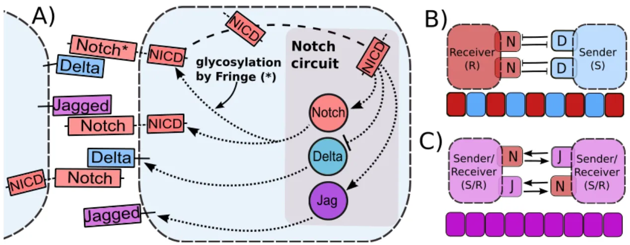
Although Delta and Jagged generate the same signal (NICD), the dynamics of Notch-Delta and Notch-Jagged signaling are quite different, because NICD regulates the production of the two ligands asymmetrically - it inhibits Delta but activates Jagged (Figure 1A). Therefore Notch and Delta form an intercellular double negative feedback loop, but Notch and Jagged form an intercellular double positive feedback loop. Consequently, the two cells interacting via Notch-Delta adopt different fates - one cell behaves as a Sender (S) with (high Delta (ligand), low Notch (receptor)) on its surface; and the other behaves as a Receiver (R) with (low Delta (ligand), high Notch (receptor)) on its surface. This cell-fate diversification mechanism is known as lateral inhibition and can lead to checkerboard-like or “salt-and-pepper” patterns (Figure 1B) as observed during bristle patterning in flies, inner ear patterning in vertebrates, and neurogenesis in both flies and vertebrates [10]. On the other hand, two cells interacting via Notch-Jagged can act as both Sender and Receiver as they have (high Jagged (ligand), high Notch (receptor)) on their surface. This cell-fate convergence mechanism is known as lateral induction (Figure 1C) and is crucial during cardiac development, inner ear development, and the formation of a smooth muscle wall around a nascent artery [11, 12, 13, 14].
vs. Notch-Jagged signaling call for elucidating their different operating principles. Notch Jag Delta Notch circuit NICD NI CD NICD NICD NICD Delta Delta Notch* Notch Notch glycosylation by Fringe (*)
Jagged Jagged D D J J
B)
A)
C)
N Sender/ Receiver (S/R) N Sender/ Receiver (S/R) N N Receiver (R) Sender (S)Figure 1: Overview of intra-cellular and inter-cellular Notch signaling pathway, and tissue patterning outcomes. A) Notch, the transmembrane receptor of one cell, binds to Delta or Jagged, the transmembrane ligands belonging to the neighboring cell. This trans-interaction leads to the cleavage and release of Notch Intracellular Domain (NICD) that migrates to the nucleus and modulates the gene expression of many Notch pathway targets. This modulation also indirectly leads to the transcriptional activation of Notch and Jagged and inhibition of Delta. Interaction between Notch receptor and ligands (Delta or Jagged) of the same cell (cis-interaction) leads to the degradation of both the receptor and the ligand. Glycosylation of Notch by Fringe modifies Notch to have higher affinity for binding to Delta and lower affinity for binding to Jagged. Due to this asymmetric regulation of Delta and Jagged by NICD. B) Notch-Delta signaling forms an intercellular toggle switch and alternate cells adopt distinct fates - Sender (low Notch (receptor), high Delta (ligand)) and Receiver (high Notch (receptor), low Delta (ligand)), giving rise to a checkerboard-like pattern (lateral inhibition), whereas C) Notch-Jagged signaling forms an intercellular double positive feedback loop and the cells adopt similar fates (lateral induction - (high Notch (receptor), high Jagged (ligand)).
on tumor progression - soluble Delta inhibits tumor growth [29, 30], while soluble Jagged strongly aggravates the malignant progression of cancer. Also, transmembrane Jagged1 can initiate metastasis by inducing cancer cells to undergo Epithelial to Mesenchymal Transition (EMT) - a dormant embryonic program that allows them to migrate and invade. It also facilitates the colonization of the Circulating Tumor Cells (CTCs) by enabling cell-cell com-munication between CTCs and the cells of the organ where they settle down and develop metastases [31, 9] that causes more than 90% of cancer-related deaths [32]. Furthermore, Notch-Jagged signaling can also maintain and/or expand the population of Cancer Stem Cells - highly plastic cancer cells that can initiate a new tumor [33, 27], thereby promoting tumor relapse. Notably, Notch-Jagged signaling also plays a crucial role in angiogenesis [34], cancer metastasis [9], and rapid development of cancer chemotherapy and radiation therapy resistance [35]. Not surprisingly, poor survival and cancer recurrence is associated with high Jagged1 levels in patients [31]. Hence, understanding Notch-Jagged signaling is essential to control tumor spread.
We devise a new theoretical framework that incorporates Jagged in addition to Delta and the asymmetric regulation of the proteins by the signal NICD that activates Notch and Jagged but represses Delta. We also included the second asymmetry between the ligands due to the effect of the glycosyltransferase Fringe and show that different outcomes are generated depending on which ligand is dominant. We elucidate the role of Jagged in cell fate determi-nation and discuss its possible implications in understanding tumor-stroma crosstalk, which frequently entails Notch-Jagged communication. We also show that inflammatory signal can expand the CSC population by augmenting Notch-Jagged signaling, presenting the first step towards understanding the molecular interplay of Notch-Jagged signaling, inflammation and CSC population.
Overview
differentiation. During angiogenesis, Jagged mediates the number of ‘stalk’ cells that may separate two ‘tip’ cells [36], and the three fates that can be obtained in the context of Notch-Delta-Jagged signaling may underlie the highly dynamic phenotypes that endothelial cells can attain that are neither entirely either tip nor stalk [37]. Both lateral inhibition and lateral induction are also critical for multistability of the pancreatic system, wherein the loss of either of them can lead to lineage switching between various cell types [38].
Chapter 1
Theoretical framework for Notch
signaling
Biological experiments are many times very difficult to interpret due to the high complexity of genetic regulatory networks which often involve counter-intuitive feedback mechanisms. Because of that, modeling approaches are fundamental to provide understanding and in-sights on how different elements operate. In silico approaches can offer complementary understanding of experimental studies as well as enable the formulation of new predictions to guide new experiments. In this chapter, we present a brief overview of previous models for Notch signaling pathway and introduce our theoretical framework.
1.1
Mathematical models for Notch-Delta signaling
1.1.1
The Collier model
dN
dt =N0H
+(D
ext)−γN (1.1)
dD
dt =D0H
−
(N)−γD (1.2)
where N0 and D0 represents the production rate of Notch and Delta, respectively and γ
represents the degradation of both Notch and Delta. The variableDextrepresents the amount
of Delta of the neighboring cells available for binding and the positive hill functionH+(D
ext)
represents the increase in the production of Notch with the amount of external Delta (Dext)
and the negative Hill function H−(N) represents the inhibition of the production of Delta
by Notch. Hill functions are defined as: H+(x) = xn
xn
0+x
n and H
−(x) = 1
xn
0+x
n.
This model brings interesting insights about patterning formation by Notch-Delta sig-naling circuit and many extensions of this model have been developed over the past years [12, 39, 40, 41]. One particularly interesting extension of Collier’s model was proposed by Petrovic et al [12]. Their model was the first framework to include both ligands Delta and Jagged, and their simulations was useful to understand some of their experimental results in the context of inner ear development [12].
1.1.2
Model of mutual inactivation of Notch and Delta
Experimental studies have suggested that the Notch receptor and the ligand Delta mutually inactivate each other when interacting in the same cell, in a mechanism called cis-inhibition [21, 42, 43, 44]. Sprinzak et al [21] measured experimentally the combined cis-trans input-output relationship for the Notch-Delta system. Their results revealed that cis and trans interactions have different responses: trans activation generate a graded response while cis-interaction is sharp and occurs at a fixed threshold. They also developed a simple mathe-matical model based on their experimental measurements that takes this feature in account. Within their framework [21], Notch receptor (N) belonging to one cell can interact with Delta (D) of the same cell - known as cis-interaction, or with those of the neighboring cell - Delta (Dext) - known as trans-interaction. The cis-interaction, also referred as cis
framework, the equation for the dynamics of Notch (N), Delta (D) and NICD (I) are given by:
dN
dt =N0−kCN D−kTN Dext−γN (1.3)
dD
dt =D0−kCDN −kTDNext−γD (1.4)
dI
dt =kTN Dext−γII. (1.5)
where γ represents the degradation rate of both Notch and Delta and γI the degradation of
NICD. kC and kT are the strengths of cis-inhibition and trans-activation respectively; and
N0 andD0 are the production rates of Notch and Delta respectively. NextandDextrepresent
the amount of protein available for binding.
Based on Sprinzak et al results, many theoretical studies have shown that cis-inhibition facilitate lateral inhibition patterning, enable faster dynamics and provide greater robustness to pattern formation [20, 45]. Recently, cis-inhibition has also been reported for interaction between Notch and Jagged [16], however its role in determining cell-fate dynamics for the combined Notch-Delta-Jagged circuit remains elusive.
1.2
A model for Notch-Delta-Jagged circuit
We generalized the theoretical framework devised by Sprinzak et al. [21] by incorporating Jagged in addition to Delta and including the feedback effects of NICD that indirectly activates Notch and Jagged, and represses Delta, thereby creating an asymmetry between Notch-Delta and Notch-Jagged interactions (Figure 1.1). The deterministic equations for the dynamics of Notch (N), Delta (D), Jagged (J) and NICD (I) are given by:
dN
dt =N0H
S+(I)
−kCN(D+J)−kTN(Dext+Jext)−γN (1.6)
dD
dt =D0H
S−(I)
−kCDN −kTDNext−γD (1.7)
dJ
dt =J0H
S+(I)
−kCJN −kTJNext−γJ (1.8)
dI
where γ represents the degradation rate of all three transmembrane proteins Notch, Jagged and Delta, and γI the degradation of NICD. kC and kT are the strengths of cis-inhibition
and trans-activation respectively; and N0, D0, and J0 are the production rates of Notch,
Delta and Jagged respectively. Next, Dext andJext represent the amount of protein available
for binding - which can be on the membrane surface of neighboring cells or in a soluble form. Experimental evidence suggest that membrane-bound ligands can generated a stronger signal when compared with soluble forms [46]. The distinction between these two forms of ligand - membrane-bound and soluble - is addressed below (see Eqs 1.16-1.19). We consider shifted Hill functions [47] to represent the effect of NICD (I) on the production rates of the proteins. Shifted Hill functions are defined as HS(I, λ) = H−(I) + λH+(I) or in simpler
notation: HS+(I) if λ > 1 and HS−(I) if λ < 1, where the weight factor λ, represents the
fold-change in production rate, therefore, for activation, λ > 1; for repression, λ < 1; and for no effect, λ = 1 (λN, λJ >1 and λD <1 in our model). For the case of two interacting
cells, the variablesNext,Dextand Jext should be replaced byN,D,J of the neighboring cell.
D
N
I
D
D
D
D
extI
N
I
N I
J
trans activation
N I
J
J
sender cellreceiver cell
cis inhibition
J
extJ
extD
extJ
extFigure 1.1: Schematic illustration of Notch signaling circuit. NICD (I) is released when the receptor (N) of the receiver cell interacts with the ligand of the sender cell (Dor J) or with external ligands in a soluble form (DextorJext) - so calledtrans-activation. The released signal activates the expression ofN andJ, and inhibit the expression ofD. Thecis-inhibition occurs between the receptor and ligand in the same cell and
leads to the degradation of both proteins.
The Notch signal (NICD) degrades rapidly as compared to N, D and J [48]. Therefore, we can assume a quasi-steady approximation for it. In this case, the equations 1.6-1.9 can be reduced to two equations by definingL=D+J and Lext=Dext+Jext, where Lrepresents
ligands available to bind. Then, the reduced system is:
dN
dt =N0H
S+(I
eq)−kCN L−kTN Lext−γN (1.10)
dL
dt =D0H
S−(I
eq) +J0HS+(Ieq)−kCLN−kTLNext−γL (1.11)
where, by quasi steady-state approximation, Ieq =kTN Lext/γI.
1.3
A model for Notch-Delta-Jagged-Fringe circuit
Glycosylation of Notch by Fringe creates additional asymmetry between Delta and Jagged by modulating the binding affinity of the two ligands to Notch - the glycosylated Notch has a higher affinity to bind to Delta, but lower affinity to bind to Jagged [18, 19]. To incorporate this mechanism within our framework, we considered two distinct sub-populations of Notch - the one modified by Fringe, and the other unmodified. Since NICD, which is represented in the model by (I), activates Fringe [15], we have taken the fraction of glycosylated Notch (denoting the effect of Fringe on Notch) to increase with (I). This glycosylated Notch has different strengths of cis-inhibition and trans-activation for Delta and for Jagged [16]. Thus, while representing effective Notch (sum of glycosylated and unglycosylated Notch), we consider the strengths of cis-inhibition and trans-activation of Notch for Delta and for Jagged to depend on (I) (see derivation of the model in the Appendix A). The resulting model for one cell is given by:
dN
dt =N0H
S+(I)
−N(kCDD+kTDDext+kCJJ+kTJJext)−γN (1.12)
dD
dt =D0H
S−
(I)−kCDN D−kTDDNext−γD (1.13)
dJ
dt =J0H
S+(I)
−kCJN J−kTJJNext−γJ (1.14)
dI
dt =N[kTDDext+kTJJext]−γII (1.15) where kC(I) and kT(I) are now functions of the signal NICD given by: k(I) = k[1 +
aH+(I)] = kHS(I, λF) where λ
F = 1 +a. The shifted Hill function HS(I, λF) represents
parameter λF represents the increase (λF > 1), decrease (λF < 1) of both trans-activation
andcis-inhibition rate due to glycosylation. Experimental evidence suggest thatλF
D >1 and
λF
J <1 [18, 19], representing the increase of the binding affinity between Notch and Delta,
and the decrease of that between Notch and Jagged.
Our models shows a good robustness with respect to changes in parameter values as discussed in the appendix B. All the codes were developed in python using the PyDSTool [49].
1.4
Considering the effect of both soluble and
membrane-bound ligands
Experimental evidence suggests that membrane-bound ligands should activate the signal strongly compared to soluble ligands. This happens because soluble ligand does not have enough mechanical pulling force to activating the signal [50]. However, alternative mech-anisms such as ligand multimerization can lead to sufficient mechanical force for ligand activation [51]. Another evidence that membrane-bound should activate the signal strongly is that lower lateral mobility of the ligand leads to higher signaling [46]. Therefore, a model that considers both soluble and membrane-bound ligands should have two terms in Notch equation for interaction with both forms of these ligands. For example, the model presented in Eqs. 1.6-1.9 would be:
dN
dt =N0H
S+(I)
−kCN(D+J)−kTmN(Dextm +Jextm)−kTsN(Dexts +Jexts )−γN (1.16)
dD
dt =D0H
S−(I)
−kCDN −kmTDNext−γD (1.17)
dJ
dt =J0H
S+(I)
−kCJN −kTmJNext−γJ (1.18)
dI
dt =N[k
m
T(Dmext+Jextm ) +kTs(Dsext+Jexts )]−γII (1.19)
where Dm
ext and Jextm represents membrane-bound Delta and Jagged respectively, and Dsext
and Js
ext represents soluble Delta and Jagged respectively. kTm and kTs represents the
trans-activation rate for membrane-bound ligands and for soluble ligands respectively, and its expected that km
1.5
Parameters’ values
Our model is based on the model proposed by Sprinzak et al [21] which well-fitted their experimental results designed to measure the interaction between Notch and Delta, both in cis and trans. Their experimental results suggest a Hill coefficient for Notch activity close to 2 and this value was chosen to represent the NICD activation of Notch (nN) and inhibition
of Delta (nD). For the case of NICD modulation of Jagged, a higher Hill coefficient (nJ = 5)
was chosen in order to represent both direct activation of Jagged by NICD and indirect modulations by miRNA - Jagged is strongly repressed by miR200 [52], however, miR200 is repressed by SNAIL [53] which in turn is activated by NICD [11], resulting in a effective strong activation of Jagged by the signal. A strong activation of Jagged is required for the maintenance of the (S/R). FornJ = 4 the range of existence of the S/R state is significantly
decreased and for nJ = 3 this state is no longer observed. Previous work [21], shows that
cis-inhibition rate (kC) is approximately ten times higher than trans-activation rate (kT)
for Notch-Delta interaction and we assumed the same values for Notch-Jagged interaction when the effect of Fringe is not taken into account. Unlike the previous model [21] in which the rates are in units of Relative Fluorescence Units, our variables represent the number of proteins in the membrane for Notch, Delta and Jagged, and the number of proteins inside the nucleus for the signal (NICD). Because of that, we scaled the values of cis-inhibition and trans-activation rate accordingly.
The rapid degradation of the signal is an important feature in the Notch signaling [48] and because of that, we considered the NICD degradation rate (γI) to be 5 times higher than the
typical protein degradation rate γ = 0.1h−1. The values of N
0, D0 and J0 which represents
the production rate of the proteins were chosen in order to keep the maximum number of proteins in the membrane up to approximately 5000 per cell. This value is consistent with experimental results where the concentration of the proteins varies up to a few hundreds
ng/ml [54] - or a few thousands of proteins per cell. Once most proteins are exported to
the membrane, we should expect a few thousands of proteins in the membrane when the expression is up-regulated. Similarly, the number of NICD inside the nucleus varies up to a few hundreds and because of that, we select the threshold of the Hill function (I0) to be 200.
assumed that glycosylation of Notch increases its affinity to Delta in a factor of 3 (λF
D = 3.0)
and decrease its affinity to Jagged in 70% (λF
J = 0.3). Experimental evidences for
modula-tion of Notch by lunatic Fringe shows a 4.4 fold reducmodula-tion for Jagged1-mediated signaling and at least two fold increase for Delta1-mediated signaling [18]. Finally,λN =λJ = 2.0 for
Notch and Jagged represents an activation of their production by the signal and λD = 0.0
for Delta represents its inhibition by the signal.
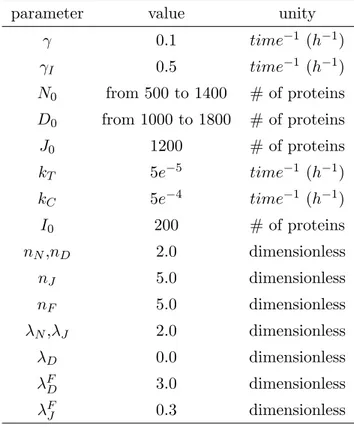
Table 1.1: Parameters values used in the simulations, unless indicated otherwise.
parameter value unity
γ 0.1 time−1 (h−1)
γI 0.5 time−1 (h−1)
N0 from 500 to 1400 # of proteins
D0 from 1000 to 1800 # of proteins
J0 1200 # of proteins
kT 5e−5 time−1 (h−1)
kC 5e−4 time−1 (h−1)
I0 200 # of proteins
nN,nD 2.0 dimensionless
nJ 5.0 dimensionless
nF 5.0 dimensionless
λN,λJ 2.0 dimensionless
λD 0.0 dimensionless
λFD 3.0 dimensionless
λFJ 0.3 dimensionless
1.6
Conclusion
Chapter 2
Notch signaling: two ligands, same
signal and different behaviours.
for example, in mammalian inner ear development [14, 12], control of epidermal stem cell clusters [57], as well as inner cardiac development [11]. Therefore, Delta and Jagged affect the collective cell fate decisions in a group of cells quite differently.
The second asymmetry between signaling through the ligands Delta and Jagged arises due to post-translational modifications of Notch that modulate the binding of Notch to Delta and to Jagged. Fringe, a glycosyltransferase, can decrease the affinity of Notch to bind to Jagged, but increase the affinity of Notch to bind to Delta [58]. Consequently, Fringe creates two distinct populations of Notch on the cell surface: one that has comparable binding affinity to both Jagged and Delta, and one that strongly prefers binding to Delta.
In this chapter, we investigate the effects of these two elements of asymmetry in Notch signaling. We first evaluate the dynamics of the circuit when only one ligand is available. In this case, the binding affinity of Notch to both ligands are the same and the only difference between Delta and Jagged is the modulation of their production by the signal (NICD), which repress Delta but activates Jagged. We also evaluate the dynamics of the combined circuit, when both ligands are available for binding. Next, we introduce the effect of Fringe, which modifies the binding affinity between Notch and the ligands. In a further analysis, we evaluate the effect of external signals which can modify the production levels of the ligands. Lastly, we evaluate the effect of soluble ligands in the dynamics of interacting cells.
2.1
Cell fate decisions in Notch-Delta, Notch-Jagged
and Notch-Delta-Jagged signaling
In order to understand the operating principles of Notch signaling, we first start evaluating the dynamics of circuit when only one ligand is available and later we evalate the combined circuit.
2.1.1
Notch-Delta (ND) circuit: a two-cell toggle switch
To evaluate the dynamics of Notch-Delta stand alone circuit, we analyzed the reduced model given by equations 1.10-1.11 whenJ0 = 0, i.e., no Jagged is produced. This simplified circuit
(high Delta (ligand), low Notch (receptor)), and (ii) Receiver (R) - in which the cell has (low Delta (ligand), high Notch (receptor)) (Figure 2.1). Therefore, Notch-Delta signaling forms an intercellular mutually inhibitory switch, which drives adjacent cells to adopt alternate fates - one cell acts as a Sender and the other as a Receiver, or vice-versa, as noted in previous studies [21, 22, 20, 45]. A canonical example of this phenomenon, also known as lateral inhibition, is the AC/VU differentiation in C. elegans, where initially the two cells are identical (i.e. both neighbors have the same levels of ligand and receptor), but then due to a random fluctuation, one cell has increased levels of ligand and/or the other has increased level of receptor; and this stochastic difference is amplified by the mutually inhibitory feedback loop - driving the cell fate diversification [59]. However, lateral inhibition need not be static, it can be dynamic too - i.e. cells can compete against their neighbors continuously, as seen during sprouting angiogenesis, where the migrating ‘stalk’ cells are continuously competing to become ‘tip’ cells [60] . Also, during neural development, lateral inhibition occurs between the dynamic clusters of neuronal precursors, in order to strike a balance between the number of cells differentiating to become neurons, and those staying as precursors for further rounds of neurogenesis [61].
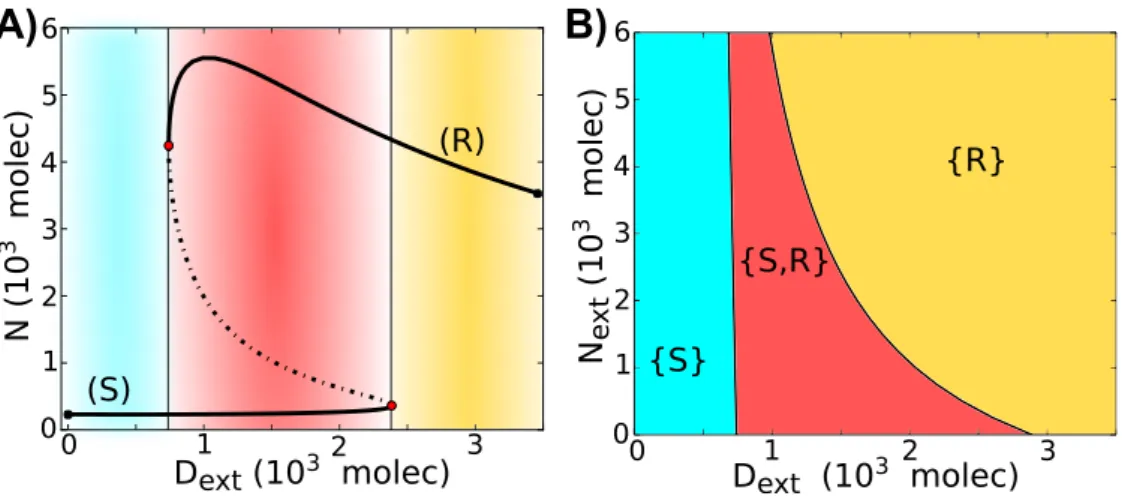
In order to better understand the dynamics of the circuit, we evaluate a bifurcation diagram when the external Delta (Dext) acts as a control parameter - the range of
exis-tence of the different Notch/Delta states of a single cell as function of (Dext). We see that
for small Dext, the cell behaves as a Sender (S); and for large Dext, the cell behaves as a
Receiver (R). We further see the existence of bistability for intermediate levels of Dext,
-the cell can ei-ther be a Sender (S) or a Receiver (R) (Figure 2.2A). Next, we present -the phase diagram (two-parameter bifurcation diagram) for a single cell driven by two control parameters, the external Notch (Next) and the external Delta (Dext) shown in Figure 2.2B.
0 2 4 6 8 0
2 4 6 8
0 2 4 6 8
D
(10
3
mo
lec
)
N (103 molec)
N (103 molec)
E
ff
ec
ti
v
e
P
o
ten
ti
a
l
Figure 2.1: Dynamical properties of Notch-Delta signaling circuit. N-D stand-alone circuit (J0 = 0,
D0= 1000 molecules/hour,N0= 500 molecules/hour,Next= 500 molecules,Dext=1100 molecules) presents two stable states: Sender (S) (high Delta, low Notch) and Receiver (R) (low Delta, high Notch). Green nullcline representsdN/dt= 0, and blue nullcline representsdD/dt= 0. Stable steady states are represented by red circles and unfilled circles represent unstable steady states. The background contours illustrate the values of the effective potential (−log(P)) where P is the probability density calculated by solving the differential equations stochastically using the Euler-Maruyama method. Most probable regions are at the red regions and least probable at the blue regions. Insets represent the one-dimensional effective potential computed along thedD/dt= 0 nullcline.
0 1 2 3 0 1 2 3 4 5 6 (R) (S)
A)
B)
0 1 2 3
0 1 2 3 4 5 6 {R} {S,R} {S}
Dext (103 molec)
N (10 3 mo lec )
Dext (103 molec) Ne x t (10 3 mo lec )
Figure 2.2: Dynamical system characteristics of the Notch-Delta circuit. A) Bifurcation, for the one-cell case, of Notch protein levels on the membrane as a function of the number of external Delta (Dext) for fixedNext= 500 molecules. Starting in the Sender (S) state, i.e.- (low Notch, high Delta) (blue region) and increasing the external Delta (Dext) at some threshold the cell undergoes a transition to the Receiver state, i.e. (high Notch, low Delta) (yellow region). The reverse transition occurs at a different number of Dext proteins that leads to a region of coexistence of both states - Sender and Receiver (red region). Solid curves represent stable steady states, while dotted curves represent unstable steady states. B) Phenotype diagram as a function of external Notch (Next) and external Delta (Dext) for one cell model. The monostable phase
{S}corresponds to the Sender state (low Notch, high Delta) and monostable phase{R} corresponds to the Receiver state (high Notch, low Delta). The bistable phase{S,R}corresponds to a region of co-existence of both states - Sender and Receiver.
2.1.2
Notch-Jagged (NJ) circuit
We next consider the dynamics of Notch-Jagged signaling by analyzing the reduced model given by equations 1.10-1.11 when D0 = 0. Notch-Jagged circuit is monostable with the
0 2 4 6 8 0
1 2 3 4
0 2 4 6 8
N (103 molec)
J
(10
3
mo
lec
)
N (103 molec)
E
ff
ec
ti
v
e
P
o
ten
ti
a
l
Figure 2.3: Dynamical properties of Notch-Delta signaling circuit. N-D stand-alone circuit (J0 = 400
molecules/hour, D0 = 0, N0 = 500 molecules/hour,Next = 500 molecules, Lext = 250 molecules) presents two stable states: Sender (S) (high Delta, low Notch) and Receiver (R) (low Delta, high Notch). Green nullcline represents dN/dt = 0, and blue nullcline representsdJ/dt= 0. Stable steady states are represented by red circles and unfilled circles represent unstable steady states. The background contours illustrate the values of the effective potential (−log(P)) where P is the probability density calculated by solving the differential equations stochastically using the Euler-Maruyama method. Most probable regions are at the red regions and least probable at the blue regions. Insets represent the one-dimensional effective potential computed along thedJ/dt= 0 nullcline.
2.1.3
Notch-Delta-Jagged (NDJ) circuit: a three-way switch.
We next study the dynamics of the combined Notch-Delta-Jagged signaling circuit, i.e., Notch signaling when driven by both its ligands - Delta and Jagged. This system can behave as a three-way switch, allowing for three states: - (i) Sender (S - high ligand, low receptor), (ii) Receiver (R - low ligand, high receptor) and (iii) hybrid Sender/Receiver (S/R - medium ligand, medium receptor) (Figure 2.4).
We further present the phase diagram (two-parameter bifurcation diagram) for a single cell driven by two control parameters, the external Notch (Next) and the external Jagged
(Jext). Note that, once Notch has the same binding affinity to both Delta and Jagged, the
model is symmetric by changingJextforDext, therefore, the following results also holds when
0 2 4 6 8 N (103 molec)
E
ff
ec
ti
v
e
P
o
ten
ti
a
l
2 4 6 8
0
D+
J
(10
3
mo
lec
)
0 2 4 6 8
N (103 molec)
Figure 2.4: Dynamical properties of Notch-Delta signaling circuit. N-D stand-alone circuit (J0 = 1000
molecules/hour,D0= 1400 molecules/hour,N0= 1500 molecules/hour,Next= 400 molecules,Lext= 1100 molecules) presents two stable states: Sender (S) (high Delta, low Notch) and Receiver (R) (low Delta, high Notch). Green nullcline representsdN/dt= 0, and blue nullcline representsdL/dt= 0, whereL=D+J. Stable steady states are represented by red circles and unfilled circles represent unstable steady states. The background contours illustrate the values of the effective potential (−log(P)) where P is the probability density calculated by solving the differential equations stochastically using the Euler-Maruyama method. Most probable regions are at the red regions and least probable at the blue regions. Insets represent the one-dimensional effective potential computed along thedL/dt= 0 nullcline.
{S/R,R}, and also a tristable phase showing the coexistence of all three possible states {S,S/R,R} (Figure 2.5A). At larger Next values, we see the hybrid S/R state can exist, for
some range of Jext, by itself, i.e. in the monostable{S/R}phase (Figure 2.5B). However, at
smaller Next values, the hybrid state always co-exists with other states in a bistable phase {S, S/R} and {S/R, R}, or in a tristable phase{S, S/R, R} (Figure 2.5C).
1 1.5 2 2.5 0 1 2 3 4
1.4 1.6 1.8 2
0 2
40.8 1 1.2 1.4
0 2 4 {S} {R} {S/R} {S/R,R} {S,R} {S ,S/R ,R} A) B) C) {S,S/R} (S) (S) (S/R) (S/R) (R) (R)
Jext (103 molec)
N
(10
3 mo
lec
)
N
(10
3 mo
lec
)
Jext (103 molec)
Ne
x
t
(10
3 mo
lec
)
Figure 2.5: Dynamical system characteristics of the Notch-Delta-Jagged circuit. A) Phenotype-diagram when the one-cell Notch-Delta-Jagged circuit is driven by both the external Notch (Next) and external Jagged (Jext), for (Dext = 0). Each phase, denoted by a different color, corresponds to a different combination of coexisting phases. Same phenotype-diagram is obtained when driven by Next andDext, forJext= 0, once Notch is considered to have the same binding affynity to Jext and Dext. B) Bifurcation of Notch protein levels on the membrane when driven by external Jagged for fixed levels ofNext= 3000 proteins. This curve shows the existence of the monostable {S/R} phase (pink region) for a large range of external ligands. C) Same as B) forNext= 1000 proteins. In this case, the hybrid S/R state co-exists with other states, i.e. seen only in bistable (blue and green regions) and tristable phases (grey region).
0 1 2 3 4 0
1 2 3 4
(S/R;S/R) (S;R)
(R;S)
N
2(10
3
mo
lec
)
N
1(103 molec)
Figure 2.6: Nullclines for the case of two cells interacting with each other through Notch-Delta-Jagged. The blue nullcline is for condition of all ODEs being set to zero except fordN1/dtand the green nullcline is
2.2
The effect of Fringe mediated asymmetric
Notch-ligand binding
Glycosylation of Notch by Fringe creates additional asymmetry between Delta and Jagged by modulating the binding affinity of the two ligands to Notch - the glycosylated Notch has a higher affinity to bind to Delta, but lower affinity to bind to Jagged [18, 19]. To incorporate this mechanism within our framework, we considered two distinct sub-populations of Notch - the one modified by Fringe, and the other unmodified. Since NICD, which is represented in the model by (I), activates Fringe [15], we have taken the fraction of glycosylated Notch (denoting the effect of Fringe on Notch) to increase with (I). This glycosylated Notch has different strengths of cis-inhibition and trans-activation for Delta and for Jagged [16]. Thus, while representing effective Notch (sum of glycosylated and unglycosylated Notch), we consider the strengths of cis-inhibition and trans-activation of Notch for Delta and for Jagged to depend on (I) (see Eq. 1.12-1.15).
Within our framework, different modulations of Notch by Fringe can be represented by different values of the parameters λF
D and λFJ, which represents either the increase (λF >1)
or decrease (λF < 1) of the cis-inhibition and trans-activation rates. When λF
D = λFJ = 1,
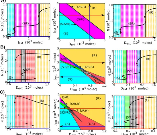
the model is the same as considered earlier without any effect of Fringe (Eqs. 1.6-1.9). In this case, the Notch-ligand binding has equal affinity for external Jagged and external Delta, as reflected in the symmetry of the phenotype diagram (two-parameter phase diagram) for external Jagged and external Delta (Figure 2.7A (center)). The bifurcation diagram for a cell driven by external Delta (Dext) and by external Jagged (Jext) presents the same
behavior - a large range of the intermediate state (S/R) in a monostable phase (Figure 2.7A). However, when the effect of Fringe is incorporated (e.g. λF
D = 3 and λFJ = 0.3), the circuit
behaves differently. The range of the existence of the 4 phases containing the hybrid S/R state, (the phases {S/R}, {S/R, R}, {S, S/R} and {S, S/R, R}), increase with the level of external Jagged (Figure 2.7B (center)). When Notch signaling is mainly mediated by Jagged (highJext), both the forward and backward transitions between (S) and (R) states require a
transitions into and from the hybrid S/R state as an intermediary step (Figure 2.7B (right)). Conversely, when the Notch signaling is mainly mediated by Delta (high Dext), the forward
0 1 2 3 0 1 2 3 {S/R} {S/R,R} {S,S/R} {R} {S} A)
0 1 2 0.2
1.0 1.8
0 1 2
0 0.4 0.8 1.2 0 1 2 3 4 5 {S/R,R} {R} {S} {S,S/R} {S ,S/R ,R} {S/R} {S ,R}
0 0.4 0.8 1.2 0 1 2 3 4 5 {S} {S,R} {S,S/R} {S,S/R,R} {S/R,R} {S/R} 0.6 1 1.4
0.1 0.2 0.3 0.4 0
1 2 3
0.2 1 1.8
0 0.2 0.4 0.6 0 1 2 3 (S) (S) (S) (S) (S) (R) (R) (R) (R) (R) {R} (R) (S/R) (S/R) B) C) 0.2 1.0 1.8 0 1 2 3 0 1 2 3 N (10
3 mo
lec
)
N
(10
3 mo
lec
)
N
(10
3 mo
lec
)
N
(10
3 mo
lec
)
N
(10
3 mo
lec
)
N
(10
3 mo
lec
)
Jext (103 molec) Dext (103 molec) Dext (103 molec)
Dext (103 molec)
Dext (103 molec) Dext (103 molec)
Dext (103 molec)
Dext (103 molec) Dext (103 molec)
Jex
t
(10
3 mo
lec
)
Jex
t (10 3 mo lec )
Jex
t (10 3 mo lec ) (S) (S/R) (S/R) (S/R)
Figure 2.7: Phenotype-diagram and bifurcation curves for the one-cell Notch-Delta-Jagged-Fringe circuit. The phenotype-diagram shows the different possible phases when the circuit is driven by variable levels of both external Jagged and external Delta. A) Phenotype-diagram (center) for λF
D = λ
F
J = 1 (no Fringe effect). In this case, the circuit response to external Jagged and external Delta is symmetric. Bifurcation curve of Notch protein levels with respect to varying external Jagged values (left) for fixed Dext = 2000 and Next = 500 molecules and (right) bifurcation curve with respect to varying external Delta values for fixed Jext = 2000 and Next = 500 molecules. B) Phenotype-diagram (center) for λ
F
D = 3 and λ F
J = 0.3
(intermediate effect of Fringe). Bifurcation curves of Notch protein levels in response to varying Dext for Jext = 1000 and Next = 500 molecules (left), i.e. Notch signaling mainly mediated through Delta and for fixedJext= 3000 andNext = 500 molecules (right), i.e. Notch signaling mainly mediated through Jagged. C) Phenotype-diagram (center) forλF
D= 5.0 andλ F
(Figure 2.7B (left)). When the effect of Fringe is considered to be too strong (λF
D = 5 and
λF
J = 0.2), the circuit is mostly bistable and therefore, the response of the circuit becomes
similar to the case of stand alone Notch-Delta signaling (Figure 2.7C).
The results above suggest that signaling through Jagged has an important role in main-taining the hybrid Sender/Receiver (S/R) state, and that Jagged makes it much more likely that transition from Sender (S) to Receiver (R) and vice-versa happen through the hybrid (S/R) state.
2.2.1
The effect of Delta/Jagged asymmetry on the cell-cell fate
modulation
Notch signaling in mammals is mediated through four types of Notch (Notch 1-4) and three types of Fringe (Lunatic, Manic and Radical Fringe) [67]. Experimental evidence suggests that most Fringe proteins act with different types of Notch, possibly leading to different forms of glycosylated Notch; thereby expanding the repertoire of responses that the Notch signaling system can mediate [18, 19]. Most experimental evidence suggests that Fringe increases the signaling mediated by Delta and decreases the signaling mediated by Jagged, resulting in λF
D >1 and λFJ <1 [18, 19]. The phenotype diagram when the circuit is driven
by different values ofλF
D andλFJ presents the response of the circuit for different combinations
of Fringe modulations (Figure 2.8).
Because Fringe is activated by NICD [15], its effect is dominant in cells with high number of Notch molecules (Receiver (R) state) that cleave to form NICD. Therefore, to analyze the effect of Fringe on Notch-Delta-Jagged signaling, we choose the external signal to the cell be composed mainly by ligands (Jext, Dext) and low values of Next; i.e. the external signal
can be considered equivalent to a Sender (S) cell. Two such different combinations are chosen - (high Dext, low Jext) and (low Dext, high Jext) (Figure 2.8). In case of (high Dext,
low Jext) and at λFD > 2 and λFJ < 1, i.e. when the external signal is mostly Delta and
Fringe increases the affinity of Notch for Delta, and decreases that for Jagged, the cell is mostly in monostable phase of the Receiver (R) state, or, in other words, the cell attains the opposite fate as that of a cell representing the external signals (Figure 2.8A). However, when the external signal is mainly Jagged, i.e. in (low Dext, high Jext), at smaller values of
1 2 3 4 5 6 {R} {S} {S,S/ R,R} {S,R} {S, S/R} {S/R,R} {S} {S,S/ R,R} {S,R} {S,S /R} 0.0
A)
B)
Fringe e
ff
ect (
λ
FJ)
F ri n g e e ff ec t ( λ F D )
0.5 1.0 1.5 2.0 0.0 0.5 1.0 1.5 2.0
Figure 2.8: Phenotype-diagram of the Notch-Delta-Jagged-Fringe model when the circuit is driven by different values of the Fringe modulation for Notch-Delta interactionλF
Dand Notch-Jagged interactionλ F
J. In
all curves the external signal represents cells in the Sender (S) state - low concentration of Notch (Next= 500) and high concentration of ligands. Each figure represents a different combinations of the number of external ligands (in number of proteins available to binding). A) Dext = 1500 and Jext = 500 molecules. B) Dext= 500 andJext= 1800 molecules.
mostly in the monostable phase of the Sender (S) state (Figure 2.8B). Therefore, the cell attains a fate similar to the cell represented by the external signal. These results suggest that while signaling through the Notch-Delta circuit, the two cells attain opposite fates, however, when signaling through the Notch-Jagged circuit, the two cells attain similar fates; thereby suggesting that Jagged helps neighboring cells to maintain similar fates.
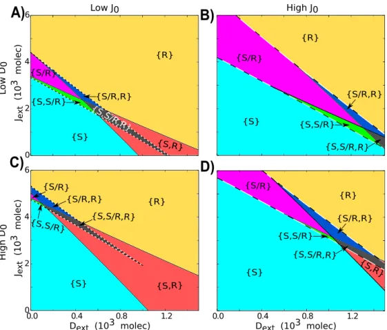
We also investigate the Fringe effect when it is driven exclusively by different levels of
Dext and Jext. We plot a bifurcation diagram for the Fringe effect (f = λFD = 1/λFJ) and
the amount of external ligand. When the cell is driven mainly by external Delta (Dext), in
presence of little or no external Jagged (Jext), increase in Fringe effect decreases the range
of existence of the phases that contain the hybrid S/R state - {S/R}, {S, S/R}, {S/R, R} and {S, S/R, R} (Figure 2.9A, C). Similar effect of Fringe levels is observed when the cell is driven by both external Delta and external Jagged (Figure 2.9D). However, when the cell is driven solely by Jagged (Dext = 0), an increase in Fringe effect does not significantly decrease
Fringe (Figure 2.9B).
1.0 2.0 3.0 4.0 5.0
0 1 2 3 4 5
Jex
t (10 3 mo lec ) {S/R} {S /R} {S,S/R} {S,S/R} {S} {S} {S,S/R,R} {R} {R} {S,R} {S/R,R} A) B)
1.0 2.0 3.0 4.0 5.0
0.0 1.0 2.0 3.0 De x t (10 3 mo lec )
Fringe Effect (λF,D = 1/λF,J = f)
1.0 2.0 3.0 4.0 5.0
0.0 1.0 2.0 3.0
Fringe Effect (λF,D = 1/λF,J = f)
Jex
t (10 3 mo lec ) D) C) {S/R} {S/R} {S,R} {S,R} {S,S/R,R} {S ,S /R ,R} {S/R,R} {S,S/R} {S,S/R} {S} {S} {R} {R}
Fringe Effect (λF,D = 1/λF,J = f)
1.0 2.0 3.0 4.0 5.0
0 1 2 3 4 5 De x t (10 3 mo lec )
Fringe Effect (λF,D = 1/λF,J = f)
Figure 2.9: Phenotype diagram when the circuit for one-cell is driven by both the effect of Fringe and external ligands. Each phase is represented by a different color and corresponds to a different combination of coexisting states. Each diagram represents different cases for external molecules available to the cell. A) External signal is mediated by Delta (Jext = 0, Next = 500 molecules). B) External signal is mediated by Jagged (Dext = 0,Next = 500 molecules). C) same as A) but for (Jext = 1000 molecules, Next = 500 molecules). D) same as B) but for (Dext= 1000 molecules,Next= 500 molecules).
2.2.2
Fringe promotes lateral inhibition patterning
As mentioned earlier, Notch-Delta signaling leads to lateral inhibition, where neighboring cells adopt alternate fates; but Notch-Jagged signaling leads to lateral induction, where adjacent cells attain similar fates [10, 12]. As Fringe can promote Notch-Delta signaling, we hereby explore its role on tissue patterning.
tissue pattern of similar fates is disrupted and the ‘salt-and-pepper’ pattern starts to emerge (Figure 2.10). Our results are consistent with experiments suggesting that Fringe promotes lateral inhibition during neurogenesis [68].
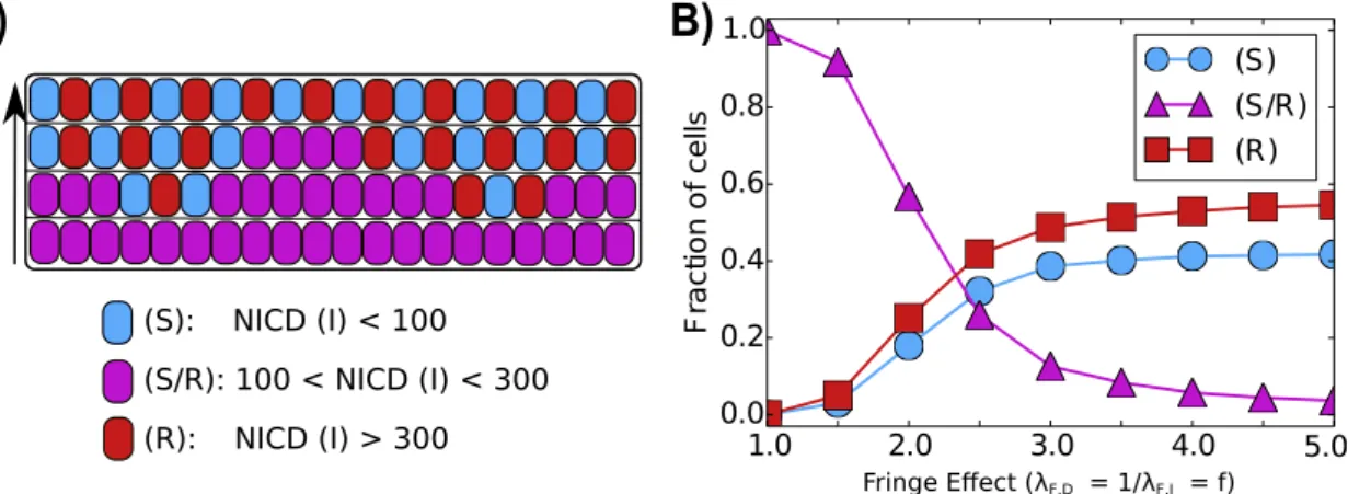
1.0 2.0 3.0 4.0 5.0
0.0 0.2 0.4 0.6 0.8 1.0 F ra c ti o n o f c e lls (S) (S/R) (R)
B)
Fringe Effect (λF,D = 1/λF,J = f)
F ri n ge E ff ec t
A)
(S): NICD (I) < 100
(S/R): 100 < NICD (I) < 300
(R): NICD (I) > 300
Figure 2.10: Fringe effect on tissue-level patterning. A) Representation of a one-dimensional layer of cells interacting through Notch signaling for different levels of Fringe. B) The average of the fraction of cells in (S), (S/R) or (R) state as a function of the effect of Fringe. Increase in Fringe effect promotes lateral inhibition - formation of ‘salt and pepper’ alternate patterns of cell fate in adjacent cells. The averages were taken over 100 simulations of a one-dimensional layer of 100 interacting cells with periodic boundary condition.
2.2.3
Fringe alters the relative stability of the steady states of
Notch-Delta-Jagged signaling
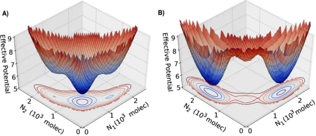
In order to evaluate the effect of noise in the N-D-J circuit, we represent the phase space by an effective potential for different values of Fringe effect. We considered two interacting cells and the phase space is presented in terms of the levels of Notch of each cells. The effective potential of the system is defined as−log(P), whereP =P(N1, N2) is the probability density
at the 2 dimensional phase space (N1xN2). Three states can be observed: first cell as a Sender
Nevertheless, when the effect of Fringe is considered, the basin of attraction of the (S/R,S/R) becomes shallower and small fluctuations can move the cells to the alternate state (one cell has high Notch and the other low Notch) which now have a deep basin of attraction (Figure 2.11B).
Figure 2.11: Three-dimensional representation of the effective potential as a function of Notch in cell 1 (N1) and in cell 2 (N2). The effective potential is defined as−log(P), whereP =P(N1, N2) is the probability
density calculated by solving the differential equations stochastically using the Euler-Maruyama method. A) represents the case of no effect of Fringe (f = 1.0). B) represents the case considering the effect of Fringe (f = 1.5).
These results indicate that Fringe promotes Notch-Delta signaling at the expense of Notch-Jagged signaling. Because Notch-Jagged signaling is more frequently implicated than Notch-Delta signaling in many aspects of tumor progression [15], our results showing the role of Fringe to inhibit Notch-Jagged signaling corroborate with Fringe being reported as a tumor suppressor in multiple cancers such as prostate, lung and basal-like breast cancer (BLBC) [69, 70, 71]. Further, loss of Fringe facilitates Notch-Jagged signaling that is critical in the tumorigenesis of BLBCs [69].
2.3
Production rates of the two ligands affect cell-fate
decision antagonistically
The two ligands - Delta and Jagged - can be produced at different rates in the same cell due to stochastic events and/or the influence of external factors such as the inflammatory cytokine TNF-αthat can promote the production of Jagged and inhibit that of Delta [36]. Therefore, we next explore the effect of varying the production rates of the ligands on cell-fate decisions mediated by Notch-Delta-Jagged (NDJ) signaling, for both one-cell and two-cell system. For the one-cell system, our results are represented as phenotype diagrams when the circuit is driven by two control parameters - external Delta (Dext) and external Jagged (Jext) (Figure
2.12). These diagrams consist of multiple phases (sets of co-existing phenotypes for the same parameter set): three monostable phases - {S}, {S/R}and {R}, three bistable phases - {S, R}, {S, S/R}and {S/R, R}, and a tristable phase -{S, S/R, R}.
Different phenotype diagrams denote different combinations of the production rates of Jagged and Delta. At low production rates of Jagged (J0), increase in production rates of
Delta (D0) decreases the parameter range for the existence of phases containing the hybrid
S/R phenotype (compare the area bounded by dashed lines in Figure 2.12C vs. that in Figure 2.12A). Similar effects of high production rates of Delta (D0) are observed at high production
rates of Jagged (J0). Therefore, high production and hence the levels of Delta inhibit
Notch-Jagged signaling and promote lateral inhibition or cell fate diversification. Conversely, high production rates of Jagged can significantly increase the parameter range for the existence of phases containing the hybrid S/R phenotype (compare the area bounded by dashed lines in Figure 2.12D vs. that in Figure 2.12B). Hence, the two ligands act antagonistically with respect to their effect on cell-fate decisions in a cell population.
Next, we investigate the effect of different production rates of Delta and Jagged (D0,J0)
in a two-cell system. Both these cells are identical and communicate with each other via the NDJ system. At low production rates of both ligands - Jagged and Delta - both cells act as Receiver (R1,R2 -low ligand, high receptor) because Notch (receptor) is still being produced
at a fixed rate. However, when Delta is produced faster than Jagged, one cell becomes the Sender (S) and the other Receiver (R) (S1, R2). Conversely, when Jagged is being produced
0 2 4 6 {R} {R} {S} {S} {S/R} {S/R} {S,S/R} {S,S/R} {S/R,R} {S/R,R} {S,S/R,R} {S ,S/R ,R} Jex
t (10 3 mo lec )
0.0 0.4 0.8 1.2
Dext (103 molec)
0.0 0.4 0.8 1.2
Dext (103 molec)
0 2 4 6
Jex
t (10 3 mo lec ) {S,S/R,R} {S,S/R,R} {S,S/R} {S/R,R} {S/R} {S,R} {S,R} {S ,R} {S/R} {R} {R} {S} {S} {S/R,R} {S,S/R}
A)
B)
C)
D)
High J0
Low J0
L o w D 0 H ig h D 0
Figure 2.12: Phenotype diagram when the circuit is driven by both external Delta (Dext) and external Jagged (Jext). Each phase is represented by a different color and corresponds to a different combination of co-existing states. Each diagram represents the case for different production rates of Jagged (J0) and Delta
(D0)- units in molecules/ hour. A) Low production rate of both Jagged and Delta (J0= 1200,D0= 1600).
B) High production rate of Jagged (J0 = 1400,D0 = 1600) C) High production rate of Delta (J0 = 1200,
D0 = 2000). D) High production rates of both Jagged and Delta (J0 = 1400, D0 = 2000). Next = 500 molecules.
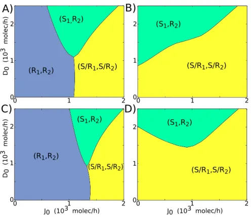
0 1 2 0 1 2 D0 (10 3 mo lec /h )
J0 (103 molec/h) (S1,R2)
0 1 2
0 0.2 0.4 0.6
J0 (103 molec/h)
NI CD (10 3 mo lec )
(S2) (R1)
(S/R1,S/R2)
A)
B)
(S/R1,S/R2)
(R1,R2)
Figure 2.13: Phenotype diagram and bifurcation curve for two interacting cells. A) Phenotype diagram when the two-cell circuit is driven by both the production rates of Jagged (J0) and Delta (D0). Each
phase is represented by a different color and corresponds to a different combination of co-existing states B) Bifurcation diagram of NICD levels with respect to the production rate of Jagged (J0) forD0 = 1000
molecules/hour (red arrow in A)
2.3.1
Ligand production rates control tissue level patterning
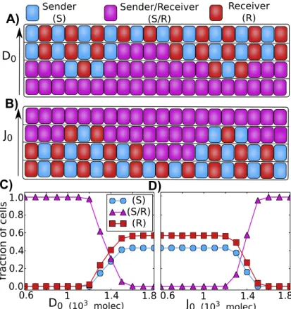
As mentioned earlier, Notch-Delta interactions lead to lateral inhibition - neighboring cells adopt alternate fates - Sender (S) and Receiver (R) [21, 20, 45, 72]. Notch-Jagged interactions lead to lateral induction [65, 64] - neighboring cells adopt similar fates - both of them can simultaneously send and receive the signal - Sender/Receiver (S/R) state. Although these mechanisms have been well-studied individually, the tissue level patterns that might emerge when both mechanisms act simultaneously have not been explored.
To address this issue, we simulate a one-dimensional layer of cells interacting via Notch pathway, for different values of D0 andJ0 - Delta and Jagged production rates, respectively.
Our results show that when the relative production rate of Delta is increased, the so-called ’salt-and-pepper’ pattern (or alternate fate pattern) at a tissue-level begins to emerge (Figure 2.14A,C). On the other hand, when the relative production rate of Jagged is increased, the ’salt-and-pepper’ pattern is disrupted, and the cells begin to adopt similar fates where they can both send and receive the signal (Figure 2.14B, D).
Sender (S)
Sender/Receiver (S/R)
Receiver (R)
D0
0.0 0.2 0.4 0.6 0.8 1.0
0.6 1 1.4 1.8 (S) (S/R)
(R)
0.6 1 1.4 1.8
fra
c
ti
o
n
o
f
c
el
ls
A)
J0
J0 (103 molec) B)
C) D)
D0 (103 molec)
Figure 2.14: Patterning at the tissue level. A) and B) Representation an 1 dimensional layer of 20 interacting cells. A) The increase of the production of Delta (D0) leads to the formation of alternate
patterns in which neighbors cells alternate between Sender and Receiver. B) The increase of the production of Jagged leads all the cells to the hybrid (S/R) state, therefore loosing the alternate (S) and (R) pattern. C) and D) Average of the fraction of cells in (S), (S/R) or (R) state as a function of ligand production. The averages were taken over 100 simulations of a 1 dimensional layer of 100 interacting cells with periodic boundary condition. C) Fraction of cells in (S), (S/R) or (R) state as a function of the production of Delta. D) Fraction of cells in (S), (S/R) or (R) state as a function of the production of Jagged. The state of the cells are defined according to the amount of signaling (I): Sender (I < 100 molecules), Sender/Receiver (100< I <300) and Receiver (I >300).
therefore driving the cells to a similar fate - hybrid Sender/Receiver (S/R) - that can promote bidirectional communication between tumor and stroma, a context where inflammation often plays a key role.
2.4
Soluble ligands affect cell-fate decision in a paracrine
manner
In addition to transmembrane ligands, Notch signaling can also be activated by soluble forms of the ligands [33, 27, 28].
Soluble Jagged1 for example has been specifically implicated in mediating long-distance communication between tumor cells and stromal cells. Jagged1 can be secreted by endothe-lial cells that can activate Notch signaling in cancer cells, inducing them to gain migratory and invasive characteristics by undergoing partial or complete Epithelial to Mesenchymal Transition (EMT) [73]. Jagged1 can also induce the expression of NF-kB [74], which can in-crease the population of CSCs and further inin-crease the secretion of Jagged1 [33], suggesting a wave-like mechanism in the tumor microenvironment to rake up the production and main-tenance of therapy-resistant CSCs. Future theoretical studies of these circuits hold promise for appreciating the key role of soluble Jagged1 in mediating two interlinked and clinically insuperable facets of cancer - metastasis (as a result of cells undergoing EMT) and tumor relapse (as a result of expanded CSC pool).
Not only soluble Jagged1, but also transmembrane Jagged1 mediates tumor progression in several ways, and has been proposed to be a therapeautic target [31]. Notch-Jagged signaling plays a crucial role in the metastasis of breast cancer cells to bone, where prostate cancer cells expressing Jagged1 communicate with Notch-expressing osteoclasts to “home” in the bone [9]. Also, overexpression of Jagged1 on cancer cells can trigger Notch activation in neighboring endothelial cells (which can possibly secrete more soluble Jagged1), promoting sprouting tumor angiogenesis [75] and thereby tumor growth. Consistent with their pro-tumor roles, high levels of Notch and Jagged1 in cancer cells often correlates with poor patient survival [31].
(two identical cells communicating via NDJ pathway), as described in equations 1.16-1.19 where Dext and Jext represents Delta and Jagged of the neighboring cell and Dexts and Jexts
represent soluble Delta and Jagged respectively.
0 1 2
0 1
2 (S1,R2)
(S/R1,S/R2)
A)
B)
0 1 2
J0 (103 molec/h) 0
1
2 (S1,R2)
(S/R1,S/R2)
0 1 2 D0 (10 3 mo lec /h )
0 1 2
(S1,R2)
(S/R1,S/R2)
(R1,R2)
0 1 2
J0 (103 molec/h) 0 1 2 D0 (10 3 mo lec /h
) (S1,R2)
(R1,R2)
(S/R1,S/R2)
C)
D)
Figure 2.15: Phenotype diagram for two-cell system (two cells interacting via NDJ circuit) when the circuit is driven by the production rates of Jagged (J0) and Delta (D0). Each phase is represented by a different
color and corresponds to a different combination of co-existing states. Each diagram represents different levels of soluble ligands Delta (Ds
ext) and Jagged (J s
ext) interacting with these identical cells. A)D s
ext= 600 molecules,Js
ext= 0. B)J s
ext= 600 molecules,D s
ext= 0. C)D s
ext= 1200 molecules,J s
ext= 0. D)J s
ext= 1200 molecules,Ds
ext= 0. The trans-activation rate for soluble ligands is considered to be 3 times lower than for membrane-bound ligands (ks
T =kT/3).
For the case of paracrine communication via soluble Delta, both the cells tend to behave as Receiver (R1, R2) for low production rate of Jagged (J0) or both of them behave as hybrid
Sender/Receiver (S/R1, S/R2) for highJ0. These cells can adopt opposite fates to each other
only for intermediate levels of J0 and high production rate of Delta (D0) (Figure 2.15A, C,