Anthracene and Pyrene Adsorbed onto Mangrove Leaves
Ping Wang1,2, Tun-Hua Wu3, Yong Zhang4*
1School of Environmental Science and Public Health, Wenzhou Medical University, Wenzhou, China,2Key Laboratory of Coastal and Wetland Ecosystems (Xiamen University), Ministry of Education, Xiamen, China,3School of Information and Engineering, Wenzhou Medical University, Wenzhou, China,4State Key Laboratory of Marine Environmental Science (Xiamen University), College of the Environment and Ecology, Xiamen University, Xiamen, China
Abstract
An established synchronous fluorimetry method was used forin situinvestigation of the photodegradation of pyrene (PYR) and anthracene (ANT) adsorbed onto fresh leaves of the seedlings of two mangrove species,Aegiceras corniculatum(L.) Blanco (Ac) andKandelia obovata (Ko) in multicomponent mixtures (mixture of the ANT and PYR). Experimental results indicated that photodegradation was the main transformation pathway for both ANT and PYR in multicomponent mixtures. The amount of the PAHs volatilizing from the leaf surfaces and entering the inner leaf tissues was negligible. Over a certain period of irradiation time, the photodegradation of both PYR and ANT adsorbed onto the leaves ofAcandKofollowed first-order kinetics, with faster rates being observed onAcleaves. In addition, the photodegradation rate of PYR on the leaves of the mangrove species in multicomponent mixtures was much slower than that of adsorbed ANT. Compared with the PAHs adsorbed as single component, the photodegradation rate of ANT adsorbed in multicomponent mixtures was slower, while that of PYR was faster. Moreover, the photodegradation of PYR and ANT dissolved in water in multicomponent mixtures was investigated for comparison. The photodegradation rate on leaves was much slower than in water. Therefore, the physical-chemical properties of the substrate may strongly influence the photodegradation rate of adsorbed PAHs.
Citation:Wang P, Wu T-H, Zhang Y (2014) Direct Observation of the Photodegradation of Anthracene and Pyrene Adsorbed onto Mangrove Leaves. PLoS ONE 9(8): e104903. doi:10.1371/journal.pone.0104903
Editor:Jie Zheng, University of Akron, United States of America
ReceivedMay 15, 2014;AcceptedJuly 15, 2014;PublishedAugust 21, 2014
Copyright:ß2014 Wang et al. This is an open-access article distributed under the terms of the Creative Commons Attribution License, which permits unrestricted use, distribution, and reproduction in any medium, provided the original author and source are credited.
Data Availability:The authors confirm that all data underlying the findings are fully available without restriction. In our previous studies, the synchronous fluorimetry has been established and utilized for investigation of the photodegradation of PYR and ANT adsorbed onto the leaves of two mangrove species as single component. In this study, the established synchronous fluorimetry was further utilized for study the photodegradation of PYR and ANT adsorbed onto the leaves of two mangrove species in multicomponent mixtures. For comparing, some data from the photodegradation of PYR and ANT adsorbed onto the leaves of two mangrove species as single component in supporting Information files have been mentioned in this paper.
Funding:The funding of our manuscript include National Natural Science Foundation of China (21177102, 21207103), Public Benefit Project of Zhejiang Province (2012C31025), Natural Science Foundation of Zhejiang Province (LY13H180012), Scientific Research Fund of Zhejiang Provincial Education Department (Y201222932), Specialized Research Fund for the Doctoral Program of Higher Education (20130121130005), Undergraduate Scientific and Technological Innovation project of Zhejiang Province (2013R413010) and WEL Visiting Fellowship Program. The funders had no role in study design, data collection and analysis, decision to publish, or preparation of the manuscript.
Competing Interests:The authors have declared that no competing interests exist.
* Email: [email protected]
Introduction
Over 80% of the earth’s terrestrial surface is covered by vegetation [1]. Vegetation plays a key role in the environmental fate of many polycyclic aromatic hydrocarbons (PAHs) [2]. Furthermore, leaf surfaces are covered with a complex lipid cuticle that can accumulate hydrophobic organic pollutants from the atmosphere [3]. PAHs are widely distributed persistent organic pollutants that are generated by natural combustion processes as well as by human activities [4,5]. These micropollutants in the environment pose a potential threat to aquatic organisms and humans because of their toxic, carcinogenic and mutagenic properties [6]. PAHs are ubiquitous in natural substances such as plants, soil, sediment, water and air [7–9]. Thus, it is important to study the transportation and transformation of PAHs in the environment. Many reports demonstrate that most PAHs in the environment biodegrade slowly and with difficulty because of their low water solubility [10]. Additionally, it has been shown that many PAHs exhibit photo-induced toxicity [11] and that the delocalizedpbond of PAHs can absorb the visible and ultraviolet
components of sunlight [12]. Therefore, photodegradation might represent an important transformation pathway for PAHs in the environment. Investigations in this field have recently intensified and have generated some solid conclusions. At present, most studies on the photodegradation of PAHs have focused on PAHs in a liquid medium or adsorbed onto solid particles [13–16]. Only a few studies have addressed the photodegradation of PAHs adsorbed onto vegetation, although the photodegradation of PAHs adsorbed onto plant surfaces (especially leaves) plays an important role in their transfer from the atmosphere to the food chain [17– 19].
behavior. Therefore, it is necessary to investigate the photochem-ical behavior of PAHs absorbed onto mangrove leaves. However, previous research on the photodegradation of PAHs adsorbed onto plant leaves has mostly been conducted by entirely destroying the sample. Thus, these traditional methods are not compatible with the direct study of the photodegradation of PAHs adsorbed onto leaves. Moreover, in a long-term study, the quantity of PAHs volatilized from the leaf surfaces and entering the inner leaf tissues may not be negligible [17,19]. Optical fibers showing high light focalization, low weights and small sizes have been used for the direct investigation of PAHs adsorbed onto solid substrates [25– 27]. In our group, a fiber-optic fluorimetry has also been established for in situ investigation of the photodegradation of fluoranthene adsorbed onto the leaves of three mangrove species [23,28]. Nevertheless, the mechanisms underlying the photodeg-radation of PAHs adsorbed onto mangrove leaves have not been fully explored. Our previous research sought to elucidate the environmental behaviors of PAHs adsorbed onto mangrove leaves using an established synchronous fluorimetry combined with a fluorescence spectrophotometer and an optical fiber to directly detect PYR and ANT adsorbed onto mangrove leaves [29]. The photodegradation of PYR and ANT adsorbed onto the leaves of two mangrove species as single component have also been studied in our laboratory [30]. However, the environmental behaviors of PAHs are very complex in the field. For example, many different types of PAHs from different sources might exist on the leaf surfaces. Because of the effects of complex factors mentioned above or others, it is difficult for in situ investigation the environmental behaviors of PAHs adsorbed on leaf surfaces in the field. In addition, only a few studies involve the photolytic behaviors of PAHs adsorbed on vegetation. Thus, preliminary experiments could be carried out in lab at first. And simple experimental conditions were set for study of the possible photolysis processes of some PAHs on the typical mangrove leaves. To further develop this line of research, synchronous fluorimetry was used forin situstudying the photodegradation of PYR and ANT adsorbed onto the leaves of two mangrove species in multicomponent mixtures. Compared with the photodegration of the PAHs adsorbed on mangrove leaves in single component, the further studies for investigation the photodegration of the PAHs adsorbed on mangrove leaves in multicomponent mixtures are more representative of real conditions for most PAHs in the environment. Seedlings ofAegiceras corniculatum(L.) Blanco (Ac) andKandelia obovata(Ko), two of the most widespread mangrove species in China, were selected for this study, and the photodeg-radation of equal quantities of PYR and ANT in water as single compounds and in multicomponent mixtures was also investigated for comparison.
Materials and Methods
Preparation of PAH solution
Stock solutions of PYR and ANT (Aldrich, purity.99%, USA) were obtained based on the method described in our previous studies [22,29]. Working solutions of PYR and ANT were prepared by diluting the stock solutions with acetone just prior to being used.
Sample collection
According to our previous studies and others, the mature viviparous hypocotyls ofKowhen off the trees could be collected for cultivation, and the non-viviparous seedlings of Ac who are least one-year old could be taken away from the forest and cultivated for further experiments [30,31]. Therefore, mature
mangrove hypocotyls of Ko and 1-year-old Ac seedlings were collected at the Longhai mangrove reserve in Zhangzhou, Fujian, China (longitude: 24u29930, latitude: 118u59590, altitude: 0 m above sea level). This mangrove forest is wild and does not belong to any individuals or organizations. Thus, no specific permissions were required for these activities. Only a few mangrove seedlings were collected for the experiment, which did not involve endangered or protected species or vertebrates. Hypocotyls of
Ko and seedlings of Ac of approximately the same size and maturity were sampled and quickly transported to the laboratory for cultivation. The hypocotyls ofAcandKowere cultivated on sand (collecting from the mangrove forest) in pots, partially submerged in nutrient Hoagland’s solution. For a 12-hour photoperiod, propagules were illuminated by 400-W Na solar lighting. The greenhouse temperature was maintained at 25– 28uC. Though, the cultivation conditions in lab were different with that in the field, our studies of past and present have attempted to simulate the living environment of the hypocotyls. The cultivation conditions in lab have their own merits. We all know that the growth of the mangrove hypocotyls in the field might be influenced by many factors. The maturity level and integrity of the hypocotyls living in the same forest might be different. Therefore, the similar maturity of hypocotyls and leaves could not be obtained in the field, which might lead to lower accuracy of the results. Thus, the possible mechanisms of environmental behaviors of the PAHs obtained in the field might be different. Taking into consideration of various factors, the cultivation conditions selected in our studies were comparatively reasonable. Ninety days later, the leaves from the two mangrove species with similar length and fresh weight were collected for the further experiment. After the collection of the leaves, experiments were carried out as soon as possible. Pretreatment of the leaves followed the steps established in our previous studies [29,30,31]. The picked fresh mangrove leaves were carefully rinsed with distilled water to remove surface silt. After air drying, six circles of 0.5 cm radius were drawn on the upper surfaces of each leaf with a pencil. The size was the same as the light circle formed by the fiber optical probe. With the use of a micropipette, a certain amount of ANT (0.25 nmol spot21) and PYR (0.25 nmol spot21) acetone solutions (5mL) were applied as homogeneous layers to the circles of upper leaf surfaces respectively. After evaporation of the acetone from the leaf surfaces at room temperature, a series of similar-sized spots were formed, each spot indicating a sample location.
An ultrasonic cleaning device (Model KQ3200, SM, Kun Shan Ultrasonic Instruments Co., Ltd., China) was utilized to extract the lipid wax from the mangrove leaves. The wax content of the leaves was subsequently quantified using a method from described in previous work [28,31,32].
Photodegradation of PAHs absorbed onto the leaves of the two mangrove species
in multicomponent mixtures. According to our previous studies and the pre-experiments, the set irradiation time interval increased gradually (5 nm, 10 nm, 15 nm and 20 nm). Thus, the optimized irradiation of the ANT and PYR adsorbed on the leaves ofKoand
Ac should be set at the same sequence. That is the optimized irradiation time for the PAHs adsorbed onto theKcleaves was set at 0 min, 1 min, 3 min, 7 min, 13 min, 20 min, 30 min, 45 min, 60 min, 80 min, and 100 min, 120 min and 140 min, respectively, and the optimized irradiation time for the adsorbed PAHs on the
Acleaves was set at 0 min, 1 min, 3 min, 7 min, 13 min, 20 min, 30 min, 45 min, 60 min, 80 min, 100 min, 120 min and 140 min, respectively [30]. Fluorescence spectra were obtained using the following instrumental setting: excitation and emission slits of 20 and 10 nm, respectively; scan speed of 120 nm min21; PMT voltage of 600 V; and the constant wavelength difference (Dl) was set at 38 nm. Finally, the fluorescence intensities of the PAHs adsorbed onto the leaves were directly determined. Replicate experiments were conducted for 3 times. To avoid interference from the scattered light, the leaf had to be kept flat, and the angle between the fiber-optic probe of the spectrophotometer and the tested leaf was 45uthroughout the experiment.
Photodegradation of PAHs dissolved in water
To compare and analyze the photodegradation behavior of each PAH in different media, the photodegradation of the same initial amount of PYR and ANT dissolved in water in single-component and multisingle-component mixtures was also studied. A photodegradation reactor constructed in our laboratory was used to study the photodegradation of each PAH in water [33]. Using a micropipette, 0.25 nmol of PYR and 0.25 nmol of ANT dissolved in water were added to the reactor, which was then immediately placed under the optical fiber of the mercury lamp. According to our previous studies and the pre-experiments [30], the optimized irradiation time for the ANT in water as single component (0 min, 1 min, 3 min, 6 min, 11 min, 16 min, 21 min, 31 min, 36 min, 41 min, and 56 min) and in multicomponent mixtures (0 min, 1 min, 3 min, 6 min, 11 min, 16 min, 21 min, 31 min, 36 min, 41 min, and 56 min, 66 min, and 76 min) was set, respectively. In addition, the optimized irradiation time for the PYR in water as
single component (0 min, 1 min, 3 min, 6 min, 11 min, 16 min, 21 min, 31 min, 36 min, 41 min, 56 min, 66 min, 86 min, 100 min, 120 min) and in multicomponent mixtures (0 min, 1 min, 3 min, 6 min, 11 min, 16 min, 21 min, 31 min, 36 min, 41 min, 56 min, 66 min, 76 min, 86 min, and 96 min) was also set, respectively. To sum up, the set irradiation time of the PAHs in water was almost fixed (mainly 5 nm), which was different with that adsorbed on mangrove leaves (5 nm, 10 nm, 15 nm and 20 nm). Thus, after a defined period of illumination mentioned above, the reactor was placed in the fluorescence spectrophotom-eter. The fluorescence intensities of the PAHs dissolved in water were ultimately obtained. Because of the small volume of the self-made reactor, the working solutions of PYR and ANT did not require stirring, and we could directly detect the PAHs during photodegradation. Replicate experiments were conducted for 6 times.
Statistical analysis
In this study, the sample preparations were the same with the method of our previous studies [29–31]. And we used four mangrove leaves from each species, yielding a total of 24 locations and 72 measurements for each species. The mean values presented represent the fluorescence intensity of the PAHs adsorbed onto different locations (24 locations from 4 leaves) in the leaves of each mangrove species. Statistical analysis of the variation in fluores-cence intensity was performed using the Statistical Package for the Social Science (SPSS), 13.0, for Windows. Significant differences in the results about the mean values of fluorescence intensity obtained by the adsorbed PAHs at different irradiation time were determined using the independent sample t-test. In addition, the confidence interval was utilized for estimate the reliability of the fluorescence intensity of the PAHs adsorbed onto different locations in the leaves of each mangrove species. And the confidence interval was stated at the 95% confidence level (p. 0.05 means that no significant difference existed).
When the concentration of a PAH is proportional to its fluorescence intensity,Ct(the PAH concentration at timet) andC0
(the initial PAH concentration) can be replaced withFt(the PAH
fluorescence intensity at time t) and F0 (the initial PAH
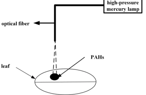
Figure 1. Schematic diagram of the photodegradation of PAHs adsorbed onto the leaves of two mangrove species.
fluorescence intensity), respectively. The data were processed based on the same equations as our previous studies [30].
Results and Discussion
Fluorescence spectra of PYR and ANT adsorbed onto mangrove leaves
In previous studies, we established the synchronous fluorimetry method for the direct determination of PYR and ANT adsorbed onto Ac and Ko leaves both as single compounds and in multicomponent mixtures. Experimental results have indicated that when the optimized scanning wavelength difference (Dl) was 38 nm, the ANT and PYR adsorbed onto the leaves of two mangrove species as single compounds and in multicomponent mixtures have their own characteristic synchronous fluorescence spectra. The fluorescence spectra of the adsorbed PAHs on mangrove leaves as single component have fluorescence maxi-mums at 369 nm and 387 nm for ANT, and 343 nm for PYR. Thus, the wavelengths of 387 nm and 343 nm were selected for the quantification of ANT and PYR as single component, respectively. In addition, the fluorescence spectra of the PAHs adsorbed on mangrove leaves in multicomponent mixtures showed peaks at 371 nm and 390 nm for ANT, and 346 nm for PYR. Thus, the wavelengths of 390 nm and 346 nm were selected for the quantification of ANT and PYR in multicomponent mixtures, respectively [31]. More recently, the photodegradation of PYR and ANT adsorbed onto the leaves of two mangrove species as a single component has been investigated by our group [30]. However, it is reported that the photodegradation of PAHs are very complex in natural environment. For example, interaction between different types of PAHs might influence the photodeg-radation of the PAH in multicomponent mixtures. Thus, compared with the PAHs as single component, studies about of the photodegradation of the PAHs in multicomponent mixtures are more representative of real conditions for most PAHs in natural environment. To better understand the photodegradation of these PAHs in multicomponent mixtures, evaluation of the
synchronous fluorimetry spectra of the two PAHs and their photodegradation during irradiation is useful. Figure 2 shows the fluorescence spectra of PYR and ANT adsorbed onto the leaves of
Ko in multicomponent mixtures, measured at different times during irradiation. The relative fluorescence intensities decreased with time. As can be observed in Figure 2, the autofluorescence of
Koleaves without PAH was weak and was insufficient to affect the determination of adsorbed PAH in further analyses. In addition, there was no overall change in the shapes of the spectra of the PAHs or the width of the half-wide spectral band. Thus, the photolytic products did not interfere with the detection of adsorbed PYR and ANT during photodegradation (Figure 2). A similar trend was observed during the photodegradation of PYR and ANT adsorbed onto the leaves ofAc. Thus, it was concluded that synchronous fluorimetry could be used for the direct study of the photodegradation of PYR and ANT adsorbed onto mangrove leaves in multicomponent mixtures.
Results of the photodegradation experiments
Volatilization and absorption losses could be the most important pathways for adsorbed PAHs on plant leaves [22]. Therefore, control experiments were carried out in which the leaves were maintained in darkness. The variations in Ft/F0 for PYR and
ANT adsorbed onto the leaves of the two mangrove species in multicomponent mixtures were determined over a certain time interval during the photodegradation experiments (Figure 3). Without illumination, Ft/F0 decreased very little as time went
on (Figure 3). Hence, photodegradation was found to be the main transformation pathway for PYR and ANT adsorbed onto the mangrove leaves, and the amount of PAH volatilizing from the leaf surfaces and entering the inner leaf tissues could be ignored in the short term. In addition, the relative fluorescence intensities of PYR and ANT as single component are also shown in Figure 3 for comparison [30]. As shown in Figure 3, the curves indicated that the photodegradation of each PAH occurred via multi-decays. Over a given period of time, the photodegradation of both PYR and ANT adsorbed onto the leaves of two mangrove species in
Figure 2. Synchronous fluorescence spectra of PYR and ANT adsorbed onto the leaves of Ko in multicomponent mixtures
(Dl= 38 nm), measured at different times during irradiation.The order of the changes in fluorescence intensity over time (at 0 min, 5 min,
20 min, 45 min and 70 min) is indicated by the arrow. Wavelengths of 390 nm and 346 nm were selected for ANT and PYR, respectively. (1): Synchronous fluorescence spectra of the uncontaminatedKoleaves.
multicomponent mixtures basically followed first-order reaction kinetics (Figure 4). In addition, the photodegradation of both PYR and ANT adsorbed onto the leaves of two mangrove species as single component basically followed first-order reaction kinetics in a given period of time (reference 30). Specifically, the given period of time for adsorbed ANT both on the leaves ofKo and Acin multicomponent mixtures was from 0 min to 45 min. The given period of time for adsorbed PYR both on the leaves of Ac in multicomponent mixtures was from 0 min to 80 min the given period of time for adsorbed PYR on the leaves of Ko in
multicomponent mixtures was from 0 min to 60 min (Figure 4). The photodegradation rate of the PAHs adsorbed ontoAcleaves was faster than that adsorbed onto Koleaves. Moreover, for a given amount of the two substances (0.25 nmol spot21), the photodegradation rate of adsorbed PYR was much lower than that of ANT. Thus, it is a reasonable assumption that the molecular structure of PAHs might be one of the important factors determining their photochemical behavior [34,35]. The experi-mental results also showed that for the same initial amount of each PAH, the photodegradation rate was in the order Ac.Ko Figure 3. Change inFt/F0with time during the photodegradation of ANT (0.25 nmol spot21) and PYR (0.25 nmol spot21) adsorbed ontoKoandAcleaves as single component and in multicomponent mixtures.
(Table 1). It is assumed that the waxes and lipids on plant leaves can absorb UV photons, which might create light-filtering effects [17,19]. Thus, the number of UV photons striking each PAH adsorbed onto the leaves decreased and thereby reduced the photodegradation rate of each PAH. In this experiment, the leaf-wax content ofAc(4.98 mg.g21) was found to be much lower than that of Ko (7.63 mg.g21). Thus, the light filtering effect of Ko
leaves might be stronger than that ofAcleaves. Consequently, for the same initial amount of each substance, the number of the UV photons striking each PAH adsorbed onto Ac leaves would be higher. Moreover, because of the lower leaf-wax content ofAc, the interactions between the adsorbed PAH andAc might become much weaker, making it much easier for the UV photons to strike the adsorbed PAH. These phenomena may act together to explain
Figure 4. First-order kinetic plots of the photodegradation of ANT (0.25 nmol spot21) and PYR (0.25 nmol spot21) adsorbed onto KoandAcleaves as single component and in multicomponent mixtures.
why the photodegradation rate of the PAHs adsorbed onto Ac
leaves was faster compared withKoleaves. (Table 1, Table 2). In addition, for both PYR and ANT, the variations in Ft/F0
remained relatively stable when the amount of the adsorbed PAHs on the leaves decreased to approximately 12% of the initial amount (Figure 3). This finding indicates that the photodegrada-tion of both PYR and ANT adsorbed onto the mangrove leaves could only result in partial degradation. In other words, photodegradation of the residual PAH adsorbed onto the mangrove leaves could no longer occur as the illumination time went on. Similar results were obtained regarding the photodeg-radation of PYR and ANT as single component in our previous work [30]. Identical results were found in studies by Xuet al.and Schuler et al., though these authors did not suggest any mechanisms for these findings [36,37]. Given previous findings by ourselves and others, a small amount of PAH might enter into the inner leaf tissues. These PAHs in the internal leaf tissues cannot absorb UV photons due to the light filtering effects of the leaves [17,19,32]. Further research will be required to better understand the possible reasons for these observations.
The experimental results also revealed that compared with PYR and ANT adsorbed onto the leaves of the same mangrove species as single component, the photodegradation rate of ANT in multicomponent mixtures was slower, while that of PYR was faster (Table 1, Table 2). Similar results were found concerning the photodegradation of PYR and ANT in water in multicomponent mixtures (Table 1, Table 2). There are several possible reasons for these results. First, for the same light intensity and irradiation time,
the total number of UV photons striking each PAH adsorbed onto the leaves as single component and multicomponent mixtures is the same. Therefore, compared with each PAH as a single compound, the number of UV photons received by each PAH in multicomponent mixtures is lower, which could slow the photodegradation rate of each PAH. Second, anthraquinone is one of the intermediate products of the photodegradation of ANT, and it has also been found to be a photosensitizer for both PYR and ANT [38]. In this experiment, the photodegradation rate of adsorbed ANT was faster compared with adsorbed PYR in multicomponent mixtures (Table 1, Table 2). Hence, in multi-component mixtures, ANT degraded first and generated some anthraquinone. This anthraquinone might accelerate the photo-degradation rate of the adsorbed PYR, and the amount of anthraquinone, as a photosensitizer of adsorbed ANT, would therefore decrease compared with that in the single-component mixture. Thus, the photodegradation rate of the adsorbed ANT would slow down in multicomponent mixtures. However, further studies are needed to confirm this hypothesis.
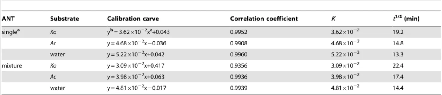
Studies show that the polarity of a solvent can significantly affect the photodegradation rate of PAHs in or on that solvent [14]. To further study the possible mechanisms underlying the photodeg-radation of PAHs adsorbed onto mangrove leaves in multicom-ponent mixtures, the photodegradation of PYR and ANT dissolved in water in multicomponent mixtures was also investi-gated for comparison (Figure 5). In this experiment, the synchro-nous fluorimetry method established in our previous studies was used to determine PYR and ANT levels in water as single Table 1.Kinetic results for the photodegradation of ANT adsorbed onto the leaves of two typical mangrove species (n = 9) and dissolved in water (n = 9) as single compounds and in multicomponent mixtures.
ANT Substrate Calibration carve Correlation coefficient K t1/2
(min)
singlea Ko yb= 3.62
61022xc+0.043 0.9952 3.62
61022 19.2
Ac y = 4.6861022x20.036 0.9908 4.6861022 14.8
water y = 5.2261022x+0.042 0.9960 5.2261022 13.3
mixture Ko y = 3.0961022x+0.417 0.9356 3.09
61022 22.4
Ac y = 3.9861022x+0.063 0.9936 3.9861022 17.4
water y = 4.8161022x20.017 0.9939 4.8161022 14.4
adata from our previous studies [30]. bthe value of ln(F
0/Ft).
cthe illumination time of ANT adsorbed onto mangrove leaves.
doi:10.1371/journal.pone.0104903.t001
Table 2.Kinetic results of the photodegradation of PYR adsorbed onto the leaves of two typical mangrove species (n = 9) and dissolved in water (n = 9) as single compounds and in multicomponent mixtures.
PYR Substrate Calibration carve Correlation coefficient K t1/2min
singlea Ko yb= 1.58
61022xc+0.114 0.9899 1.5861022 43.9
Ac y = 2.1361022x+0.125 0.9865 2.1361022 32.5
water y = 2.8861022x+0.088 0.9937 2.88
61022 24.1
mixture Ko y = 2.6161022x+0.357 0.9393 2.6161022 26.6
Ac y = 2.9561022x+0.203 0.9815 2.9561022 23.5
water y = 3.4661022x20.045 0.9878 3.4661022 20.0
adata from our previous studies [30]. bthe value of ln(F
0/Ft).
cillumination time of the PYR adsorbed onto mangrove leaves.
component and in multicomponent mixtures [33,39]. Experimen-tal results showed that the photodegradation of PYR and ANT dissolved in water as single component and multicomponent
mixtures followed first-order reaction kinetics (Figure 5). The photodegradation rate of each PAH in water as single component and in multicomponent mixtures was faster compared with the
Figure 5. Photodegradations of ANT (0.25 nmol) and PYR (0.25 nmol) in water as single component and in multicomponent mixtures.(1): Change inFt/F0over time during the photodegradation of the two PAHs adsorbed onto the leaves of the two mangrove species. (2):
PAHs adsorbed onto mangrove leaves (Table 1, Table 2). There are two possible reasons for this phenomenon. First, the wax layer on the mangrove leaves mainly consists of saturated fatty acids and unsaturated fatty acids, which could reflect or adsorb UV light. Therefore, the number of UV photons striking the adsorbed PAH might decrease and thereby reduce the photodegradation rate [17,18,37]. Second, it has been shown that the more polar the solvent, the faster photodegradation is [40,41]. In the present study, we assessed the PAHs in water, which is polar, and on the mangrove leaf surface, which is nonpolar. Thus, the photodegra-dation of the PAHs adsorbed onto the leaves was slower than when they were dissolved in water. In addition, it was clearly observed that the relative fluorescence intensities of PYR and ANT in water in multicomponent mixtures decreased almost to zero as the illumination time went on (Figure 5). In other words, PYR and ANT in homogeneous water could be thoroughly degraded within a certain period of time. This result differed from the photodegradation of PYR and ANT adsorbed onto the heterogeneous mangrove leaves. We can speculate that the photodegradation mechanisms for PAHs adsorbed onto mangrove leaves and dissolved in water may differ. Therefore, the photochemical behaviors of PAHs were dependent not only on their molecular structure but also on the physical-chemical properties of the substrate. Further experimental verification is needed.
Conclusions
In this work, synchronous fluorimetry was shown to be a simple, rapid and easy method for direct investigation of the photodeg-radation of PYR and ANT adsorbed onto the leaves ofAcandKo
in multicomponent mixtures. This method may provide a new way to study the mechanisms underlying the phytoremediation of
PAHs in mangrove wetlands or in other contaminated media. The photodegradation of both PYR and ANT on the leaves of the studied species followed first-order reaction kinetics. However, the photodegradation processes were different for the same PAHs adsorbed onto the leaves of different mangrove species. In addition, for the same amount of substance, the photodegradation rate of PYR was much slower than that of ANT. Compared with the photodegradation rates in single-component mixtures, the rates in multicomponent mixtures were different. The photodeg-radation mechanisms of the PAHs adsorbed onto the mangrove leaves, whether in single or multicomponent mixtures, might also be different than the mechanisms for the PAHs is dissolved in water. However, further research is needed to address this conjecture. In this experiment, only the PAHs adsorbed onto the leaf surfaces could be studied, and the PAHs entering into the internal leaf tissues could not be quantified or investigated for further photodegradation. Thus, there are still detailed and comprehensive studies to be carried out in the future. For example, in our group, two-photon laser confocal scanning microscopy has been used for direct visualization of PAHs on and within mangrove leaves. Combining these two methods with some traditional approaches may be helpful for better under-standing the environmental behaviors of PAHs in real environ-ments. Therefore, the synchronous fluorimetry method might be valuable in theoretical and practical applications.
Author Contributions
Conceived and designed the experiments: PW YZ THW. Performed the experiments: PW THW YZ. Analyzed the data: PW THW YZ. Contributed reagents/materials/analysis tools: PW THW YZ. Contributed to the writing of the manuscript: PW YZ THW.
References
1. Simonich SL, Hites A (1994) Importance of vegetation in removing polycyclic aromatic hydrocarbons from the atmosphere. Nature 370: 49–51.
2. Collins C, Fryer M, Grosso A (2006) Plant uptake of non ionic organic chemicals. Environ Sci Technol 40: 45–52.
3. Sojinu OS, Sonibare OO, Gayawan E (2013) Investigating polycyclic aromatic Hydrocarbons profiles in higher plants using statistical models. Int J Phytor-emediat 15: 439–451.
4. Al-Saleh I, Alsabbahen A, Shinwari N, Billedo G, Mashhour A, et al. (2013) Polycyclic aromatic hydrocarbons (PAHs) as determinants of various anthropo-metric measures of birth outcome. Sci Total Environ 444: 565–578. 5. Kwon HO, Choi SD (2014) Polycyclic aromatic hydrocarbons (PAHs) in soils
from a multi-industrial city, South Korea. Sci Total Environ 470: 1494–1501. 6. Pongpiachan S, Tipmanee DW, Muprasit J, Feldens P, Schwarzer K (2013) Risk assessment of the presence of polycyclic aromatic hydrocarbons (PAHs) in coastal areas of Thailand affected by the 2004 tsunami. Mar Poll Bull 76: 370– 378.
7. Augusto S, Ma´guas C, Matos J, Pereira MJ, Branquinho C (2010) Lichens as an integrating tool for monitoring PAH atmospheric deposition: a comparison with soil, air and pine needles. Environ Pollut 158: 483–489.
8. Luiz FF, Mirian ACC, Alberto GFJ, Elisamara SS, Frederico SS, et al. (2012) Characterization and distribution of polycyclic aromatic hydrocarbons in sediments from Suruı´ Mangrove, Guanabara Bay, Rio de Janeiro, Brazil. J Coastal Res 28: 156–162.
9. Wang XT, Miao Y, Zhang Y, Li YC, Ming-Hong Wu MH, et al. (2013) Polycyclic aromatic hydrocarbons (PAHs) in urban soils of the megacity Shanghai: occurrence, source apportionment and potential human health risk. Sci Total Environ 447: 80–89.
10. Bamforth SM, Singleton I (2005) Bioremediation of polycyclic aromatic hydrocarbons: current knowledge and future directions. J Chem Technol Biot 80: 723–736.
11. Wang Y, Chen JW, Li F, Qin H, Qiao XL, et al. (2009) Modeling photoinduced toxicity of PAHs based on DFT-calculated descriptors. Chemosphere 76: 999– 1005.
12. Dittmar T, Koch BP (2006) Thermogenic organic matter dissolved in the abyssal ocean. Mar Chem 102: 208–217.
13. Debestani R, Ellis KJ, Sigman ME (1995) Photodecomposition of anthracene on dry surface: products and mechanism. J Photoch Photobio A 86: 231–239.
14. Kahan TF, Donaldson DJ (2007) Photolysis of polycyclic aromatic hydrocarbons on water and ice surfaces. J Phys Chem A 111: 1277–1285.
15. Lehto KM, Vuorimaa E, Lemmetyinen H (2000) Photolysis of polycyclic aromatic hydrocarbons (PAHs) in dilute aqueous solutions detected by fluorescence. J Photoch Photobio A 136: 53–60.
16. Reyes CA, Medina M, Crespo-Hernandez C, Cedeno MZ, Arce R, et al. (2000) Photochemistry of pyrene on unactivated and activated silica surfaces. Environ Sci Technol 34: 415–421.
17. Niu JF, Chen JW, Martens D, Quan X, Yang FL, et al. (2003) Photolysis of polycyclic aromatic hydrocarbons adsorbed on spruce [Picea abies (L.) Karst.] needles under sunlight irradiation. Environ Pollut 123: 39–45.
18. Huang XD, Dixon DG, Greenberg BM (1993) Impact of UV radiation and photo-modification on the toxicity of PAHs to the higher plantLemna gibba (Duckweed). Environ Toxicol Chem 12: 1067–1077.
19. Wang DG, Chen JW, Zhen X, Qiao XL, Huang LP (2005) Disappearance of polycyclic aromatic hydrocarbons sorbed on surfaces of pine [Pinua thunbergii] needles under irradiation of sunlight: volatilization and photolysis. Atmos Environ 39: 4583–4591.
20. Henry KM, Twilley RR (2013) Soil development in a coastal Louisiana wetland during a climate-induced vegetation shift from salt marsh to mangrove. J Coastal Res 29: 1273–1283.
21. Tansel B, Lee M, Tansel DZ (2013) Comparison of fate profiles of PAHs in soil, sediments and mangrove leaves after oil spills by QSAR and QSPR. Mar Poll Bull 73: 258–262.
22. Wang P, Zhang Y, Wu TH (2010) Novel method for in situ visualization of polycyclic aromatic hydrocarbons in mangrove plants. Toxicol Environ Chem 92: 1825–1829.
23. Chen L, Zhang Y, Liu BB (2010)In situ simultaneous determination the photolysis of multi-component PAHs adsorbed on the leaf surfaces of living Kandelia candelseedlings. Talanta 83: 324–331.
24. Ke L, Wong TWY, Wong YS, Tam NF (2002) Fate of polycyclic aromatic hydrocarbon (PAH) contamination in a mangrove swamp in Hong Kong following an oil spill. Mar Pollut Bull 45: 339–347.
26. Panne U, Niessner R (1993) A fiber-optical sensor for polynuclear aromatic hydrocarbons based on multidimensional fluorescence. Sensors Actuators B -Chem 13: 288–292.
27. Rogers KR, Poziomek EJ (1996) Fiber optic sensors for environmental monitoring. Chemosphere 33:1151–1174.
28. Chen L, Wang P, Liu JB, Liu BB, Zhang Y, et al. (2011) In situ monitoring the photolysis of fluoranthene adsorbed on mangrove leaves using fiber-optic fluorimetry. J Fluoresc 21: 765–773.
29. Wang P, Wu TH, Zhang Y (2012) Monitoring and visualizing of PAHs into mangrove plant by two-photon laser confocal scanning microscopy. Mar Poll Bull 64: 1654–1658.
30. Wang P, Wu TH, Zhang Y (2014) In situ investigation the photodegradation of the PAHs adsorbed on mangrove leaf surfaces by synchronous solid surface fluorimetry. Plos One 9: e84296.
31. Wang P, Wu TH, Wang XD, Zhang Y (2012) Novel method for in situ investigation of PAH adsorption onto mangrove leaves. J Coast Res 28: 499– 504.
32. Wang P, Du KZ, Zhu YX, Zhang Y (2008) A novel analytical approach for investigation of anthracene adsorption onto mangrove leaves. Talanta 76:1177– 1182.
33. Xiao X (2009) Photodegradation of Dissolved Anthracene and Pyrene in a Mixture and the Effect of Surfactant on the Photodegradation of Dissolved Anthracene or Pyrene [D]. Xiamen: Xiamen University, Ph.D. thesis, 18p.
34. Feilberg A, Nielsen T (2001) Photodegradation of nitro-PAHs in viscous organic media used as models of organic aerosols. Environ Sci Technol 35: 108–113. 35. Mallakin A, Dixon DG, Bruce MG (2000) Pathway of anthracene modification
under simulated solar radiation. Chemosphere 40: 1435–1441.
36. Xu Z (2004) Investigation on the Chemical and Photochemical Transformation of the Some Organic Compounds in the Atmosphere. Dalian, China: Dalian University of Technology, Ph.D. thesis, 53p.
37. Schuler F, Schmid P, Schlatter CH (1997) Photodegradation of polychlorinated dibenzo-P- dioxins and dibenzofurans in cuticular waxes of laurel cherry (Prunus laurocerasus). Chemosphere 36: 21–34.
38. Ehrhardt M, Weber RR (1991) Formation of low molecular weight carbonyl compounds by sensitized photochemical decomposition of aliphatic hydrocar-bons in seawater. Fresenius J Anal Chem 339: 772–776.
39. Cai ZQ, Lu ZB, Zhu YX, Hong LY, Zhang Y (2006) Simultaneous determination of dissolved anthracene and pyrene in an aqueous solution by synchronous fluorimetry. J Clin Lab Anal 25: 61–64.
40. Low GKC, Batley GB, Brockbank CI (1987) Solvent-induced photodegradation as a source of error in the analysis of polycyclic aromatic hydrocarbons. J Chromatogr A 392: 199–210.