ASSESSMENT OF URODYNAMIC BLADDER BEHAVIOR ON FILLING
WITH SOLUTIONS REPRESENTING PHYSIOLOGICAL EXTREMES OF
URINARY OSMOLARITY
JOSE C. TRUZZI, HOMERO BRUSCHINI, MIGUEL SROUGI, VALDEMAR ORTIZ
Department of Urology , Paulista School of Medicine, Federal University of Sao Paulo, UNIFESP, Sao Paulo, SP, Brazil
ABSTRACT
Objective: Verify if there is any difference in sensitive and motor bladder response in the presence of solutions with different osmolarities, simulating physiological extremes of urinary osmo-larity.
Materials and Methods: Thirty-three patients (24 men and 9 women) with mean age of 46.4 years (8 to 87 years) took part in this study. They were all subjected to 2 consecutive urodynamic examinations. In each exam, the vesical filling was accomplished by using a hyperosmolar (1000 mOsm/L) or hypo-osmolar (100 mOsm/L) sodium chloride solution in similar speed. The sequence in which each solution was instilled was determined by a double blind draw. The urodynamic results obtained from the infusion of both solutions were compared, regardless the sequence of administration.
Results: Fifteen patients (45.4%) showed detrusor hyperactivity, 12 of whom with neuro-logical antecedents. The mean age of those with detrusor hyperactivity was 45.8 years, against 46.9 for those without hyperactivity. The infusion of the hyperosmolar/hypo-osmolar solution generated the following results, when comparing patients without vs. with detrusor hyperactivity: initial sen-sation of vesical filling (mL): 167.5 / 159.2 vs. 134.9 / 157.3 (p > 0.05); volume of occurrence of the first involuntary bladder contraction (mL): 163.9 / 151.9 (p > 0.05); detrusor micturition pressure (cm H2O): 24.0 / 24.4 vs. 13.8 / 27.5 (p > 0.05).
Conclusion: The vesical filling with solutions simulating extreme urinary osmolarities, ac-complished with similar speed and without previous identification, did not likewise alter the sensitive and motor urodynamic behavior in the current study.
Key words: bladder; urodynamics; hypertonic solutions; hypotonic solutions; overactive bladder
Int Braz J Urol. 2005; 31: 569-78
INTRODUCTION
There is no consensus in the literature that factors such as pH and urinary osmolarity can con-tribute to bladder behavior variations and daily uri-nary pattern. Few are the works devoted to the study of vesical responses using solutions with different
con-tractions in patients without previous vesical insta-bility (2).
After studying the effects of urinary pH in bladder sensitivity in asymptomatic voluntary pa-tients, we have concluded that the more acid the urine more precociously the sensation of vesical plenitude is manifested (3). There are still evidence of the oc-currence of urinary osmolarity variations and increase of sodium excretion in nocturnal diuresis in enuretic patients that can potentially relate the sensitive and motor cystometric behavior to change in bladder con-tents and osmolarity (4).
The urothelium is the structure that keeps an intimate contact with the urine and its participation in bladder sensitive and motor response up to now is not completely defined. The classic concept of urothelial impermeability prevailed for decades, but has been questioned by many studies, such as the one that dem-onstrated a variation in urine composition from the renal pelvis to the bladder (5). This interaction between the urothelium and the urine can be explained by the transport of sodium that occurs through the apical cells, or yet, by the hypothetical existence of a countercur-rent mechanism, as the ones found in nephrons, con-ferring a larger participation of urine in the bladder functioning. (6). More recently, a synthesis of a sub-stance by the urothelium was demonstrated, with an inhibitor role of the detrusor activity (7). Such a fact can correspond to at least one of the physiopathologi-cal processes responsible for the idiopathic detrusor hyperactivity, enuresis, and interstitial cystitis among other affections of the lower urinary tract.
The aim of this study is to verify if hyper- or hypo-osmolar solutions, representing the extreme of urinary osmolarity, promote different responses in urodynamic parameters, when instilled inside the bladder during the cystometry.
MATERIALS AND METHODS
Sample
Thirty-three patients in an outpatient clinic follow-up took part in this study, all with indication of an urodynamic study performed as a subsidiary exam in the diagnostic investigation. This study was performed in patients with different ages, both sexes,
either with different bladder functional states and the presence or not of neuropathies (Table-1). By adopt-ing this method, we tried to verify if some of these conditions could change the cystometric responses when performed with extreme osmolarity solutions. Were excluded from this study patients that presented acute infection of the urinary tract, vesical neoplasia,
Initials Age Sex Primary Disease (years)
VLSA 38 F Diabetes mellitus
FP 38 M Rachimedullar trauma (T6-7)** AGC 77 M Parkinson disease + HPB INS 16 F Transverse myelitis ESA 14 F Nocturnal enuresis
MAAP 29 F Urinary urgency and incontinence WRD 72 F Urinary urgency and incontinence NA 69 M Lower urinary tract symptoms VSG 22 M Medullar dysraphia (S1)**
IMS 34 F Effort related urinary incontinence VM 39 F Effort related urinary incontinence MBB 66 M Medullary vascular accident MMMF 49 F Effort related urinary incontinence MCA 26 M Rachimedullar trauma (C5-6)** OOA 74 M Prostatic hyperplasia
JCL 47 M Lower urinary tract symptoms VJT 50 M Tropical espastic paraparesis PPS 75 M Vascular cerebral accident VHS 26 M Cervical and lumbar ependymoma EPM 37 M Rachimedullar trauma (T10)** JFM 80 M Vascular cerebral accident GLL 49 M Disk hernia
RAG 34 M Rachimedullar trauma (T?)** JASI 54 M Rachimedullar trauma (L4)** AFL 08 M Vesico-ureteral reflux EPLN 11 M Repeated urinary infections FS 87 M Prostatic hyperplasia
JFC 61 M Lower urinary tract symptoms CN 73 M Lower urinary tract symptoms ATL 41 M Rachimedullar trauma (T6)** GLC 62 F Effort related urinary incontinence VAM 07 M Cerebral palsy
RCO 68 M Lower urinary tract symptoms Table 1 – General characteristics of patients studied.
stenosis of the urethra, in postoperative period of uri-nary tract or nervous system diseases in the last 2 months before the exam, patients in use of drugs that could interfere in the functioning of the vesico-sphinc-teric apparatus and presenting psychiatric disturbances. All patients have received a detailed expla-nation of the procedure to be performed and an in-formed consent was signed. In the case of patients with less than 18 years old, the authorization for the performance of the exam was supplied by his/her le-gal representative. The study protocol was approved by the university’s medical ethics commission.
To compare and analyze the results the pa-tients were classified into 4 groups according to: a) age group, patients aged more than 40 and less than 40 years of age was defined as divisor, taking as a base the age median, by statistical approximation, thus constituting 2 similar subgroups, b) sex, male, female, c) presence or absence of detrusor hyperactivity: the criteria for the inclusion in each group was urodynamic. The patients that took part in the first group were those that presented presence of detrusor hyperactivity either in the first or in the second cystometry performed. The definition adopted for detrusor hyperactivity was the one established by the International Continence Society in 1988, i.e., the existence of detrusor contractions registered during the filing phase, that can be involuntary or provoked and that the patient is not able to suppress it com-pletely (8). This was the detrusor hyperactivity defi-nition in use in the beginning of the study. Those that did not fulfill such requirements took part on the group of patients without detrusor hyperactivity, d) bearers and non-bearers of neuropathies: were considered neuropaths the patients that presented any form or neurological disease, with potential repercussion for the urinary tract, already diagnosed or being investi-gated, independently from the primary etiology, time of evolution of the disease and intensity of the com-mitment, all the other ones were classified as non bearers of neuropathies.
Procedures
The osmolarities of the solutions were de-fined as 1000 mOsm for hypertonic or hyperosmolar and 100 mOsm for hypotonic or hypo-osmolar, being
those values close to the extremes possible to be found naturally in human urine (9).
The solutions were manipulated in the neph-rology and the uneph-rology labs in our institution by means of a sodium chloride dissolution P.A. (Merck) in distilled water. In order to reach the desired osmolarity, the quantity of solute necessary for such was calculated. Thus, the hyperosmolar solution, with 1000 mOsm, contained 528 mEq/L of sodium chloride that corresponded to the addi-tion of 30.89 grams of the salt in one litter of dis-tilled water. The hyposmolar solution, with 100 mOsm, contained 52.8 mEq/L of sodium chloride that corresponded to the addition of 3.089 grams of the salt in one litter of distilled water.
After the preparation, the solutions were dis-tributed in the bottles and labeled as “solution A” or “solution B”. The designation of a certain osmolarity was always the same. The biochemist responsible for the manipulation of the solutions denominated solu-tion A, the hyperosmolar, and solusolu-tion B, the hypo-osmolar. These denominations were kept secret until the end of the study.
The urodynamic exams were always per-formed by the same urologist. The urodynamic equip-ment used was Aquarius - Laborie(Canada), with 3 channels for data obtainment and digital decoding of traced graphs. The urodynamic studies were only performed after the patient’s physical exam, anam-nesis and verification of subsidiary lab and radiologic exams.
Cystometry was performed with patients in the lithotomy position, in the conventional way us-ing a double-channel urethral catheter 7F (Cook), one for the liquid infusion, other for intravesical pres-sure registration and rectal balloon for simultaneous abdominal pressure registration. The solutions used in the cystometry were at room temperature and the speed of vesical filling was of 50 mL/min., controlled by an infusion pump.
order was alternated (“quasi randomization”). In this way the sequence for patient 1 as solution A, for tient 2 solution B, in the second cystometry for pa-tient 2, solution B in the first and solution A in the second and thus successively up to the last patient. The objective of alternating the solution sequence was to avoid being tendentious or having interferences be-tween the 2 successive exams. Once the phase of vesi-cal filling due to imperious desire to urinate or supra-pubic pain referred by the patient was finished, a flow-pressure micturition study was performed. The pa-tient assumed either orthostatic or sitting position and presented spontaneous micturition registered in the flowmeter. The ones that presented detrusor hyper-activity accompanied by involuntary urine loss be-fore they could reach and keep a trustable vesical fill-ing for registration (at least 150 mL), were not sub-mitted to the flow-pressure study. The urethral cath-eter was kept in position and allowed the measure-ment of the final residual urinary volume. After a 5-minute interval to change solutions, the second cystometry was performed with the other solution.
The urodynamic parameters studied during the vesical filling were: volume and detrusor pres-sure at the moment that the initial sensation of vesi-cal filling and the habitual micturition desire mani-fested, functional bladder capacity (maximum vol-ume reached at the end of filling), pressure at the end of the vesical filling, volume in which involuntary bladder contractions occurred, the maximum pressure reached during these events and vesical complacency. In the comparative study between neuropathy bear-ers and non-bearbear-ers, vesical filling sensation variables were not assessed due to the error factor in interpret-ing these variables on the part of those patients with sphincteric neurogenic affection. In the phase of vesi-cal voiding or urinary study the following urodynamic parameters: maximum urinary flow, mean urinary flow, detrusor pressure at the maximum flow, urinary volume and residual volume. All parameters studied are in consonance with metric units and definitions established by the International Continence Society.
Statistical Analysis
The results obtained for each urodynamic pa-rameter studied in each group of patients were
com-pared according to the osmolarity of the solution uti-lized and with the sequence of the exams performed (first or second exam). With this measurement, we could establish whether a difference in the results was due to an osmolar characteristic of the solution or whether it represented only a tendency result by the sequence of the 2 urodynamic studies. It was only considered of statistical significance the difference that occurred independently from the sequence of the instilled solutions.
In the statistical analysis of the results, the variables were submitted to logarithmic transforma-tion aiming at stabilizing the variance (10). For the assessment of the means the multivariate method of analyzing mean profile, being fixed in 0.05 or 5% the rejection level of nullity hypothesis.
RESULTS
Basic Parameters of the Population Studied
The mean ages of the patients that partici-pated in this study was 46.4 years and the median 47 years. Sixteen patients presented age inferior to 40 years and 17 patients more than 40 years, Twenty four patients were male with ages varying from 7 to 87 years (mean 49.2 years) and 9 female, with ages between 14 and 72 years (mean 39.2 yeas). Detru-sor hyperactivity was found in 15 patients. The mean age of patients with detrusor hyperactivity was 45.8 years and of those without detrusor hyperactivity it was 46.9 years. Twelve bearers of detrusor hyper-activity were male and 3 were female. Neuropathy was present in 10 patients with detrusor hyperactiv-ity and in 9 patients without hyperactivhyperactiv-ity. Only 2 patients did not present involuntary bladder contrac-tions in the first cystometry and presented during the second. Both were male and the sequence of the instilling solutions was hypo-osmolar/ hyperosmolar for the first and hyperosmolar/hypo-osmolar for the second.
Comparison of the Hyper- and Hypo-osmolar
Solutions According to the Parameters Studied
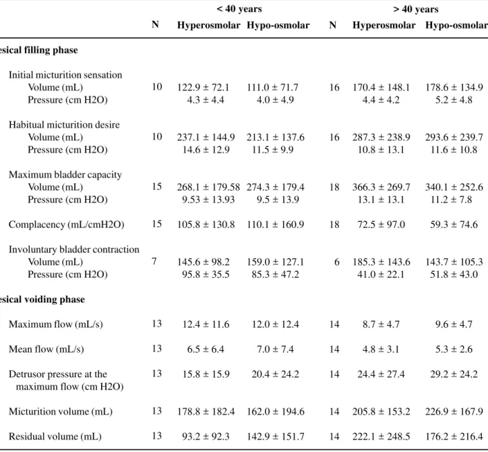
hypo-and hyperosmolar solutions there was no statistical difference in the results in both age groups (Table-2). No difference was found in any of the urodynamic parameters studied when exams utiliz-ing hyper- and hypo-osmolar solutions are compared in female and male sexes (Table-3).
There was no significant difference in the cystometric and flow values in the presence of
hyper-and hypo-osmolar solutions, independently from the presence of detrusor hyperactivity or from the order in which the solutions were instilled. The volume in which involuntary bladder contractions occurred and the detrusor pressure reached during the same did not suffer any interference due to the intravesical osmo-larity change. Aiming at minimizing error factors aris-ing from the obtainment of only the group mean of
Table 2 – Values of urodynamic variables when vesical filling is accomplished with hyper- and hypo-osmolar solutions in patients with age inferior and superior to 40 years-old (mean ± standard deviation).
Vesical filling phase
Initial micturition sensation Volume (mL)
Pressure (cm H2O)
Habitual micturition desire Volume (mL)
Pressure (cm H2O)
Maximum bladder capacity Volume (mL)
Pressure (cm H2O)
Complacency (mL/cmH2O)
Involuntary bladder contraction Volume (mL)
Pressure (cm H2O)
Vesical voiding phase
Maximum flow (mL/s)
Mean flow (mL/s)
Detrusor pressure at the maximum flow (cm H2O)
Micturition volume (mL)
Residual volume (mL)
N
10
10
15
15
7
13
13
13
13
13
Hyperosmolar
122.9 ± 72.1
004.3 ± 4.4
237.1 ± 144.9
014.6 ± 12.9
268.1 ± 179.58
09.53 ± 13.93
105.8 ± 130.8
145.6 ± 98.2
095.8 ± 35.5
012.4 ± 11.6
006.5 ± 6.4
015.8 ± 15.9
178.8 ± 182.4
093.2 ± 92.3
Hypo-osmolar
111.0 ± 71.7
004.0 ± 4.9
213.1 ± 137.6
011.5 ± 9.9
274.3 ± 179.4
009.5 ± 13.9
110.1 ± 160.9
159.0 ± 127.1
085.3 ± 47.2
012.0 ± 12.4
007.0 ± 7.4
020.4 ± 24.2
162.0 ± 194.6
142.9 ± 151.7
N
16
16
18
18
06
14
14
14
14
14
Hyperosmolar
170.4 ± 148.1
004.4 ± 4.2
287.3 ± 238.9
010.8 ± 13.1
366.3 ± 269.7
013.1 ± 13.1
072.5 ± 97.0
185.3 ± 143.6
041.0 ± 22.1
008.7 ± 4.7
004.8 ± 3.1
024.4 ± 27.4
205.8 ± 153.2
222.1 ± 248.5
Hypo-osmolar
178.6 ± 134.9
005.2 ± 4.8
293.6 ± 239.7
011.6 ± 10.8
340.1 ± 252.6
011.2 ± 7.8
059.3 ± 74.6
143.7 ± 105.3
051.8 ± 43.0
009.6 ± 4.7
005.3 ± 2.6
029.2 ± 24.2
226.9 ± 167.9
176.2 ± 216.4
< 40 years > 40 years
Table 3 – Values of urodynamic variables when vesical filling is accomplished with hyper- and hypo-osmolar solutions in female and male patients (mean ± standard deviation).
Vesical filling phase
Initial micturition sensation Volume (mL)
Pressure (cm H2O)
Habitual micturition desire Volume (mL)
Pressure (cm H2O)
Maximum bladder capacity Volume (mL)
Pressure (cmH2O)
Complacency (mL/cm H2O)
Involuntary bladder contraction Volume (mL)
Pressure (cm H2O)
Vesical voiding phase
Maximum flow (mL/s)
Mean flow (mL/s)
Detrusor pressure at the maximum flux (cm H2O)
Micturition volume (mL)
Residual volume (mL)
N
9
9
9
9
3
7
7
7
7
7
Hyperosmolar
110.8 ± 83.5
002.2 ± 1.6
187.4 ± 140.3
011.2 ± 12.1
261.7 ± 176.1
05.56 ± 4.1
115.0 ± 148.5
127.3 ± 77.4
082.0 ± 25.6
018.3 ± 9.9
009.9 ± 5.5
018.7 ± 11.9
234.0 ± 123.7
071.1 ± 68.9
Hypo-osmolar
109.6 ± 78.7
03.67 ± 3.0
186.4 ± 134.3
008.9 ± 8.9
259.7 ± 153.6
005.2 ± 4.2
080.6 ± 54.8
125.3 ± 83.1
059.0 ± 50.2
018.1 ± 10.4
010.4 ± 6.5
028.0 ± 27.6
232.3 ± 148.0
071.3 ± 66.0
N
17
17
24
24
10
20
20
20
20
20
Hyperosmolar
174.0 ± 139.3
005.5 ± 4.7
310.6 ± 225.6
012.8 ± 13.7
344.2 ± 253.2
013.7 ± 15.0
077.4 ± 98.4
174.9 ± 128.6
067.1 ± 44.6
007.7 ± 6.7
004.1 ± 3.8
020.8 ± 25.5
178.3 ± 177.9
191.2 ± 219.3
Hypo-osmolar
175.4 ± 130.5
005.3 ± 5.6
302.9 ± 230.2
013.0 ± 10.9
329.2 ± 242.3
012.6 ± 11.6
083.0 ± 140.5
159.9 ± 123.2
073.1 ± 48.0
008.2 ± 7.3
004.7 ± 4.3
024.0 ± 23.5
182.8 ± 192.6
191.3 ± 204.1
Female Male
* p > 0.05 for all variables in the comparison between hyper- and hypo-osmolar solutions and the sequence in which they were instilled.
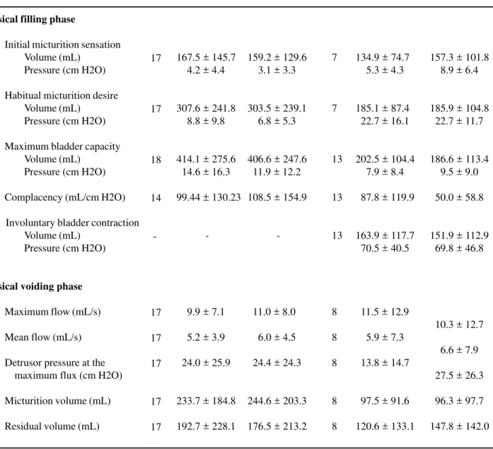
the triggering volume of the first involuntary vesical contraction, we made an individual comparison for each patient. In 54% of the cases there was a reduc-tion of the volume in which the involuntary bladder contraction occurred (the reduction varied from 3 to 40%) when compared hypo- and hyperosmolar solu-tions. Thirty eight percent of the patients presented
and in the maximum detrusor pressure reached, inde-pendently from the fact that the solution is hyper- or hypo-osmolar, of the presence of neuropathy or not and of the sequence of instilling of solutions. Due to the small number of patients without neuropathy and with involuntary bladder contractions (n = 3), these
data were not considered in the analysis of this study. The use of the hyper- and hypo-osmolar solutions did not promote any significant change in vesical compla-cency, in the detrusor pressure at the end of the filling and in the functional bladder capacity in patients with or without neurological diseases (Table-5).
Table 4 – Values of urodynamic variables when vesical filling is accomplished with hyper- and hypo-osmolar solutions in patients with and without vesical hyperactivity (mean ± standard deviation).
Vesical filling phase
Initial micturition sensation Volume (mL)
Pressure (cm H2O)
Habitual micturition desire Volume (mL)
Pressure (cm H2O)
Maximum bladder capacity Volume (mL)
Pressure (cm H2O)
Complacency (mL/cm H2O)
Involuntary bladder contraction Volume (mL)
Pressure (cm H2O)
Vesical voiding phase
Maximum flow (mL/s)
Mean flow (mL/s)
Detrusor pressure at the maximum flux (cm H2O)
Micturition volume (mL)
Residual volume (mL)
N
17
17
18
14
-17
17
17
17
17
Hyperosmolar
167.5 ± 145.7
004.2 ± 4.4
307.6 ± 241.8
008.8 ± 9.8
414.1 ± 275.6
014.6 ± 16.3
99.44 ± 130.23
-009.9 ± 7.1
005.2 ± 3.9
024.0 ± 25.9
233.7 ± 184.8
192.7 ± 228.1
Hypo-osmolar
159.2 ± 129.6
003.1 ± 3.3
303.5 ± 239.1
006.8 ± 5.3
406.6 ± 247.6
011.9 ± 12.2
108.5 ± 154.9
-011.0 ± 8.0
006.0 ± 4.5
024.4 ± 24.3
244.6 ± 203.3
176.5 ± 213.2
N
7
7
13
13
13
8
8
8
8
8
Hyperosmolar
134.9 ± 74.7
005.3 ± 4.3
185.1 ± 87.4
022.7 ± 16.1
202.5 ± 104.4
007.9 ± 8.4
087.8 ± 119.9
163.9 ± 117.7
070.5 ± 40.5
011.5 ± 12.9
005.9 ± 7.3
013.8 ± 14.7
097.5 ± 91.6
120.6 ± 133.1
Hypo-osmolar
157.3 ± 101.8
008.9 ± 6.4
185.9 ± 104.8
022.7 ± 11.7
186.6 ± 113.4
009.5 ± 9.0
050.0 ± 58.8
151.9 ± 112.9
069.8 ± 46.8
010.3 ± 12.7
006.6 ± 7.9
027.5 ± 26.3
096.3 ± 97.7
147.8 ± 142.0
Without Hyperactivity With Hyperactivity
COMMENTS
The concept of impermeability of the urothelium, being attributed to it the simple role of hemato-urinary barrier, has been reassessed the last few years (6,11,12). Some studies about the urothelium physiology have demonstrated the ex-istence of an ion transport through its apical cells, which remain in direct contact with the urine (13). On the other hand, it is not yet well understood the mechanism involved in the conversion of this transcellular ion transport into nerve impulses (14). It was recently demonstrated that there are changes in various physicochemical characteristics of the urine along its trajectory from the kidney until the bladder (5). One of the most notorious is the increase in the vesical urinary osmolarity when compared to the urine obtained directly from the renal pelvis. It is possible that this osmolar variation have implica-tions in the process that involves either vesical sen-sitivity or the triggering of detrusor contraction. It was experimentally demonstrated in rats that the instilling of hyperosmolar solutions with acid pH lead to a reduction of the bladder capacity. The op-posite occurred with the administration of alkaline and hypo-osmolar solutions (15). In this line of re-search the existence of a parallelism between the
oscillations of the urinary pH in the different phases of the menstrual cycle in normal women and the stan-dard micturition behavior presented by them was evidenced. The lesser the urinary pH the more pre-cocious and with lesser intravesical volumes the micturition desire was manifested (3). From the re-port of an inverse relation between urinary osmo-larity and cystometric capacity, it was proposed that the degree of hydration and the hydro-electrolytic balance are factors that possibly interfere in the set-tings of micturition urgency (16).
By developing the present study we have tried to objectively demonstrate through the performance of an urodynamic exam, the existence of a direct ac-tion of the osmolar urine composiac-tion in the vesical filling sensation and in the contractile mechanism of the urinary bladder. Observing the answer of one pa-tient to the hypo- and hyperosmolar solutions and with the same instilling speed, we have minimized the in-terference of individual characteristics in the com-parison of the results obtained. Another very impor-tant aspect in this work’s method was the alternate instilling of solutions in each patient, removing thus the possibility of an error arising from their adminis-tration always in a similar sequence. Therefore, the urodynamic response to the hyperosmolar solution can be differentiated from the one obtained with the
Table 5 – Values of urodynamic variables when vesical filling is accomplished with hyper- and hypo-osmolar solutions in patients with and without neuropathy (mean ± standard deviation).
Vesical filling phase
Maximum bladder capacity Volume (mL)
Pressure (cm H2O)
Complacency (mL/cm H2O)
Involuntary bladder contraction Volume (mL)
Pressure (cm H2O)
N
19
14
03
Hyperosmolar
266.9 ± 173.4
008.6 ± 11.9
099.4 ± 130.2
127.7 ± 149.2
055.7 ± 36.0
Hypo-osmolar
260.7 ± 128.5
008.4 ± 8.2
072.6 ± 84.4
122.3 ± 154.0
046.0 ± 29.0
N
14
19
10
Hyperosmolar
362.0 ± 269.1
013.6 ± 14.4
078.9 ± 101.3
174.8 ± 113.7
075.0 ± 42.5
Hypo-osmolar
346.7 ± 268.4
012.2 ± 12.1
089.5 ± 145.9
160.8 ± 106.6
077.0 ± 49.8
Without Neuropathy With Neuropathy
hypo-osmolar solution, independently from the se-quence in which they were instilled.
However, the differences obtained in this study when compared to the results of instilling hyper-and hypo-osmolar solutions during vesical filling did not reach statistic significance.
As to the detrusor motricity, it was experi-mentally demonstrated in rats, the largest incidence of bladder contractions after instilling a sodium chlo-ride hypertonic solution, when compared to the iso-tonic solution of the same salt (6). In this sense it was already demonstrated that extracellular hyperosmolarity directly depolarize smooth muscu-lar cells generating an increase in the detrusor activ-ity whereas hypo-osmolaractiv-ity promotes opposite re-sponses (18). In our study, when we compared urodynamic manifestations in patients with detrusor hyperactivity, we have verified a lack of difference in urodynamic parameters utilizing both hyper- and hypo-osmolar solutions. The same lack of statistical difference in motor vesical behavior was observed in the micturition phase, compared to the results of the experimental studies described above.
Aiming at establishing if the presence of neu-ropathy could act as an interfering factor in detrusor hyperactivity development, as well as in the other urodynamic variables to both extreme osmolarity so-lutions, we have compared the results between pa-tients bearing a neurological disease and those who did not have such commitment. There was no signifi-cant difference between both groups when exposed to different solutions, independently from the se-quence in which each one was instilled.
We did not obtain different urodynamic re-sponses when we compared both solutions with os-molarity extremes for all analyzed variables in the present study. Such fact made us precociously inter-rupt the study without the inclusion of new patients when the level of significance for statistical analysis was reached. We know, from what was exposed, that the variety of clinical situations of the patients in-volved in this study could have acted as a limiting factor in the interpretation of the results. Thus, little differences in the response during the urodynamic exam may have not reached statistical significance. On the other hand, the clinical and even the
urodynamic value in those cases are questionable. It is also possible that the permanence con-tact time of each solution with the vesical urothelium, i.e., the necessary time to reach vesical plenitude, will have been insufficient for the apical cells to be adapted to the environment they were exposed to and thus, the receptors responsible for the absorption of sodium. Furthermore it is correct to suppose that hyperosmolar solutions derive from oliguria and thus, with longer contact with the urothelium, in normal physiological conditions. Ferguson emphasized in his revision on the urothelial function that the mean time to have a change in the distribution of amiloride-sensitive so-dium concentration receptors is of approximately 15 minutes (14). Evidences published in the last years on a more effective action of urothelium on vesical sensitive behavior; the alteration in urinary osmolar-ity along the excretory system, are strong indications that a more profound research is necessary in this field of urology.
CONCLUSIONS
Vesical fillings with solutions that simulate extreme urinary osmolarities, performed with simi-lar speed and without previous identification do not alter sensitive or motor urodynamic behavior in pa-tients of the present study. To our knowledge, it is the first double-blind study in the literature where 2 so-lutions that simulate extreme urinary osmolarities (hyper- and hypo-osmolarities), were tested during an urodynamic study.
CONFLICT OF INTEREST
None declared.
REFERENCES
1. Higson RH, Smith JC, Hills W: Intravesical lignocaine and detrusor instability. Br J Urol. 1979; 51: 500-3. 2. Aslund K, Rentzhogh L, Sandstromb G: Effects of ice
3. Lavin JM, Hosker GL, Smith AR: Does urinary pH influence micturition desire? Neurourol Urodyn. 1997; 16: 396-7.
4. Rittig S, Matthiesen TB, Pedersen EB, Djurhuus JC: Sodium regulating hormones in enuresis. Scand J Urol Nephrol Suppl. 1999; 202: 45-6.
5. Cahill DJ, Fry CH, Foxall PJ: Variation in urine com-position in the human urinary tract: evidence of urothelial function in situ? J Urol. 2003; 169: 871-4. 6. Hohlbrugger G: Changes of hypo- and hypertonic
so-dium chloride induced by the rat urinary bladder at vari-ous filling stages. Evidence for an increased transurothelial access of urine to detrusor nerve and muscle cells with distension. Eur Urol. 1987; 13: 83-9. 7. Chaiyaprasithi B, Mang CF, Kilbinger H, Hohenfellner M: Inhibition of human detrusor contraction by a urothelium derived factor. J Urol. 2003; 170: 1897-900.
8. Mark SD, Webster GD: Detrusor hyperactivity. In: Raz S (ed.), Female Urology, 2nd ed. Philadelphia, Saunders. 1996; pp. 231-43.
9. Gerber GS, Brendler CB: Evaluation of the urologic patient: history, physical examination, and urinalysis. In Walsh PC, Retik AB, Vaughan Jr ED, Wein AJ (ed.), Campbell’s Urology, 8th ed. Philadelphia, Saunders. 2002; pp. 83-110.
10. Timm NH: Profile Analysis. In: Timm, Multivariate Analysis with Applications in Education and
Psychol-ogy. 1st ed. Monterey, Brooks / Cole Publishing Co. 1975; p. 444.
11. Ekstrom B: Intravesical instillation of drugs in patients with detrusor hyperactivity. Scand J Urol Nephrol Suppl. 1992; 149: 1-67.
12. Shafik A, El Sibai O, Shafik AA, Ahmed I: Do vesical and voided urine have identical compositions? Scand J Urol Nephrol. 2004; 38: 243-6.
13. Lewis SA, Diamond JM: Active sodium transport by mammalian urinary bladder. Nature. 1975; 253: 747-8.
14. Ferguson DR: Urothelial function. BJU Int. 1999; 84: 235-42.
15. Hohlbrugger G, Lentsch P: Intravesical ions, osmo-lality and pH influence the volume pressure response in the normal rat bladder, and this is more pro-nounced after DMSO exposure. Eur Urol. 1985; 11: 127-30.
16. Lee F, Oliver SE, Susser J, Mundy AR, Craggs MD, Foxall PJ: The effect of urine composition on sensa-tions of urinary urge. BJU Int. 2001; 88: 287. 17. Ohshima K: Effects of various osmotic solutions on
membrane properties of smooth muscle cells of the guinea pig ureter. Invest Urol. 1981; 19: 79-84. 18. Uvelius B: Effects of variations in extracellular
osmo-lality on spontaneous contractile activity and response to nerve stimulation in rat detrusor muscle in vitro. Urol Int. 1985; 40: 196-200.
Received: March 3, 2005 Accepted after revision: August 31, 2005
Correspondence address:
Dr. José Carlos Truzzi
Rua Dr. Oscar Monteiro de Barros, 617 / 141 São Paulo, SP, 05641-010, Brazil