Structure-Based Vaccines Provide Protection in a Mouse
Model of Ehrlichiosis
Sunil Thomas1,2*, Nagaraja R. Thirumalapura1, Patricia A. Crocquet-Valdes1, Bruce A. Luxon3, David H. Walker1,2,4*
1Department of Pathology, University of Texas Medical Branch, Galveston, Texas, United States of America,2Center for Biodefense and Emerging Infectious Diseases, University of Texas Medical Branch, Galveston, Texas, United States of America,3Institute of Human Infections and Immunity, Institute for Translational Science, Department of Biochemistry and Molecular Biology, University of Texas Medical Branch, Galveston, Texas, United States of America, 4Sealy Center for Vaccine Development, University of Texas Medical Branch, Galveston, Texas, United States of America
Abstract
Background:Recent advances in bioinformatics have made it possible to predict the B cell and T cell epitopes of antigenic proteins. This has led to design of peptide based vaccines that are more specific, safe, and easy to produce. The obligately intracellular gram negative bacteriaEhrlichiacause ehrlichioses in humans and animals. As yet there are no vaccines to protect againstEhrlichiainfection.
Methodology/Principal Findings:We applied the principle of structural vaccinology to design peptides to the epitopes of Ehrlichia murisouter membrane P28-19 (OMP-1/P28) andEhrlichiaHeat shock protein 60 (Hsp60/GroEL) antigenic proteins. Both P28-19 andEhrlichiaHsp60 peptides reacted with polyclonal antibodies againstE. canisandE. chaffeensisand could be used as a diagnostic tool for ehrlichiosis. In addition, we demonstrated that mice vaccinated withEhrlichiaP28-19 and Hsp60 peptides and later challenged withE. muriswere protected against the pathogen.
Conclusions/Significance:Our results demonstrate the power of structural vaccines and could be a new strategy in the development of vaccines to provide protection against pathogenic microorganisms.
Citation:Thomas S, Thirumalapura NR, Crocquet-Valdes PA, Luxon BA, Walker DH (2011) Structure-Based Vaccines Provide Protection in a Mouse Model of Ehrlichiosis. PLoS ONE 6(11): e27981. doi:10.1371/journal.pone.0027981
Editor:Mauricio Martins Rodrigues, Federal University of Sa˜o Paulo, Brazil
ReceivedAugust 18, 2011;AcceptedOctober 28, 2011;PublishedNovember 17, 2011
Copyright:ß2011 Thomas et al. This is an open-access article distributed under the terms of the Creative Commons Attribution License, which permits unrestricted use, distribution, and reproduction in any medium, provided the original author and source are credited.
Funding:This study was supported by grant AI31431 from the National Institute of Allergy and Infectious Diseases. The funder had no role in study design, data collection and analysis, decision to publish, or preparation of the manuscript.
Competing Interests:The authors have declared that no competing interests exist. * E-mail: [email protected] (ST); [email protected] (DHW)
Introduction
Vaccines are considered as one of the most successful medical intervention against infectious diseases. Vaccines include killed or attenuated organisms or purified products derived from them. One of the drawbacks of killed or attenuated vaccines is the potential side effect of some of the antigenic proteins. This led to the design of recombinant vaccines based on whole antigens. As whole antigenic proteins are not essential in inducing immunity, it led to the emergence of a new branch of vaccine design termed structural vaccinology [1,2]. Structure based vaccines have the rationale that protective epitopes are enough to induce immune responses and provide protection against pathogens [3]. Structure-based peptide antigens induce antibodies which recognize the denatured form of a protein from which their sequences are derived [4].
The obligate intracellular bacterium Ehrlichia chaffeensis that resides in mononuclear phagocytes is the etiologic agent of human monocytotropic ehrlichiosis (HME). HME is an emerging and often life-threatening zoonotic, tick-transmitted infectious disease in the United States [5–7]. Lack of early diagnosis and treatment of HME are the main factors that lead to severe and fatal disease.
Ehrlichia also causes diseases in companion animals and
domes-ticated ruminants. E. chaffeensis and E. canis cause canine ehrlichioses in dogs, whereasE. ruminantiumcauses heartwater in cattle, sheep and goats. Vaccines are needed for these tick transmitted pathogens, but are hindered by many obstacles that exist in their development. These include knowledge of genetic and antigenic variability, identification of the ehrlichial antigens that stimulate protective immunity or elicit immunopathology, development of animal models that reflect the immune responses of the hosts and understanding molecular host-pathogen interac-tions involved in immune evasion or that may be blocked by the host immune response. As yet there are no commercially available vaccines to protect against ehrlichiosis [8].
Development of a murine model of persistent ehrlichiosis has greatly facilitated our understanding of the pathogenesis and mechanisms of host defenses against ehrlichial infections. Mildly virulentEhrlichia muris infection in immunocompetent C57BL/6 mice results in persistent infection and mimics E. chaffeensis
induction of strong cell-mediated CD4 and CD8 type 1 responses and humoral immunity [12]. T cell independent humoral immunity has also been reported to be sufficient for protection against fatal infection with intracellular ehrlichial pathogens [16]. We recently demonstrated that Ehrlichia Hsp60 (GroEL) and P28 (OMP-1) are the major antigenic proteins ofE. murisand they are also post-translationally modified [17]. Based on these observations, we bioinformatically identified the predicted hydro-philic epitope sequences of Ehrlichia Hsp60 and P28-19 and synthesized peptides to use as diagnostic probes to detect antibodies against Ehrlichia. In this paper we demonstrate that both Ehrlichia muris Hsp60 and P28 peptides reacted with antibodies againstE. chaffeensisandE. canisand could be used in diagnosis of infected animals. We also demonstrated that the structure-based peptides provided protection againstEhrlichiaand hence could be used as a vaccine against ehrlichioses.
Results
Peptides based on the epitopes of EhrlichiaHsp60 and outer membrane protein P28-19 detected pathogen-specific antibodies
The outer membrane proteins P28 andEhrlichiaHsp60 (GroEL) are the major antigenic proteins ofEhrlichia[17]. Previous studies in our laboratory and elsewhere have demonstrated that P28-19 is the most efficient and protective P28 paralog which provides protection againstEhrlichia[18,19].
We identified theEhrlichiaHsp60 and P28-19 epitopes based on the predicted hydrophilicity [20]. Three regions of theE. muris
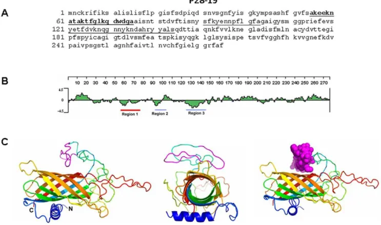
P28-19 protein sequence had substantial predicted hydrophilicity as determined by the Lasergene software. The peptides correspond to amino acids 55–75, 91–103, and 124–145 (Fig. 1). Initial studies demonstrated that the peptide corresponding to the amino acids 55–75 of the P28-19 protein is more sensitive in detection of
Ehrlichiaspecific antibodies. Based on the information we injected P28-1955275peptide to induce antibody generation or function as
a vaccine candidate.
Three regions of the Ehrlichia Hsp60 protein sequence had substantial predicted hydrophilicity as determined by the Laser-gene software (DNAStar, WI, USA). The peptides correspond to amino acids 43–63, 179–200, and 386–406 (Fig. 2). The peptide corresponding to amino acid 43–63 ofEhrlichiaHsp60 was found to be more reactive with variousEhrlichia- specific antibodies and also induced production of antibodies (Fig. 2). Based on the information we injectedEhrlichia Hsp60 43–63 peptide to induce
antibody generation or function as a vaccine candidate.
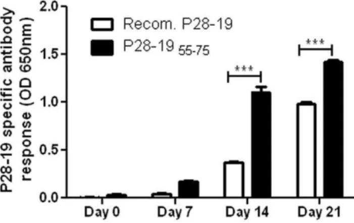
We probed for Ehrlichia-specific antibodies in sera of mice infected withE. muris. As a control we used recombinant P28-19 protein. The peptide P28-1955–75was found to be more sensitive
in detectingEhrlichia-specific antibody than recombinant P28-19. The peptide detected theEhrlichia-specific antibody even from sera of mice obtained as early as seven days after E. murisinfection (Fig. 3). In subsequent studies we used P28-1955-75as a diagnostic
probe or vaccine candidate.
Figure 1. Amino acid sequence of P28-19.(A) P28-19 peptides corresponding to the underlined predicted hydrophilic sequence were synthesized. The peptide corresponding to the bold underlined (55–75) sequence was found to react with antibodies toEhrlichiaas well as to induce antibody production. (B) Hydrophobicity plot of P28-19. The sequences underlined (in red and blue) were used for synthesizing peptides; however the best peptide sequence selected is underlined in red. (C) (Left) Predicted 3D structure of P28-19 (side view), (Middle) predicted 3D structure of P28-19 (basal view), (Right) predicted 3D structure of P28-19 with the Van der Waals radii of the heavy atoms highlighting the region of interest (P28-1955275).
We probed sera of dogs with ehrlichioses to determine the efficacy of both P28-19 55–75 and Hsp60 43–63 peptides as
diagnostic tools. We coated the peptides on an ELISA plate and assayed forE. chaffeensisandE. canisantibody from sera of infected dogs. The ELISA plate coated with the Hsp60 43–63 peptide
detectedEhrlichia-specific antibody in the sera of dogs infected with
E. canis or E. chaffeensis(Fig. 4A). The assay confirmed that the peptide corresponding to theEhrlichiaHsp6043–63epitope could
be used in diagnostics. Similarly, P28-19 55–75 peptide detected
antibodies to both E. chaffeensis and E. canis from infected dog serum samples, whereas, there was no antibody binding from sera of uninfected dogs (Fig. 4B). To confirm that P28 and Ehrlichia
Hsp60 peptides are specific to detect Ehrlichia but not other pathogens we probed sera of mice infected withRickettsia australis,
R. conorii, R. akariandOrientia tsusugamushi. The P28-1955-75and Ehrlichia Hsp60 43–63 peptides did not react with sera of mice
infected with Rickettsia australis, R. conorii, R. akari and Orientia tsusugamushi (Fig. 4C, 4D). The experiments demonstrated that bothEhrlichiaHsp6043–63and P28-1955–75epitope peptides could
be used as antigens in diagnostic applications.
Antibodies against the epitopes ofEhrlichiaHsp60 and the outer membrane protein P28-19 detected pathogen-specific antigen
Antibodies are used extensively as diagnostic tools in a wide array of different analyses. We injected different amounts of
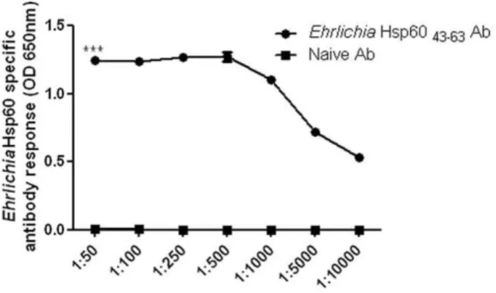
Ehrlichia Hsp60 43–63 to induce polyclonal antibody. Fifty
micrograms (0.02mM) of Ehrlichia Hsp60 43–63 peptide induced
the optimum amount of antibody for our studies (data not shown). Using ELISA we determined the detectable level of antibody which could bind toEhrlichiaHsp6043–63peptide. The antibody to EhrlichiaHsp60 detected EhrlichiaHsp60 43–63peptide even at a
Figure 2. Amino acid sequence ofEhrlichiaHsp60.(A) Hsp60 peptides corresponding to the underlined predicted hydrophilic sequence were synthesized. The peptide corresponding to the bold underlined (43–63) sequence was found to react with antibodies toEhrlichiaas well as to induce antibody production. (B) Hydrophobicity plot ofEhrlichiaHsp60. The sequences underlined (in red and blue) were used for synthesizing peptides; however, the best peptide sequence selected is underlined in red. (C) (Left) Predicted 3D structure ofEhrlichiaHsp60, (Right) predicted 3D structure ofEhrlichiaHsp60 with the Van der Waals radii of the heavy atoms highlighting the region of interest (Hsp6043–63).
doi:10.1371/journal.pone.0027981.g002
high dilution (Fig. 5) (p,0.001 as determined by two way ANOVA). We also used the antibody to probe forE. murisand
E. chaffeensisin DH82 monocytic cells [21]. To determine whether the antibodies against the epitope of P28-1955–75could be used as
a probe we immunized mice with P28-1955–75peptide and used
the sera as a probe to detectE. murisin DH82 cells. The P28-1955–75
peptide induced antibody production that detected E. muris in DH82 cells as determined by fluorescence microscopy (Fig. 6A).
The peptides of P28-1955–75andEhrlichiaHsp6043–63
functioned as vaccines to protect against the pathogen Since the P28-19 55–75 and Ehrlichia Hsp60 43–63 epitope
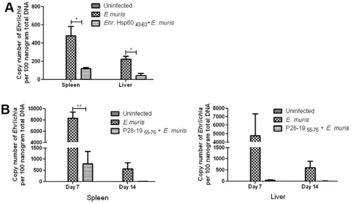
peptides induced antibodies, we reasoned that they also could provide protection against Ehrlichia thereby functioning as potential vaccine candidates. To prove our hypothesis mice were injected with P28-1955–75orEhrlichiaHsp6043–63epitope peptides
and challenged 30 days later withE. muris. The spleen and liver were collected at different days after bacterial challenge and the bacterial copy number determined by quantitative real time PCR. We observed lower bacterial load in both spleen and liver on days 7 and 14 after bacterial infection in the vaccinated mice compared to unvaccinated controls (Fig. 7). The results demonstrated that P28-19 55–75 and Ehrlichia Hsp60 43–63 peptides functioned as
vaccine candidates and provided protection against Ehrlichia
infection.
Immunization with vaccines stimulates the immune system to produce a robust antibody response that can provide protection against pathogens. To determine the antibody responses against theEhrlichiaHsp6043–63peptide vaccine, we collected blood from
vaccinated mice on days 7 and 14 and performed ELISA. There was a significant difference in the antibody response between unvaccinated and Ehrlichia Hsp60 43–63 vaccinated mice after
challenge withE. muris. However, there was no difference between the antibody levels in vaccinated mice between days 7 and 14. The
EhrlichiaHsp6043–63-specific antibody levels in infected
unvacci-nated mice were highest on day 14 compared to day 7 (Fig. 8A). To determine the antibody responses against the P28-19 55–75
peptide vaccine, we collected blood from immunized mice on days 7 and 14 and subjected the samples to ELISA. There was a significant difference in the antibody response between unvacci-nated and P28-1955–75 vaccinated mice after challenge withE.
muris. Antibody levels were higher on day 14 compared to day 7 (Fig. 8B).
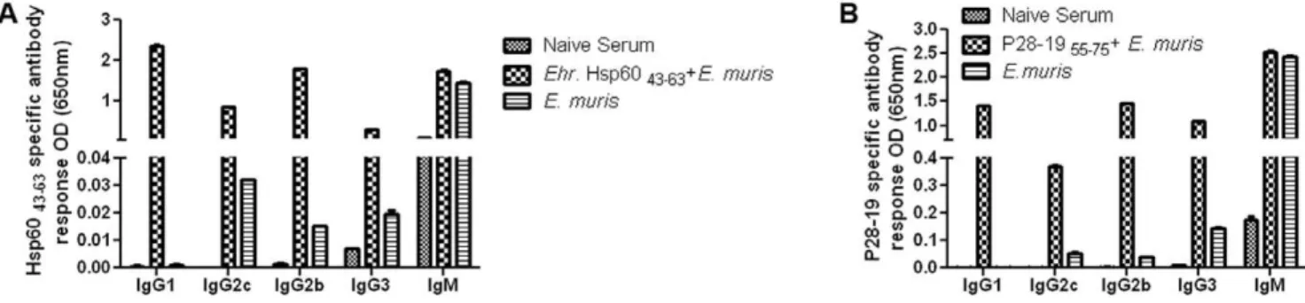
As antibody isotype responses can be useful indicators of immune bias during infection [22], we determined the antibody isotypes after vaccination with the peptide epitopes. The level of antibody isotypes increased by day 14 compared to day 7 after bacterial challenge (data not shown). TheEhrlichia Hsp6043–63
-vaccinated mice had higher levels of IgG1, IgG2c, IgG2b, IgG3 and IgM after bacterial challenge compared to unvaccinated mice on day 14 (Fig. 9A). By ELISA we analyzed the isotypes of the antibodies of P28-19 peptide in vaccinated and unvaccinated mice after challenge withE. muris(day 14 post challenge). The P28-1955–75
vaccinated mice challenged withE. murishad higher levels of IgG1, IgG2b, IgG3 and IgM compared to unvaccinated mice infected with the pathogen (Fig. 9B).
EhrlichiaHsp60 and P28-19 specific memory CD4+Th1 responses are induced duringE. murisinfection
We determined by flow cytometry ifEhrlichiaHsp6043–63and
P28-19 specific memory T cells are induced during E. muris
infection. Splenocytes fromE. muris-infected mice were harvested on day 45 post-infection and stimulatedin vitrowith theEhrlichia
Hsp6043–63and P28-1955–75 for 18h. Compared to uninfected
naı¨ve mice, E. muris-infected mice had significantly higher frequencies and absolute numbers of Ehrlichia Hsp60 43–63 and
P28-1955–75 specific IFN-c-producing CD4+ Th1 cells in their spleen (Figure 10).
Discussion
The genetic diversity of microorganisms presents a challenge in developing broadly effective vaccines [23]. Vaccine design is progressing from empiricism towards the increasingly rational presentation of the targets of protective immunity [1]. Using structural information of proteins, it is possible to engineer optimized antigens that are more stable, homogeneous, and efficiently produced, making immunization more practical and affordable [1]. Thus structural vaccinology is an emerging strategy for the rational design of vaccine candidates [2]. Structural vaccines based on epitopes are more specific, safe, and easier to produce [24], and peptide vaccines based on the epitopes represent a potential strategy for the prevention and treatment of pathogenic diseases, cancers and autoimmune disorders. Their low cost, ease of synthesis and inherent safety are all attractive features [25]. Structure-based design has been employed to develop vaccines against Group B Streptococcus infections [2],
Neisseria [25], and influenza virus [26,27]. Scarselli et al. [28] combined atomic level structural information with genomics and classical vaccinology to design a single immunogen that elicits protective immunity against more than 300 natural variants of the bacterial pathogen meningococcus group B. This accomplishment provides a glimpse of the power of structure-based vaccine design to create immunogens capable of eliciting protective responses against genetically diverse pathogens.
Ehrlichiaare host defense-evasive and cell function-manipulative, vector-transmitted pathogens and are responsible for serious diseases of agricultural, veterinary and human importance [8].
E. chaffeensis, E. ewingii, and anE. muris-like organism are the known species ofEhrlichiainfecting humans [5,29,30]. Interestingly, recent studies demonstrated E. muris-like organisms infecting deer in Wisconsin [31]. In dogs,E. canis,E. chaffeensisandE. ewingiiare the major pathogens; whereas in livestock of sub-Saharan Africa E. ruminantiumcause serious disease. As yet there are no commercially available vaccines to protect against ehrlichioses.
Figure 3. P28-1955-75peptide reacted withE. murisantibody.
The peptide corresponding to the predicted hydrophilic sequence of amino acids 55–75 of P28-19 reacted with Ehrlichia antibody. The peptide was found to be more sensitive in reacting with theEhrlichia
antibody than the recombinant P28-19 protein (***p,0.001 as determined by Studentttest).
Successful trials with live and attenuated vaccines against E. ruminantium indicate that vaccine-induced immunity is feasible for
Ehrlichia spp. containing defined immunoprotective proteins. Some ehrlichial proteins targeted by the host immune response are well defined, such as major OMPs (MAP1, MAP2 and OMP-1) [8]. Studies from our laboratory and others have previously demonstrated the importance of antibodies in protection againstEhrlichia. Passive transfer of polyclonal immune sera or mAbs confers protection in SCID mice againstE. murisandE. chaffeensis, respectively [18,32]. Prior studies in mouse models have demonstrated that vaccination with P28 leads to immunity and clearance ofEhrlichiainfection.
Usingin silicotechniques, we identified the predicted hydrophilic epitopes of P28-19 andEhrlichiaHsp60 which induce an antibody response. We selected epitope regions which did not have any hydrophobic sequences. Of the three epitopes selected from each of the two antigenic proteins only the peptide corresponding to epitope at the N-terminal sequence of the protein was found to be highly reactive with the sera. One of the reasons the N-terminus sequence of proteins exhibits immunogenicity is that these regions have a higher tendency to be exposed in the protein’s native conformation [33]. We also confirmed by BLAST analysis (http:// blast.ncbi.nlm.nih.gov/Blast.cgi) that the sequences of P28-19 and Figure 4.EhrlichiaHsp6043–63and P28-1955–75peptides reacted withEhrlichia-specific antibody from dogs infected withE. canisand E. chaffeensis.(A)EhrlichiaHsp6043–63peptide reacted with antibodies from five dogs infected withE. chaffeensisand five dogs infected withE.
canis.(B) P28-1955–75peptide reacted with antibodies from five dogs infected withE. chaffeensisand five dogs infected withE. canis. Each bar
represents the mean of three replicates. The horizontal line in the graphs represents Mean+3 SD of negative samples. The positive samples are significantly different from negative samples. (C) P28-1955–75peptide did not react with antibodies from mice infected withRickettsiaorOrientia. (D)
EhrlichiaHsp6043–63peptide did not react with antibodies from mice infected withRickettsiaorOrientia.
doi:10.1371/journal.pone.0027981.g004
Ehrlichia Hsp60 selected for peptide generation are highly conserved in the genusEhrlichia(data not shown).
Zhang et al. [34] used recombinant proteins and peptides corresponding to the hypervariable region of P28 to detect
Ehrlichia specific antibody in infected dogs. In our studies we designed peptides based on hydrophilicity. The P28-19 peptide corresponding to the amino acid sequence 55–75 had the highest reactivity withEhrlichiaantibodies, and it also induced antibodies. A previous study showed that mice immunized with the recombinantE. chaffeensisP28-19 (rP28) enhanced the spontaneous clearance of the infection in BALB/c mice [35]. A study by Li et al. [18] indicated that a mAb directed against theE. chaffeensis P28-19 (OMP-1g) reduced the bacterial burden in SCID mice. Though they reported that the region spanning amino acids 68–90 of
P28-19 ofE. chaffeensiscontains a B cell eptiope, subsequent studies by the group [19] utilized the 77–92 amino acid sequence to generate peptides. The peptides were found to be weak in inducing immunogenicity compared to peptides from other regions of P28-19. The high specificity of our peptide may be related to the hydrophilicity of the sequence.
Ehrlichia lacks many genes involved in metabolism, and it requires various nutrients and metabolic compounds facilitated through pores or channels in the bacterial outer membrane [36]. Kumagai et al. [36] demonstrated that the Ehrlichia outer membrane P28-19 has porin activities and the structure of the protein was consistent with a beta-barrel structure, but the authors did not show the 3D structure of P28. We confirm based on the predicted 3D structure that the P28-19 has a beta-barrel structure and the hydrophilic epitope of our interest is located in the the external loop. Bioinformatics analyses demonstrated thatEhrlichia
Hsp60 43–63 and P28-19 55–75 are highly conserved in Ehrlichia
(.95%). The peptide corresponding toEhrlichiaHsp60 amino acid sequence 43–64 cross reacted with E. canis and E. chaffeensis
antibodies. As the peptides induce an antibody response, we reasoned that the peptide could function as an immunogen, and the induced antibodies could clear the pathogens. Our studies demonstrated that both Ehrlichia Hsp60 43–63 and P28-19 55–75
detectedEhrlichiaspecific antibodies in mice and dogs infected with differentEhrlichiaspecies, whereas, the peptides did not react with any non-specific antibodies. A preliminary experiment demon-strated that the peptides could also detectEhrlichiaantibodies from human sera infected withEhrlichia(data not shown). The results demonstrated that both the peptides could be used in diagnosis of
Ehrlichiain animals and humans.
As peptidesper seare poor in inducing immunogenicity KLH is used as a career protein. It has been demonstrated that use of peptides in ELISA in the absence of KLH career protein was found to be less sensitive [37]. Roberts et al. [37] also demon-strated that KLH was also necessary for a strong and specific antibody response. Vaccination with theEhrlichiaHsp6043–63and
P28-1955–75peptides did not fully reduce the bacterial load after 7
days of Ehrlichia challenge. By day 14 after bacterial challenge Figure 5. Reactivity of Ehrlichia Hsp60 antibody. TheEhrlichia
Hsp6043–63peptide was sensitive in detecting different dilutions of the
EhrlichiaHsp60 antibody. TheEhrlichiaHsp60 (250ng) was probed with different dilutions ofEhrlichiaHsp60 specific sera or sera from naı¨ve mice. Finally they were probed with goat anti-mouse-AP (1:2000) and the OD measured at 650 nm after the addition of substrate. Each value is the mean of 3 replicates (***p,0.001 as determined by two way ANOVA).
doi:10.1371/journal.pone.0027981.g005
Figure 6. Detection ofE. murisby fluorescence microscopy.(A) Antibodies produced against peptide P28-1955–75detectedE. murisin infected
DH82 cells. Left: Infected DH82 cell probed with P28-19 antibody, followed by FITC conjugated goat anti-mouse antibody. Middle: DAPI staining. Right: Merge. Arrows indicateE. muris.(B) Naı¨ve serum did not detectE. murisin DH82 cells. Left: Infected DH82 cell probed with naive serum, followed by FITC-conjugated goat anti-mouse antibody. Middle: DAPI staining. Right: Merge. Arrows indicateE. muris.
there was no detectable Ehrlichia load, the antibody levels also increased by day 14 compared to day 7. The low reduction in bacterial load by day 14 may be due to the high levels of antibody and its isotypes on day 14 after bacterial challenge.
Hsps are highly conserved in nature from bacteria to humans [38]. They are molecular chaperones essential for maintaining cellular functions by preventing misfolding and aggregation of nascent polypeptides and by facilitating protein folding [39]. Bacterial Hsp proteins are immunomodulatory and are known to stimulate monocytes and macrophages [40]. Furthermore, Hsps have been reported to be dominant antigens for the host immune response to various pathogens and, thus, have great potential as vaccine candidates [41]. Hsp60 has been an effective subunit vaccine against Yersinia enterocolitica [42], and Woo et al. [43]
demonstrated that Hsp60 is a highly antigenic protein in
Burkholderia pseudomallei. Patients infected with B. pseudomallei
develop a strong antibody response against Hsp60, suggesting that the recombinant protein may be useful for serodiagnosis and vaccine development [43]. Hsp60 of Francisella tularensis was evaluated as a vaccine candidate by Noah et al. [44], which elicited secretion of the chemokines CXCL8 and CCL2 through a TLR4-dependent mechanism. A combination of LPS and Hsp60 increased immunity against the pathogen compared to immuni-zation with LPS alone. Analysis of antibody isotypes of mice vaccinated with GroEL of Salmonella enterica serovar Typhi suggested predominance of the Th2 type immune response in animals immunized with GroEL and adjuvant, whereas immuni-zation of animals with GroEL-alone shifted the immune response Figure 7. Immunization with EhrlichiaHsp60 43–63and P28-19 55–75 peptides protected mice fromEhrlichia infection. (A) Mice
immunized withEhrlichiaHsp6043–63were protected againstE. murischallenge as determined by the bacterial load measured by quantitative real
time-PCR on day 14 afterE. murischallenge (*p,0.05 as determined by t test). (B) Mice immunized with P28-1955–75peptide was protected againstE.
murischallenge as determined by the bacterial load measured by quantitative real time-PCR on days 7 and 14 afterE. murischallenge (**p,0.01 as determined byttest).
doi:10.1371/journal.pone.0027981.g007
Figure 8. Protection induced byEhrlichiaHsp6043–63and P28-1955–75peptides was associated with induction ofEhrlichia- specific IgG antibody.(A)EhrlichiaHsp6043–63vaccinated mice induced higher IgG antibody levels after challenge withE.muris compared to unvaccinated
E. muris-infected mice (***p,0.001 as determined by t test). (B) P28-1955–75peptide vaccinated mice induced higher IgG antibody levels afterE. muris
challenge compared to unvaccinatedE. muris-infected mice (***p,0.001 as determined by Studentttest). doi:10.1371/journal.pone.0027981.g008
toward the Th1 phenotype. Mice immunized with Hsp60 showed a significant increase in lymphocyte proliferation and had higher IFN-cand IL-2 levels [40], and demonstrated effective protection by Hsp60 against human typhoid. Hsp60 also provided protection againstB. anthracisby stimulating both humoral and cell mediated immunity [45].
In addition to their use as indicators of cytokine bias during infection, antibody isotypes have direct functional relevance to disease severity in helminth-malaria co-infection. Antibodies are absolutely required for the ultimate clearance of malaria parasites. In mice, antibodies of the cytophilic isotype IgG2a have been shown to recognize infected erythrocytes and facilitate their destruction by phagocytes. Similarly, in humans IgG1 and IgG3 antibodies are associated with enhanced parasite clearance (reviewed by Fairlie-Clarke et al. [22]).
We observed an increase in spleen size (splenomegaly) after infection (data not shown). The size of the spleen was largest on day 14 and was gradually reduced after day 21, and by day 28 the size corresponded to that of a normal mouse. E. murisinfection resulted in an increase in antibody levels on day 14 after bacterial infection; interestingly the vaccinated mice also cleared the bacteria completely on day 14. It has been demonstrated that antibodies against Bordetella parapertussis have no effect until B. parapertussis-specific T cell responses are generated around day 14 [46]. TheEhrlichiaHsp60 and P28-19 peptides are likely to induce CD4+ T cell responses as we detected antigen-specific IFN-c -producing memory CD4+T cells in mice infected withE. murison day 45 post-infection. Our study suggestsEhrlichiaHsp6043–63and
P28-1955–75peptides controlledEhrlichiaby stimulating B and T
cells.
Overall, our studies demonstrate the power of structural vaccinology. Peptides designed based on the hydrophilicity of P28-19 andEhrlichiaHsp60 could act as diagnostic antigens and could also function as a vaccine. Rudra et al. [47] demonstrated a self-assembling peptide acting as an immune adjuvant. Future studies will involve the design and development of structural vaccines, which could also include peptide sequences that could act as adjuvants.
Materials and Methods
Design of P28-19 andEhrlichiaHsp60 peptides
To determine a protein sequence for potential antigenic epitopes, sequences that are hydrophilic, surface-oriented, and flexible are selected. Most naturally occurring proteins in aqueous solutions have their hydrophilic residues on the protein surface and hydrophobic residues buried in the interior. Three regions of
the E. muris P28-19 and Hsp60 protein sequence had good hydrophilicity predicted by the Lasergene software (DNAStar, WI, USA). We selected hydrophilic sequences of both the
Ehrlichia P28-19 and Hsp60 proteins with no hydrophobic residues. The hydrophilic regions of P28-19 correspond to amino acids 55–75, 91–103, and 124–145 (Fig. 1). The hydrophilic regions ofEhrlichiaHsp60 correspond to amino acids 43–63, 179– 199, and 387–406 (Fig. 2). The sequences showed homology to otherEhrlichiaspecies. The peptides (underlined) were synthesized and conjugated to KLH (Biosynthesis, Lewisville, TX) and used as probes to detect antibodies toE. canisandE. chaffeensis or to raise antibodies.
3D structure prediction
The 3D structure of P28-19 in Fig. 1 was modeled using the online I-TASSER (iterative threading assembly refinement) server [48,49]. I-TASSER builds 3D models from an amino acid sequence using fold recognition and multiple-threading alignments by LOMETS, a meta-threading server in the Zhang lab at the Univ. of Michigan which combines seven
state-of-the-art threading programs (FUGUE, HHsearch, MUSTER,
PROSPECT, PPA, SP3 and SPARK) then performs iterative structural assembly simulations. The function of the predicted models is then inferred by structurally matching the 3D models with known proteins using protein function databases. The best predicted model from I-TASSER (Fig. 1) gave a C-score of 23.338, a TM-score of 0.3460.12, and an Exp. RMSD of 14.163.8. The C-score is a confidence value for estimating the quality of the model and generally ranges from [25, 2] with a higher score being better; TM-scores measure structural similarity and are used to measure the accuracy of structural modeling with a TM-score.0.5 indicating a model having the correct topology and a TM-score ,0.17 showing random similarity. RMSD is simply the average distance of all amino acid pairs between two structures. Protein segments that are relatively unstructured such a loops and coils can result in high RMSD scores. Based on these results the beta-barrel portion of the model in Fig. 1 is likely to be a reasonable representation of the 3D structure of body of the protein. The coils however, which were modeledab initio, are likely idiosyncratic and there is no way to verify their structure without doing x-ray crystallog-raphy or NMR, which are well-beyond the scope of this study. We double checked this model against the best model produced by Phyre2 with similar results. Thus, this model should be approached with caution and care taken not to over-interpret the structure of the loops and coils.
Figure 9. Antibody isotypes in mice immunized withEhrlichiaHsp6043–63and P28-1955–75peptides.(A) Mice vaccinated withEhrlichia
Hsp6043–63peptide had higher levels of IgG1, IgG2c, IgG2b, and IgG3 compared to unvaccinated mice after bacterial challenge. (B) Mice vaccinated
with P28-1955–75peptide had higher levels of IgG1, IgG2b, IgG2c, and IgG3 compared to unvaccinated mice after bacterial challenge. The data were
The 3D structure of Ehrlichia Hsp60 in Fig. 2 was modeled using the online Phyre2 server [50]. Phyre2 aligns hidden Markov models via HHsearch to improve alignment accuracy and detection rate. In ‘‘intensive’’ mode, which was used here, Phyre2 also incorporates Poing [51], a new ab initio folding simulation based on Langevin dynamics, to model regions of the protein that have no detectable homology with known structures. For our Hsp60 sequence, 100% of the residues were modeled at .90% confidence level. The top three PDB models, all GroEL chaperone proteins, had 100% confidence levels and sequence ids of 51–56%. While the model presented in Fig. 2 is likely to be a reasonable estimate of the true 3D structure of this protein, there
is no way to validate this so caution should be used in its interpretation.
Detection of anti-Ehrlichiaantibodies using theEhrlichia Hsp6043–63 and P28-1955–75peptides
We used ELISA to detect Ehrlichia antibodies in the sera of infected mice and dogs. 250 nanograms of the peptides were coated on an ELISA plate (MaxiSorp, Nunc, Denmark) for 1 hour at room temperature. After washing, the plates were blocked with 5% FCS (in PBS-Tween) for 1 hour. The plates were further incubated with sera of infected mice or dogs for 1 hour at room temperature. Washing was followed by incubation with secondary Figure 10.EhrlichiaHsp6043–63and P28-1955–75-specific memory CD4+T cells develop duringE. murisinfection.We determined by
flow cytometry the frequencies and absolute numbers ofEhrlichiaHsp6043-63- and P28-19–specific IFN-c-producing CD4+T cells in the spleen of mice infected withE. muris. (A) Mice infected withE. murishad higher frequency ofEhrlichiaHsp6043–63- and P28-1955–75-specific IFN-c-producing
CD4+T cells in the spleen on day 45 after infection compared to naı¨ve uninfected mice. Representative dot plots were gated on live cells followed by CD3+T cells (B) Absolute numbers ofE. muris-specific IFN-c-producing CD4+T cells in the spleen of the same mice detected followingin vitro stimulation with theEhrlichiaHsp6043–63,P28-1955–75peptides; rP28-19 andE. muriswhole cell lysate are shown for comparison. Horizontal bars
represent the mean; data are representative of two independent experiments (n = 3 animals per group). doi:10.1371/journal.pone.0027981.g010
antibody conjugated to alkaline phosphatase (AP) (Kirkegaard and Perry Laboratories, Gaithersburg, MD) for 1 hour. After the addition of substrates (Blue PhosTM phosphatase substrate, Kirkegaard and Perry Laboratories, Gaithersburg, MD), optical densities were measured using an ELISA plate reader (Molecular Devices, Sunnyvale, CA) at 650 nm after 30 min. incubation at room temperature. All assays were performed in triplicate wells, and the average values were calculated.
Mice
Six to eight-week old female C57BL/6 mice were used in all experiments. Mice were purchased from the Jackson Laboratory (Bar Harbor, ME) and housed and cared for in the Animal Research Center at the University of Texas Medical Branch in accordance with the Institutional Animal Care and Use Commit-tee guidelines under whose review and approval the experiments were conducted (Protocol No. 95-09-066).
Immunizations andEhrlichia murischallenge
Mice were immunized i.p., with two doses of 50 micrograms (0.02mM) of each P28-1955–75 peptide or EhrlichiaHsp60 43–63
peptides conjugated to KLH 15 days apart (the first immunization with complete Freund’s adjuvant and the second immunization with incomplete Freund’s adjuvant) (3 mice per group). Thirty days after the first immunization mice were challenged intraper-itoneally (i.p.) with a high dose of E. muris (,16104 bacterial genomes) and observed daily. Controls included unchallenged naı¨ve mice as well as unvaccinated mice injected with E. muris
alone. Mice were sacrificed on days 7, 14 and 21 after ehrlichial challenge, and spleen and liver were harvested and sera collected. The ehrlichial load in spleen and liver was determined by quantitative RT-PCR. Sera were assayed for determination of antibody titers.
Measurement of antibody subclasses
ELISA was performed to measure the concentration ofE. muris -specific IgG subclass antibodies as described previously [13,52]. Briefly, the ELISA plates were coated with 50ml of peptide (Ehrlichia Hsp60 43–63) or recombinant P28-19 protein at a
concentration of 4mg/ml in PBS. Serum samples were diluted 1:100, and 100ml of each sample was added to peptide-coated wells and incubated at 25uC for 1 h. Alkaline phosphatase-conjugated goat anti-mouse IgG1, IgG2c, IgG2b, IgG3, or IgM antibodies (SouthernBiotech, Birmingham, AL) were added at a dilution of 1:300, and color was developed using Blue PhosTM phosphatase substrate (Kirkegaard and Perry Laboratories, Gaithersburg, MD). Optical densities were measured using an ELISA plate reader (Molecular Devices, Sunnyvale, CA) at 650 nm after 30 min. incubation at room temperature. All assays were performed in triplicate wells, and the average values were calculated. When peptides conjugated to KLH were used as a probe in ELISA, KLH was used as control and the results subtracted from positive values.
Fluorescence microscopy
P28-1955–75peptides were injected (i.p. - two times, 15 days
apart) into C57BL/6 mice. Antibody was obtained 40 days after the first injection.E. muris infected DH82 cells were fixed in 50% methanol-acetone for 5 minutes and later incubated with the anti-EhrlichiaP28-19 55–75antibody (or naı¨ve antibody
as control) (1:125) (45 min). After three washes in PBS they were reacted with anti-mouse immunoglobulin G conjugated to Alexa 488. Finally they were mounted in mounting medium
containing DAPI (Vectashield, Vector Labs, Burlingame, CA). Experiments were repeated three times. The cells were viewed by epifluorescence microscopy (Olympus BX51, Japan).
Assessment of ehrlichial load in organs by quantitative real-time PCR
The bacterial burdens in the organs were determined by quantitative real-time PCR. Ehrlichia-specific dsb gene, which encodes a disulfide bond-forming protein (GenBank accession#
AY236484 and AY236485) was selected as the target gene for amplification ofE. muris.The sequences of the primers and probes and thermal cycle conditions were described previously [15]. PCR analyses were considered negative for ehrlichial DNA if the critical threshold values (Ct) exceeded 40 cycles. Expression of the ehrlichial load was normalized relative to the total DNA (Figure 6). Each sample was run in duplicate.
Assessment of Hsp60- and P28-19-specific memory CD4+ T cell responses inE. muris-infected mice
The frequencies of antigen specific IFN-c-producing T cells in the spleens were determined by flow cytometric analysis. Splenocytes of individual mice were culturedin vitroin a 12-well plate at a concentration of 56106 cells per well in complete medium (RPMI 1640 medium containing 10% heat-inactivated fetal bovine serum, 10 mM HEPES buffer, 50mM
2-mercapto-ethanol, and antibiotics [penicillin 100 units/ml and streptomycin 100mg/ml]) in the presence of Hsp6043-63peptide, P28-1955–75
peptide, recombinant P28-19 orE. muriswhole cell lysate antigen (5mg/ml).. Positive and negative control wells contained conca-navalin A at a concentration of 5mg/ml or medium, respectively. The cells were harvested after 18 hours of in vitro antigen stimulation (100 microgram per well) followed by 4 hour incubation with Brefeldin A (BD GolgiPlug, BD Biosciences, San Diego, CA) and stained with specific antibodies as described below.
After blocking Fc receptors with anti-Fc II/III receptor mAbs (BD PharMingen, San Diego, CA) in FACS buffer (Dulbecco’s PBS without Mg2+
or Ca2+
containing 1% fetal calf serum and 0.09% sodium azide) at 4uC for 15 minutes, cells were labeled with fluorochrome-conjugated mAbs (BD Biosciences Pharmin-gen, San Diego, CA) specific for mouse CD3 (APC; clone 17A2), and CD4 (FITC; clone RM4-5), and CD8 (PerCP-Cy 5.5; clone 53–6.7). Later, the cells were fixed, permeabilized and stained for intracellular IFN-c(PE; clone XMG1.2) using BD Cytofix/ Cytoperm Fixation/Permeabilization kit following the manufac-turer’s instructions. Flow cytometric data were collected using FACSCanto (BD Immunocytometry Systems, San Jose, CA). Live cells were gated based on a vital dye (Near-IR Live/dead fixable dead cell stain; Invitrogen, Carlsbad, CA), and a total of 200,000 events were collected. Data were analyzed using FCS Express software (De Novo Software, Los Angeles, CA). Dot plots were gated on CD3+ T cells and the frequencies and absolute numbers of antigen-specific IFN-c-producing CD4+ T cells in the spleens were determined after subtracting the background staining of unstimulated cells in wells containing medium only.
Statistical analysis
Acknowledgments
We thank Dr. Xu-Jie Yu for helpful discussions, and Mark Griffin for assistance with the flow cytometry. We are grateful to Dr. Edward Breitschwerdt and Barbara Hegarty, College of Veterinary Medicine, North Carolina State University, for providing serum samples of Ehrlichia-infected dogs. We are also grateful to Thomas Shelite for providing sera of mice infected withRickettsiaandOrientia.
Author Contributions
Conceived and designed the experiments: ST. Performed the experiments: ST NT PC-V BL. Analyzed the data: ST BL DHW. Contributed reagents/materials/analysis tools: DHW BL. Wrote the paper: ST BL DHW.
References
1. Dormitzer PR, Ulmer JB, Rappuoli R (2008) Structure-based antigen design: a strategy for next generation vaccines. Trends Biotechnol 26: 659–667. 2. Nuccitelli A, Cozzi R, Gourlay LJ, Donnarumma D, Necchi F, et al. (2011)
Structure-based approach to rationally design a chimeric protein for an effective vaccine against Group BStreptococcusinfections. Proc Natl Acad Sci USA 108: 10278–10283.
3. Koide S, Yang X, Huang X, Dunn JJ, Luft BJ (2005) Structure-based design of a second-generation Lyme disease vaccine based on a C-terminal fragment of Borrelia burgdorferiOspA. J Mol Biol 350: 290–299.
4. Rowlands DJ (1992) How can peptide vaccines work? FEMS Microbiol Lett. 79: 479–481.
5. Paddock CD, Childs JE (2003) Ehrlichia chaffeensis: a prototypical emerging pathogen. Clinical Microbiol Rev 16: 37–64.
6. Walker DH, Ismail N, Olano JP, McBride JW, Yu XJ, et al. (2004)Ehrlichia chaffeensis: a prevalent, life-threatening, emerging pathogen. Trans Am Clin Climatol Assoc 115: 375–382.
7. Walker DH (2005)Ehrlichiaunder our noses and no one notices. Arch Virol Suppl 19: 147–156.
8. McBride JW, Walker DH (2010) Progress and obstacles in vaccine development for the ehrlichioses. Expert Rev Vaccines 9: 1071–1082.
9. Olano JP, Wen G, Feng HM, McBride JW, Walker DH (2004) Histologic, serologic, and molecular analysis of persistent ehrlichiosis in a murine model. Am J Pathol 165: 997–1006.
10. Sotomayor EA, Popov VL, Feng HM, Walker DH, Olano JP (2001) Animal model of fatal human monocytotropic ehrlichiosis. Am J Pathol 158: 757–769. 11. Bitsaktsis C, Huntington J, Winslow G (2004) Production of IFN-cby CD4 T cells is essential for resolvingEhrlichiainfection. J Immunol 172: 6894–6901. 12. Feng HM, Walker DH (2004) Mechanisms of immunity toEhrlichia muris: a
model of monocytotropic ehrlichiosis. Infect Immun 72: 966–971.
13. Ismail N, Soong L, McBride JW, Valbuena G, Olano JP, et al. (2004) Overproduction of TNF-alpha by CD8+type 1 cells and down-regulation of IFN-cproduction by CD4+Th1 cells contribute to toxic shock-like syndrome in an animal model of fatal monocytotropic ehrlichiosis. J Immunol 172: 1786–1800.
14. Ismail N, Stevenson HL, Walker DH (2006) Role of tumor necrosis factor alpha (TNF-alpha) and interleukin-10 in the pathogenesis of severe murine mono-cytotropic ehrlichiosis: increased resistance of TNF receptor p55- and p75-deficient mice to fatal ehrlichial infection. Infect Immun 74: 1846–1856. 15. Stevenson HL, Jordan JM, Peerwani Z, Wang HQ, Walker DH, et al. (2006) An
intradermal environment promotes a protective type-1 response against lethal systemic monocytotropic ehrlichial infection. Infect Immun 74: 4856–4864. 16. Bitsaktsis C, Nandi B, Racine R, MacNamara KC, Winslow G (2007)
T-Cell-independent humoral immunity is sufficient for protection against fatal intracellular ehrlichia infection. Infect Immun 75: 4933–4941.
17. Thomas S, Thirumalapura NR, Crossley EC, Ismail N, Walker DH (2009) Antigenic protein modifications inEhrlichia. Parasite Immunol 31: 296–303. 18. Li JS, Yager E, Reilly M, Freeman C, Reddy GR, et al. (2001) Outer membrane
protein-specific monoclonal antibodies protect SCID mice from fatal infection by the obligate intracellular bacterial pathogenEhrlichia chaffeensis. J Immunol 166: 1855–1862.
19. Nandi B, Hogle K, Vitko N, Winslow GM (2007) CD4 T-cell epitopes associated with protective immunity induced following vaccination of mice with an ehrlichial variable outer membrane protein. Infect Immun 75: 5453–5459. 20. Kyte J, Doolittle RF (1982) A simple method for displaying the hydropathic
character of a protein. J Mol Biol 157: 105–132.
21. Thomas S, Popov VL, Walker DH (2010) Exit mechanisms of the intracellular bacteriumEhrlichia. PLoS One 5(12): e15775.
22. Fairlie-Clarke KJ, Lamb TJ, Langhorne J, Graham AL, Allen JE (2010) Antibody isotype analysis of malaria-nematode co-infection: problems and solutions associated with cross-reactivity. BMC Immunol 11: 6.
23. Kwong PD, Shapiro L (2011) Vaccine design reaches the atomic level. Sci Transl Med 3: 91ps29.
24. Barh D, Misra AN, Kumar A, Vasco A (2010) A novel strategy of epitope design inNeisseria gonorrhoeae. Bioinformation 5: 77–85.
25. Croft NP, Purcell AW (2011) Peptidomimetics: modifying peptides in the pursuit of better vaccines. Expert Rev Vaccines 10: 211–226.
26. Wang TT, Tan GS, Hai R, Pica N, Ngai L, et al. (2010) Vaccination with a synthetic peptide from the influenza virus hemagglutinin provides protection against distinct viral subtypes. Proc Natl Acad Sci USA 107: 18979–18984.
27. Akarsu H, Iwatsuki-Horimoto K, Noda T, Kawakami E, Katsura H, et al. (2011) Structure-based design of NS2 mutants for attenuated influenza A virus vaccines. Virus Res 155: 240–248.
28. Scarselli M, Arico` B, Brunelli B, Savino S, Di Marcello F, et al. (2011) Rational design of a meningococcal antigen inducing broad protective immunity. Sci Transl Med 3: 91ra62.
29. Karpathy SE, Levin ML, Munderloh UG, Goldsmith C, Grant D, et al. (2010) Mouse model and morphology of anEhrlichia muris-like human pathogen. 24th
Meeting of ASR, Abstract 102.
30. Pritt BS, Sloan LM, Johnson DK, Munderloh UG, Paskewitz SM, et al. (2011) Emergence of a new pathogenicEhrlichiaspecies, Wisconsin and Minnesota, 2009. N Engl J Med 365: 422–429.
31. Telford SR, Goethert HK, Cunningham JA (2011) Prevalence ofEhrlichia muris in Wisconsin deer ticks collected curing the mid 1990s. Open Microbiol J 5: 18–20.
32. Li JS, Chu F, Reilly A, Winslow GM (2002) Antibodies highly effective in SCID mice during infection by the intracellular bacteriumEhrlichia chaffeensisare of picomolar affinity and exhibit preferential epitope and isotype utilization. J Immunol 169: 1419–1425.
33. Van Regenmortel MHV (1986) Which structural features determine protein antigenicity? Trends in Biochemical Sciences 11: 36–39.
34. Zhang JZ, Guo H, Winslow GM, Yu XJ (2004) Expression of members of the 28-kilodalton major outer membrane protein family ofEhrlichia chaffeensisduring persistent infection. Infect Immun 72: 4336–4343.
35. Ohashi N, Zhi N, Zhang Y, Rikihisa Y (1998) Immunodominant major outer membrane proteins of Ehrlichia chaffeensis are encoded by a polymorphic multigene family. Infect Immun 66: 132–139.
36. Kumagai Y, Huang H, Rikihisa Y (2008) Expression and porin activity of P28 and OMP-1F during intracellularEhrlichia chaffeensisdevelopment. J Bacteriol 190: 3597–3605.
37. Roberts WK, Livingston PO, Agus DB, Pinilla-Ibarz J, Zelenetz A, et al. (2002) Vaccination with CD20 peptides induces a biologically active, specific immune response in mice. Blood 99: 3748–3755.
38. Harboe M, Quayle AJ (1991) Heat shock proteins: friend and foe? Clin Exp Immunol. 86: 2–5.
39. Tsan MF, Gao B (2009) Heat shock proteins and immune system. J Leukoc Biol 85: 905–910.
40. Galdiero M, de l’Ero GC, Marcatili A (1997) Cytokine and adhesion molecule expression in human monocytes and endothelial cells stimulated with bacterial heat shock proteins. Infect Immun 65: 699–707.
41. Bansal A, Paliwal PK, Sagi SS, Sairam M (2010) Effect of adjuvants on immune response and protective immunity elicited by recombinant Hsp60 (GroEL) of Salmonella typhiagainstS. typhiinfection. Mol Cell Biochem 337: 213–221. 42. Noll A, Autenrieth IB (1996) Yersinia-hsp60-reactive T cells are efficiently
stimulated by peptides of 12 and 13 amino acid residues in a MHC class II (I-Ab)-restricted manner. Clin Exp Immunol 105: 231–237.
43. Woo PC, Leung PK, Wong SS, Ho PL, Yuen KY (2001) GroEL encodes a highly antigenic protein inBurkholderia pseudomallei. Clin Diagn Lab Immunol 8: 832–836.
44. Noah CE, Malik M, Bublitz DC, Camenares D, Sellati TJ, et al. (2010) GroEL and lipopolysaccharide fromFrancisella tularensislive vaccine strain synergistically activate human macrophages. Infect Immun 78: 1797–1806.
45. Sinha K, Bhatnagar R (2010) GroEL provides protection againstBacillus anthracis infection in BALB/c mice. Mol Immunol 48: 264–271.
46. Kirimanjeswara GS, Mann PB, Harvill ET (2003) Role of antibodies in immunity toBordetellainfections. Infect Immun 71: 1719–1724.
47. Rudra JS, Tian YF, Jung JP, Collier JH (2010) A self-assembling peptide acting as an immune adjuvant. Proc Natl Acad Sci USA 107: 622–627.
48. Roy A, Kucukural A, Zhang Y (2010) I-TASSER: a unified platform for automated protein structure and function prediction. Nature Protocols 5: 725–738.
49. Zhang Y (2007) Template-based modeling and free modeling by I-TASSER in CASP7. Proteins 69(Suppl 8): 108–117.
50. Kelley LA, Sternberg MJE (2009) Protein structure prediction on the web: a case study a case study using the Phyre study using the Phyre server. Nature Protocols 4 : 363–371.
51. Jefferys BR, Kelley LA, Sternberg MJE (2010) Protein folding requires crowd control in a simulated cell. J Mol Biol 397: 1329–1338.
52. McBride JW, Corstvet RE, Gaunt SD, Boudreaux C, Guedry T, et al. (2003) Kinetics of antibody response toEhrlichia canisimmunoreactive proteins. Infect Immun 71: 2516–2524.