Evaluation of the
Leptospira interrogans
Outer Membrane Protein OmpL37 as a
Vaccine Candidate
Thaís Larré Oliveira1, André Alex Grassmann1, Rodrigo Andrade Schuch1, Amilton Clair Pinto Seixas Neto1, Marcelo Mendonça1, Daiane Drawanz Hartwig1,2, Alan John
Alexander McBride1, Odir Antônio Dellagostin1*
1Programa de Pós-Graduação em Biotecnologia, Centro de Desenvolvimento Tecnológico, Universidade
Federal de Pelotas, Pelotas, RS, Brazil,2Departamento de Microbiologia e Parasitologia, Instituto de Biologia, Universidade Federal de Pelotas, Pelotas, RS, Brazil
Abstract
The identification of potential vaccine candidates against leptospirosis remains a challenge. However, one such candidate is OmpL37, a potentially surface-exposed antigen that has the highest elastin-binding ability described to date, suggesting that it plays an important role in host colonization. In order to evaluate OmpL37’s ability to induce a protective immune response, prime-boost, DNA and subunit vaccine strategies were tested in the hamster model of lethal leptospirosis. The humoral immune response was evaluated using an indirect ELISA test, and the cytokine profile in whole blood was determined by quantita-tive real-time PCR. Unlike the DNA vaccine, the administration of recombinant OmpL37 induced a strong IgG antibody response. When individually administrated, both formula-tions stimulated a TNF-αmediated inflammatory response. However, none of the OmpL37 formulations or vaccination strategies induced protective immunity. Further studies are required towards the identification of new vaccine targets against leptospirosis.
Introduction
Leptospirosis is a tropical, neglected, zoonotic disease caused by pathogenic spirochetes of the Leptospiragenus. It is typically associated with inadequate sanitation, poverty, and recreational or professional activities that involve exposure to known risk factors [1–2]. Transmission occurs mainly through direct or indirect contact with the contaminated urine of reservoir ani-mals [3]. The global burden of leptospirosis is estimated to be 890,000 annual cases [4], with approximately 4,000 confirmed cases in Brazil [5]. However, laboratory diagnosis remains a challenge and is unavailable in many countries therefore the actual global prevalence is likely underestimated. Leptospirosis ranges from a mild influenza-like illness to a severe disease that can cause multiple organ failure. The mortality rate varies from 10% (Weil’s disease) to 70% (leptospirosis-associated pulmonary haemorrhage syndrome) [6]. There are approximately 49,000 deaths per year worldwide from Leptospirosis [4].
OPEN ACCESS
Citation:Oliveira TL, Grassmann AA, Schuch RA, Seixas Neto ACP, Mendonça M, Hartwig DD, et al. (2015) Evaluation of theLeptospira interrogansOuter Membrane Protein OmpL37 as a Vaccine Candidate. PLoS ONE 10(11): e0142821. doi:10.1371/journal. pone.0142821
Editor:Yung-Fu Chang, Cornell University, UNITED STATES
Received:August 28, 2015
Accepted:October 27, 2015
Published:November 20, 2015
Copyright:© 2015 Oliveira et al. This is an open access article distributed under the terms of the Creative Commons Attribution License, which permits unrestricted use, distribution, and reproduction in any medium, provided the original author and source are credited.
Data Availability Statement:All relevant data are within the paper.
Funding:This work was supported by Brazilian funding agencies CAPES, CNPq (grant # 407180/ 2013-3), and FAPERGS. The funders had no role in study design, data collection and analysis, decision to publish, or preparation of the manuscript.
More than 260 serovars ofLeptospiraspp. have been described, and this antigenic diversity is related to variations in their lipopolysaccharides (LPS) [7]. Commercially available vaccines are bacterin-based (killed whole cells), do not provide cross-protection against different sero-vars, confer only short-term immunity and usually fail to prevent the transmission of the dis-ease [8–9]. Efforts to develop recombinant vaccines against leptospirosis have focused on conserved outer membrane proteins (OMPs) that represent potential targets for immunologi-cal defence mechanisms. Several leptospiral proteins have been evaluated as vaccine candidates; however, to date, no broadly conserved antigen has been able to induce sterilizing and long-term protective immunity. As such, there is a distinct need to characterize new targets [10–11].
OmpL37 is a potential OMP fromL.interrogansand fulfils several requirements for a potential vaccine candidate. It was recently characterized as a cell surface-exposed protein [12], that binds to several extracellular matrix components and has an increased affinity for elastin. Elastin-rich tissues (e.g., skin, lungs, arteries and bladder) are highly relevant to the pathogene-sis and transmission of leptospiropathogene-sis [13]. Furthermore, one study reported that the expression of OmpL37 was up-regulated during infection [14] and within dialysis membrane chambers (DMCs) implanted in rat peritoneum to mimicin vivoconditions [15], suggesting that this protein plays an important role in pathogenesis. OmpL37 is highly conserved among patho-genicLeptospiraspp. and is recognized by patient convalescent sera [13].
DNA and subunit vaccines represent potential intervention strategies for leptospirosis and have been extensively evaluated [16–20]. Both are able to induce a humoral immune response, and DNA vaccines stimulate cell-mediated immunity. The aim of this study was to characterize and evaluate the immunoprotective potential of OmpL37 fromL.interrogansserovar Copen-hageni strain Fiocruz L1-130 using prime-boost, DNA, and recombinant protein-based vacci-nation strategies.
Materials and Methods
Bacterial strains and culture conditions
L.interrogansserogroup Icterohaemorrhagiae serovar Copenhageni strain Fiocruz L1-130 was cultured in Ellinghausen-McCullough-Johnson-Harris (EMJH) liquid medium (Difco; BD, Franklin Lakes, NJ, USA), and was supplemented withLeptospiraenrichment EMJH (Difco) at 30°C [28]. All procedures withL.interroganswere conducted using a low passage strain,i.e. eight passages in hamsters followed by three passagesin vitro. TheEscherichia colistrains TOP10 and BL21 (DE3) Star (Invitrogen, São Paulo, Brazil) were grown at 37°C in Luria-Ber-tani (LB) medium (with the addition of ampicillin to 100μg.ml-1as required).
Presence and conservation of
ompL37
in
Leptospira
spp
L.licerasiaesv. Varillal strain VAR010 [25], andL.biflexasv. Patoc strain Patoc1 Ames [26]. A BlastP analysis was conducted excludingLeptospiragenus (taxid: 171) in order to determine the presence of OmpL37 orthologues in other bacteria.
Cloning of
ompL37
TheompL37PCR product amplified fromL.interroganssv. Copenhageni strain Fiocruz L1-130 genome, as described above, was cloned into theE.coliexpression vector pAE [27] for expression of an OmpL37 recombinant protein (rOMPL37) with an N-terminal 6×His tag. For construction of the DNA vaccine,ompL37was amplified using the primers OmpL37-pTar-geT-For (5ˈ-ACCATGGGAGATCAGATCAACTTAG) and OmpL37-Rev and cloned into the mammalian expression vector pTargeT (Promega, Madison, WI, USA). Both plasmid con-structs were confirmed by PCR, restriction digestion and DNA sequencing.
Production of recombinant OmpL37 and Western blot
The recombinant vector pAE/ompL37was used to transformE.coliBL21 Star (DE3) cells (Invitrogen). The 6×His-tagged rOmpL37 protein was expressed, purified by affinity chroma-tography under denaturing conditions, and characterized by Western blot, as previously described [28]. A pool of convalescent sera collected from severe leptospirosis patients (diluted 1:300) and an anti-human IgG peroxidase conjugate (diluted 1:2,000) were used to confirm that native OmpL37 stimulated the host immune system during infection. The concentration of rOmpL37 was determined using a BCA protein assay kit (Pierce; Thermo Scientific, Rock-ford, IL, USA). Following this, rOmpL37 (50μg/dose) was used to produce a mouse
anti-rOmpL37 serum, as previously described [29], and the serum was characterized by Western blot.
In vitro
expression of rOmpL37 in mammalian cells
CHO-K1 cells were grown on microscope coverslips in six-well plates and transfected with pTargeT/ompL37or pTargeT (control), using Nanofect Transfection Reagent (Qiagen) accord-ing to the manufacturer’s protocol. Briefly, CHO-K1 cells were maintained in serum-free Dul-becco’s modified Eagle’s medium (DMEM). When 80% confluence was achieved, the CHO-K1 cells were transfected with 2μg of plasmid DNA mixed with Nanofect reagent in serum-free
DMEM. Twenty-four hours after transfection, the functionality and expression of rOmpL37 were evaluated by indirect immunofluorescence assay (IFA) using mouse anti-OmpL37 serum (1:100 in PBS) and anti-mouse IgG-FITC conjugate (1:200 in PBS). CHO-K1 DNA was stained with Hoechst 33258. The reading was performed with a fluorescence microscope at 400× magnification.
Immunization and challenge of hamsters
Five groups of six female hamsters, aged between 4 and 6 weeks, were immunized in the quad-riceps muscle twice, with a 21-day interval between each immunization, as follows: rOm-pL37-Alhydrogel (2 × 100μg), pTargeT/ompL37(2 × 100μg), prime-boost pTargeT/ompL37
(100μg) plus rOmpL37 (100μg), pTargeT (2 × 100μg) and PBS-Alhydrogel. A positive control
group of four hamsters were immunized with 109heat-killed whole-leptospires. Forty-two days after the first immunization, hamsters were challenged intraperitoneally with a dose of 103leptospires, equivalent to five times the 50% lethal dose (LD50) of theL.interroganssv.
anesthetic drops before each immunization and challenge, and the sera were stored at -20°C. The hamsters were observed daily for end-point criteria, including loss of appetite, gait diffi-culty, prostration, dyspnea, ruffled fur and weight loss of10% of the animal's maximum weight. The animals that reached end-point criteria and animals that survived through the end of the experiment were humanly euthanized by deep anesthesia using pentobarbital. Survivors were euthanized 30 days post challenge. Following euthanasia, kidney samples were collected for evaluation of leptospiral colonization byin vitroculture in EMJH medium, as previously described [28]. All animal procedures were approved by the Ethics Committee in Animal Experimentation of the Federal University of Pelotas, under protocol number 2348.
Antibody response determination using ELISA
The humoral immune response induced by the different vaccine formulations was evaluated by indirect enzyme-linked immunosorbent assay (ELISA) using rOmpL37 as the antigen. Briefly, rOmpL37 diluted in carbonate-bicarbonate buffer, pH 9.6, was used in a concentration of 50 ng per well. The ELISA plates were washed three times with PBST (PBS with 0.05% [v/v] Tween 20) and blocked with 5% fat-free dry milk. Hamster’s sera were added at a 1:400 dilution in PBS for 1 h at 37°C, and then the plates were washed three times with PBST. Peroxidase-conjugated anti-golden Syrian hamster IgG antibody (Rockland) was added at a 1:6,000 dilution. After incubation at 37°C for 1 h, plates were washed five times with PBST, and the reaction was developed by addingo-phenylenediamine dihydrochloride (Sigma-Aldrich) and hydrogen peroxide. The reaction was stopped with 25μl of 4 N H2SO4, and the absorbance was
read at 492 nm.
Blood RNA isolation and cDNA synthesis
Total RNA was isolated from pooled blood samples using the RiboPure-Blood Kit (Ambion), according to the manufacturer’s instructions. cDNA synthesis was performed using the High-Capacity cDNA Reverse Transcription Kit (Applied Biosystems) as per the process described by the manufacturer.
Quantitative real-time PCR assay
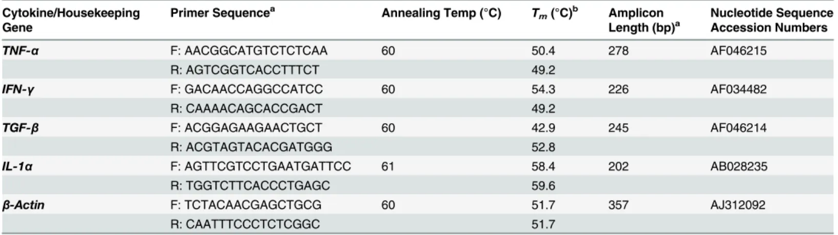
Real-time PCR (qRT-PCR) reactions were run on a Stratagene Mx3005P Real-Time PCR Sys-tem (Agilent Technologies, Santa Clara, CA, USA). The qPCR using SYBR Green PCR Master Mix (Applied Biosystems) and primers (Table 1) was carried out in a 25μl reaction volume
(50 ng cDNA, 12.5μl Master Mix, 0.5μM of each primer). The cycling conditions consisted of
95°C for 10 min (denaturation), followed by target DNA amplification for 45 cycles (95°C for 5 s, 60°C or 61°C for 30 s, and a variable extension time at 72°C). The melting curves were ana-lysed immediately after amplification at a linear temperature transition rate of 0.1°C/s from 55 to 95°C, with continuous fluorescence acquisition. The relative CT(ΔΔCT) method [31] was
used to quantify cytokine gene expression. Briefly, the fold change of each target gene was nor-malized to theβ-actin housekeeping gene CT (ΔCT), and compared to a calibrator sample, the
same normalized gene in the pre-immune sera sample (ΔΔCT). The final value represents the
relative fold between immunized and non-immunized hamsters.
Statistical analysis
considered significant at aPvalue of0.05. The analyses were carried out with GraphPad Prism 4 and QuickCalcs software.
Results
Distribution of
ompL37
among
Leptospira
spp
PCR analysis showed that theompL37gene is present inL.interrogans(nine serovars),L. borg-petersenii(six serovars),L.kirschneri(two serovars), andL.santarosai(one serovar).In silico protein sequence analysis showed that the predicted OmpL37 sequence inL.interroganssv. Copenhageni was 100% identical to the LA1495 sequence inL.interroganssv. Lai. Compared to OmpL37 inL.borgpeterseniisv. Hardjo,L.santarosaisv. Shermani andL.licerasiaesv. Var-illal, the identity was 88, 87 and 63% with a query coverage of 100%, 100% and 99%, respec-tively. A less conserved protein orthologue was observed in saprophyticL.biflexawith an identity of 46% (94% coverage). OutsideLeptospiragenus, proteins with 32% and 28% of iden-tity, with over 90% of coverage, were identified in spirochetesTurneriella parvaandLeptonema illini(Leptospiraceae), respectively.
Subunit and DNA vaccine preparation
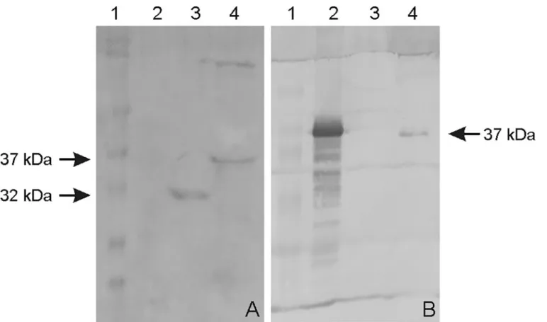
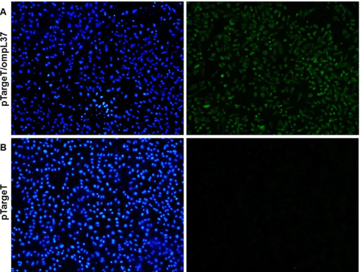
TheompL37plasmid sequences were confirmed by sequencing. The OmpL37 recombinant protein was expressed in inclusion bodies inE.coliwith the expected size of 37 kDa. After dial-ysis against PBS, the protocol for solubilization in urea and purification of the recombinant protein resulted in a yield of 10.4 mg.L-1. The rOmpL37 was recognized by convalescent sera evaluated by Western blot. Mouse anti-rOmpL37 serum recognized the native OmpL37 pro-tein inL.interroganssv. Copenhageni strain Fiocruz L1-130 whole cell lysate (WCL),Fig 1. The expression of OmpL37 in the pTargeT construct was confirmed through the detection of rOmpL37 in transfected CHO-K1 cells (Fig 2).
Humoral immune response in vaccinated hamsters
In order to assess the specific antibody response in groups of hamsters immunized with the OmpL37 vaccine preparations, an indirect ELISA was performed with the sera from each
Table 1. Detailed primers and conditions used for qRT-PCR assays.
Cytokine/Housekeeping Gene
Primer Sequencea Annealing Temp (°C) T
m(°C)b Amplicon
Length (bp)a
Nucleotide Sequence Accession Numbers
TNF-α F: AACGGCATGTCTCTCAA 60 50.4 278 AF046215
R: AGTCGGTCACCTTTCT 49.2
IFN-γ F: GACAACCAGGCCATCC 60 54.3 226 AF034482
R: CAAAACAGCACCGACT 49.2
TGF-β F: ACGGAGAAGAACTGCT 60 42.9 245 AF046214
R: ACGTAGTACACGATGGG 52.8
IL-1α F: AGTTCGTCCTGAATGATTCC 61 58.4 202 AB028235
R: TGGTCTTCACCCTGAGC 59.6
β-Actin F: TCTACAACGAGCTGCG 60 51.7 357 AJ312092
R: CAATTTCCCTCTCGGC 51.7
aVernel-Pauillac and Merien, 2006; Vernel-Pauillac and Goarant, 2010. b
Melting temperature.
animal collected on days 0 (pre-immune), 21 and 42 post-immunization (pi), using rOmpL37 as the immobilized antigen (Fig 3). The rOmpL37 vaccine induced significantly higher anti-body levels than the control group (P<0.05). A significant response was observed in the
prime-boost group following the rOmpL37 boost on Day 42, while the DNA vaccine failed to induce a significant immune response.
Cytokine expression profile in hamsters immunized with OmpL37
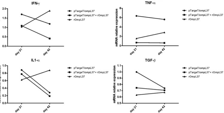
The induction of IFN-γ, IL-1α, TNF-αand TGF-βwas evaluated by qRT-PCR and only a 2-fold or greater change in mRNA levels was considered significant [32]. IFN-γ(ratio = 0.41) and IL-1α(ratio = 0.19) mRNA expression levels were down-regulated in the prime-boost group at Day 42. In contrast, TNF-αexpression increased at Day 42 in animals immunized with rOmpL37 (ratio = 2.84). Following immunization with the DNA vaccine, TNF-αwas up-regulated at Days 21 (ratio = 6.4) and 42 (ratio = 5.6), while IL-1α(ratio = 0.28) was down-reg-ulated at Day 42. TGF-βwas expressed at basal levels in the vaccinated hamsters (Fig 4).Efficacy of the OmpL37 vaccine preparations
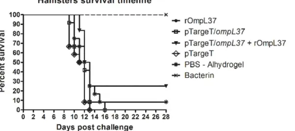
The protective efficacy of the OmpL37 vaccine preparations was determined in two indepen-dent experiments. The statistical analyses are presented inTable 2, and the survival rates are shown inFig 5. Considering both protection against mortality and increased survival, none of
Fig 1. Western blot of recombinant and native OmpL37 proteins.A: rOmpL37 characterization with convalescent human sera. (1) Full-Range Rainbow Molecular Weight Marker (GE Healthcare); (2) Negative control (BSA); (3) Positive control (rLipL32); (4) rOmpL37. B: Anti-rOmpL37 serum characterization. (1) Full-Range Rainbow Molecular Weight Marker (GE Healthcare); (2) rOmpL37; (3) Negative control (BSA); (4)L.interrogansserovar Copenhageni Fiocruz L1-130 WCL.
Fig 2. Immunofluorescence (IFA) analysis of the expression of recombinant OmpL37 protein in CHO-K1 cells 24 h after transfection with pTargeT/
ompL37(A) or pTargeT alone (B).The IFA was based on a mouse anti-rOmpL37 antibody and FITC conjugated anti-mouse IgG. Panels on the left shows
Hoechst 33258 DNA staining and on the right, antibody reactions.
doi:10.1371/journal.pone.0142821.g002
Fig 3. Specific IgG response in hamsters inoculated with different vaccine formulations.Recombinant rOmpL37 expressed byE.coliwas used as the antigen in ELISA. Mean values were calculated from serum samples assayed in triplicate. Results are expressed as the mean absorbance±standard
deviation. OD492, optical density at 492 nm. Significant differences, at aPvalue of 0.001 in comparison to the control group, are shown by an asterisk.
the OmpL37 vaccine formulations induced a protective immune response. The maximum effi-cacy of 25% was observed in the prime-boost group. In addition, leptospires were present in the kidneys of the surviving animals, including those in the bacterin group, indicating a lack of sterilizing immunity.
Discussion
Traditional bacterin vaccine preparations used in humans and animals present several limita-tions including severe side effects, short-term immunity and restricted-serovar protection. Sev-eral studies have evaluated different formulations of recombinant vaccine candidates with the intention of improving leptospirosis vaccines. To date, the most promising results were obtained using the Lig (Leptospiral immunoglobulin-like) proteins. Mice immunized with LigA or LigB survived lethal challenge, showing 90–100% of protection [33]. A vaccine using
Fig 4. Relative mRNA expression levels of IFN-γ, TNF-α, IL1-αand TGF-βin pooled hamster blood samples.The relative CT(ΔΔCT) method was used
to quantify cytokine gene expression: CTs were normalized against theβ-actin gene CT(ΔCT) and then compared to the same normalized gene in the
pre-immune sera sample (calibrator). A 2-fold or greater change in mRNA levels was considered significant. The control groups were set to 1. The values represent grouped results of two independent experiments.
doi:10.1371/journal.pone.0142821.g004
Table 2. Effect of immunization with OmpL37 vaccines in hamsters.
Treatment Group No. Survivors/Total (% protection) % Culture
#Exp 1 #Exp 2 #Total Positive
rOmpL37 0/6 (0) 0/6 (0) 0/12 ND
pTargeT/ompL37 0/6 (0) 0/6 (0) 0/12 ND
pTargeT/ompL37+ rOmpL37 3/6 (50) 0/6 (0) 3/12 (25) 3/3 (100%)
pTargeT 0/6 (0) 0/6 (0) 0/12 ND
PBS-Alhydrogel 1/6 (16.6) 0/6 (0) 1/12 (8.3) 1/1 (100%)
Killed-whole leptospires 4/4 (100) 4/4 (100) 8/8 (100) 4/8 (50%)
the C-terminal portion of the LigA protein, induced protection ranging from 63% to 100% in hamsters [28]. Of note, sterilizing immunity has not yet been achieved [11,34] and LigA is not present inLeptospiraspp., highlighting the need for new conserved antigens for vaccine devel-opment. This study evaluated, for the first time, the outer membrane protein OmpL37 fromL. interroganssv. Copenhageni strain Fiocruz L1-130 as a vaccine antigen against leptospirosis. In addition to the localization of OmpL37 on the surface of the outer membrane of the leptospiral cell and a possible role during infection, orthologues of theompL37gene were present in differ-ent serovars, suggesting it is a potdiffer-ential candidate for a cross-protective vaccine [12–14].
One limitation that hinders the development of vaccines against leptospirosis is the lack of correlates of protection, immune markers that have contributed to many vaccinology studies [35–36]. To date, no markers have been identified for leptospirosis and the hamster model of lethal leptospirosis remains the preferred method of assessing vaccine efficacy [37–38]. Both humoral and cellular immune responses have been reported to play roles in the response against leptospirosis [39]; as such, this study included a protein-boost strategy that aimed to improve the immunogenicity of the OmpL37 DNA vaccine, as previously demonstrated for other antigens [18–19]. Independent experiments using subunit, DNA vaccine, and prime-boost immunization strategies were performed. A non-protective humoral immune response was stimulated by rOmpL37 (Fig 2) and survival among animals in the prime-boost group was not associated with increased IgG levels (data not shown). Although the functionality of the DNA vaccine was confirmed by the transfection assay, it did not induce an IgG response and this was in agreement with previous observations [19].
Due to the lack of reagents to measure serum concentrations of Syrian hamster immune effectors, studies have been limited to determining cytokine profiles at the transcriptional level [32,40]. This analysis revealed increased circulating TNF-αin hamsters vaccinated with rOmpL37 (Day 42) or the DNA vaccine (Days 21 and 42) compared to the control groups. Delayed and sustained TNF-αproduction has been associated with a poor prognosis during infection [41]. High levels of IL1-αseems to be related to severe disease [32]. In our experi-ments, IL1-αwas down-regulated in the OmpL37 prime-boost and the DNA vaccine groups. Previous research has indicated that levels of TNF-αand IL1-αare higher in hamsters infected with virulent leptospires than those infected with an avirulent strain [32]. IFN-γproduction
Fig 5. Survival of hamsters immunized with rOmpL37 vaccines after lethal challenge.Survival curves were compared using log-rank analysis. The results are a summary of two independent experiments (Table 2).
was related to protection in cattle vaccinated with monovalent serovar Hardjo vaccines [42]. In the present study, however, increased IFN-γmRNA levels were found in animals vaccinated with rOmpL37, and this vaccine preparation did not protect against lethal infection. The role of the cytokine profile in protecting against leptospirosis remains poorly understood and needs further investigation.
E.coli-based expression systems are widely used, despite the fact that they are unable to carry out post-translational modifications such as glycosylation, methylation and acetylation [43]. This is usually irrelevant for bacterial diseases vaccines; however, there is evidence that pathogenicLeptospiraspp. have protein modification systems [44]. Furthermore, lipidation improved the immunogenicity of recombinant antigens, such as OspA fromBorrelia burgdor-feriand LigANI fromL.interrogans[45–46]. Altered immunogenic properties may have affected the folding of rOmpL37.
Although three animals survived in the prime-boost group, none of the OmpL37 vaccine formulations evaluated in this study induced significant protection against lethal leptospirosis. Even though it is a recurring issue in the animal models of leptospirosis [47–48], this was somewhat unexpected due to the many attractive features inherent in OmpL37, including the induction of immune responses following vaccination, as shown here. Any attempt to explain the lack of protection in this study would be highly speculative. Further experiments are neces-sary to confirm the distribution of OmpL37 across the outer membrane, the exposed regions and any redundancy of function. Once distribution across the outer membrane is confirmed, additional studies employing different approaches, including alternative adjuvants and vaccine immunization strategies, would be viable. Our group is currently applying different rational approaches, ranging from bioinformatics to serological investigation of leptospiral surface exposed OMPs, to identify new antigens for the development of an effective vaccine against leptospirosis.
Conclusions
OmpL37, potentially a surface-exposed outer membrane protein expressed during host infec-tion, induced strong immune responses, but failed to stimulate protection against leptospirosis when presented as a recombinant protein in Alhydrogel and DNA vaccine preparations. Fur-ther studies are necessary to identify new potential vaccine candidates against lethal
leptospirosis.
Acknowledgments
We are grateful to Michele dos Santos for the technical assistance provided during this study. This work was supported by Brazilian funding agencies CAPES, CNPq and FAPERGS.
Author Contributions
Conceived and designed the experiments: TLO AAG OAD. Performed the experiments: TLO AAG RAS ACPSN MM. Analyzed the data: TLO AAG DDH AJAM. Wrote the paper: TLO AAG DDH AJAM OAD.
References
1. Adler B, de la Peña Moctezuma A.Leptospiraand leptospirosis. Veterinary Microbiology. 2010 1/27/; 140(3–4): 287–96. doi:10.1016/j.vetmic.2009.03.012PMID:19345023
3. Reis RB, Ribeiro GS, Felzemburgh RD, Santana FS, Mohr S, Melendez AX, et al. Impact of environ-ment and social gradient onLeptospirainfection in urban slums. PLoS Negl Trop Dis. 2008; 2(4): e228. doi:10.1371/journal.pntd.0000228PMID:18431445
4. Picardeau M, Bertherat E, Jancloes M, Skouloudis AN, Durski K, Hartskeerl RA. Rapid tests for diagno-sis of leptospirodiagno-sis: current tools and emerging technologies. Diagn Microbiol Infect Dis. 2014 Jan; 78 (1): 1–8. doi:10.1016/j.diagmicrobio.2013.09.012PMID:24207075
5. Health Surveillance Secretary. Leptospirosis case notification records, Brazil. 2013.
6. Gouveia EL, Metcalfe J, de Carvalho AL, Aires TS, Villasboas-Bisneto JC, Queirroz A, et al. Leptospiro-sis-associated Severe Pulmonary Hemorrhagic Syndrome, Salvador, Brazil. Emerg Infect Dis. 2008 Mar; 14(3): 505–8. doi:10.3201/eid1403.071064PMID:18325275
7. Bulach DM, Kalambaheti T, de la Pena-Moctezuma A, Adler B. Lipopolysaccharide biosynthesis in Leptospira. J Mol Microbiol Biotechnol. 2000; 2(4): 375–80. PMID:11075908
8. McBride AJ, Athanazio DA, Reis MG, Ko AI. Leptospirosis. Current opinion in infectious diseases. 2005 Oct; 18(5): 376–86. PMID:16148523
9. Ko AI, Goarant C, Picardeau M.Leptospira: the dawn of the molecular genetics era for an emerging zoonotic pathogen. Nat Rev Microbiol. 2009 Oct; 7(10): 736–47. doi:10.1038/nrmicro2208PMID: 19756012
10. Haake DA, Matsunaga J.Leptospira: a spirochaete with a hybrid outer membrane. Mol Microbiol. 2010 Jun 28.
11. Dellagostin OA, Grassmann AA, Hartwig DD, Felix SR, da Silva EF, McBride AJ. Recombinant vac-cines against leptospirosis. Hum Vaccin. 2011 Nov; 7(11): 1215–24. doi:10.4161/hv.7.11.17944 PMID:22048111
12. Pinne M, Haake DA. A comprehensive approach to identification of surface-exposed, outer membrane-spanning proteins ofLeptospira interrogans. PLoS One. 2009; 4(6): e6071. doi:10.1371/journal.pone. 0006071PMID:19562037
13. Pinne M, Choy HA, Haake DA. The OmpL37 surface-exposed protein is expressed by pathogenic Lep-tospiraduring infection and binds skin and vascular elastin. PLoS Negl Trop Dis. 2010; 4(9): e815. doi: 10.1371/journal.pntd.0000815PMID:20844573
14. Matsui M, Soupe ME, Becam J, Goarant C. Differential in vivo gene expression of majorLeptospira pro-teins in resistant or susceptible animal models. Appl Environ Microbiol. 2012 Sep; 78(17): 6372–6. doi: 10.1128/AEM.00911-12PMID:22729538
15. Caimano MJ, Sivasankaran SK, Allard A, Hurley D, Hokamp K, Grassmann AA, et al. A model system for studying the transcriptomic and physiological changes associated with mammalian host-adaptation byLeptospira interrogansserovar Copenhageni. PLoS pathogens. 2014 Mar; 10(3): e1004004. doi: 10.1371/journal.ppat.1004004PMID:24626166
16. Branger C, Chatrenet B, Gauvrit A, Aviat F, Aubert A, Bach JM, et al. Protection againstLeptospira interroganssensu lato challenge by DNA immunization with the gene encoding hemolysin-associated protein 1. Infect Immun. 2005 Jul; 73(7): 4062–9. PMID:15972494
17. Faisal SM, Yan W, Chen CS, Palaniappan RU, McDonough SP, Chang YF. Evaluation of protective immunity ofLeptospiraimmunoglobulin like protein A (LigA) DNA vaccine against challenge in ham-sters. Vaccine. 2008 Jan 10; 26(2): 277–87. PMID:18055070
18. Feng CY, Li QT, Zhang XY, Dong K, Hu BY, Guo XK. Immune strategies using single-component LipL32 and multi-component recombinant LipL32-41-OmpL1 vaccines againstLeptospira. Braz J Med Biol Res. 2009 Sep; 42(9): 796–803. PMID:19649391
19. Hartwig DD, Forster KM, Oliveira TL, Amaral M, McBride AJ, Dellagostin OA. A prime-boost strategy using the novel vaccine candidate, LemA, protects hamsters against leptospirosis. Clin Vaccine Immu-nol. 2013 May; 20(5): 747–52. doi:10.1128/CVI.00034-13PMID:23515012
20. He HJ, Wang WY, Wu ZD, Lv ZY, Li J, Tan LZ. Protection of guinea pigs againstLeptospira interrogans serovar Lai by LipL21 DNA vaccine. Cell Mol Immunol. 2008 Oct; 5(5): 385–91. doi:10.1038/cmi.2008. 48PMID:18954563
21. Morey RE, Galloway RL, Bragg SL, Steigerwalt AG, Mayer LW, Levett PN. Species-Specific Identifica-tion of Leptospiraceae by 16S rRNA Gene Sequencing. J Clin Microbiol. 2006 Oct; 44(10): 3510–6. PMID:17021075
22. Ren SX, Fu G, Jiang XG, Zeng R, Miao YG, Xu H, et al. Unique physiological and pathogenic features ofLeptospira interrogansrevealed by whole-genome sequencing. Nature. 2003 Apr 24; 422(6934): 888–93. PMID:12712204
24. Chou LF, Chen YT, Lu CW, Ko YC, Tang CY, Pan MJ, et al. Sequence ofLeptospira santarosaiserovar Shermani genome and prediction of virulence-associated genes. Gene. 2012 Dec 15; 511(2): 364–70. doi:10.1016/j.gene.2012.09.074PMID:23041083
25. Ricaldi JN, Fouts DE, Selengut JD, Harkins DM, Patra KP, Moreno A, et al. Whole genome analysis of Leptospira licerasiaeprovides insight into leptospiral evolution and pathogenicity. PLoS Negl Trop Dis. 2012; 6(10): e1853. doi:10.1371/journal.pntd.0001853PMID:23145189
26. Picardeau M, Bulach DM, Bouchier C, Zuerner RL, Zidane N, Wilson PJ, et al. Genome Sequence of the SaprophyteLeptospira biflexaProvides Insights into the Evolution of Leptospira and the Pathogen-esis of Leptospirosis. PLoS ONE. 2008; 3(2): e1607. doi:10.1371/journal.pone.0001607PMID: 18270594
27. Ramos CR, Abreu PA, Nascimento AL, Ho PL. A high-copy T7Escherichia coliexpression vector for the production of recombinant proteins with a minimal N-terminal His-tagged fusion peptide. Braz J Med Biol Res. 2004 Aug; 37(8): 1103–9. PMID:15273812
28. Silva EF, Medeiros MA, McBride AJ, Matsunaga J, Esteves GS, Ramos JG, et al. The terminal portion of leptospiral immunoglobulin-like protein LigA confers protective immunity against lethal infection in the hamster model of leptospirosis. Vaccine. 2007 Aug 14; 25(33): 6277–86. PMID:17629368 29. Fernandes CP, Seixas FK, Coutinho ML, Vasconcellos FA, Seyffert N, Croda J, et al. Monoclonal
anti-bodies against LipL32, the major outer membrane protein of pathogenicLeptospira: production, char-acterization, and testing in diagnostic applications. Hybridoma (Larchmt). 2007 Feb; 26(1): 35–41. 30. Grassmann AA, Felix SR, dos Santos CX, Amaral MG, Seixas Neto AC, Fagundes MQ, et al.
Protec-tion against lethal leptospirosis after vaccinaProtec-tion with LipL32 coupled or coadministered with the B sub-unit ofEscherichia coliheat-labile enterotoxin. Clin Vaccine Immunol. 2012 May; 19(5): 740–5. doi:10. 1128/CVI.05720-11PMID:22379066
31. Vernel-Pauillac F, Merien F. Proinflammatory and immunomodulatory cytokine mRNA time course pro-files in hamsters infected with a virulent variant ofLeptospira interrogans. Infect Immun. 2006 Jul; 74 (7): 4172–9. PMID:16790792
32. Vernel-Pauillac F, Goarant C. Differential cytokine gene expression according to outcome in a hamster model of leptospirosis. PLoS Negl Trop Dis. 2010; 4(1): e582. doi:10.1371/journal.pntd.0000582 PMID:20076757
33. Koizumi N, Watanabe H. Leptospiral immunoglobulin-like proteins elicit protective immunity. Vaccine. 2004 Mar 29; 22(11–12): 1545–52. PMID:15063580
34. Koizumi N, Watanabe H. Leptospirosis vaccines: Past, present, and future. J Postgrad Med. 2005 Jul-Sep; 51(3): 210–4. PMID:16333195
35. Pizza M, Scarlato V, Masignani V, Giuliani MM, Arico B, Comanducci M, et al. Identification of vaccine candidates against serogroup B meningococcus by whole-genome sequencing. Science. 2000 Mar 10; 287(5459): 1816–20. PMID:10710308
36. Etz H, Minh DB, Henics T, Dryla A, Winkler B, Triska C, et al. Identification of in vivo expressed vaccine candidate antigens fromStaphylococcus aureus. Proc Natl Acad Sci U S A. 2002 May 14; 99(10): 6573–8. PMID:11997460
37. Haake DA. Hamster model of leptospirosis. Curr Protoc Microbiol. 2006 Sep;Chapter 12: Unit 12E 2. 38. Evangelista KV, Coburn J.Leptospiraas an emerging pathogen: a review of its biology, pathogenesis
and host immune responses. Future Microbiol. 2010 Sep; 5(9): 1413–25. doi:10.2217/fmb.10.102 PMID:20860485
39. Fraga TR, Barbosa AS, Isaac L. Leptospirosis: Aspects of Innate Immunity, Immunopathogenesis and Immune Evasion from the Complement System. Scand J Immunol. 2011 Jan 3; 73(5): 408–19. doi:10. 1111/j.1365-3083.2010.02505.xPMID:21204903
40. Cavaillon JM, Munoz C, Fitting C, Misset B, Carlet J. Circulating cytokines: the tip of the iceberg? Circ Shock. 1992 Oct; 38(2): 145–52. PMID:1423923
41. Chirathaworn C, Kongpan S. Immune responses toLeptospirainfection: roles as biomarkers for dis-ease severity. Braz J Infect Dis. 2014 Jan-Feb; 18(1): 77–81. doi:10.1016/j.bjid.2013.08.002PMID: 24275371
42. Zuerner RL, Alt DP, Palmer MV, Thacker TC, Olsen SC. ALeptospira borgpeterseniiserovar Hardjo vaccine induces a Th1 response, activates NK cells, and reduces renal colonization. Clin Vaccine Immunol. 2011 Apr; 18(4): 684–91. doi:10.1128/CVI.00288-10PMID:21288995
43. Sorensen HP. Towards universal systems for recombinant gene expression. Microb Cell Fact. 2010; 9: 27. doi:10.1186/1475-2859-9-27PMID:20433754
45. del Rio B, Seegers JF, Gomes-Solecki M. Immune response toLactobacillus plantarumexpressing Borrelia burgdorferiOspA is modulated by the lipid modification of the antigen. PLoS One. 2010; 5(6): e11199. doi:10.1371/journal.pone.0011199PMID:20585451
46. Lourdault K, Wang LC, Vieira A, Matsunaga J, Melo R, Lewis MS, et al. Oral immunization with Escheri-chia coliexpressing a lipidated form of LigA protects hamsters against challenge withLeptospira inter-rogansserovar Copenhageni. Infect Immun. 2014 Feb; 82(2): 893–902. doi:10.1128/IAI.01533-13 PMID:24478102
47. Lucas DS, Cullen PA, Lo M, Srikram A, Sermswan RW, Adler B. Recombinant LipL32 and LigA from Leptospiraare unable to stimulate protective immunity against leptospirosis in the hamster model. Vac-cine. 2011 Apr 18; 29(18): 3413–8. doi:10.1016/j.vaccine.2011.02.084PMID:21396409