Pituitary glyco pro te in ho rm o ne
a
-subunit se cre tio n by cirrho tic patie nts
Departamentos de 1Endocrinologia and 2Gastroenterologia, Fundação Faculdade Federal de Ciências Médicas de Porto Alegre, Porto Alegre, RS, Brasil
Departamento de 3Endocrinologia, Escola Paulista de Medicina, Universidade Federal de São Paulo, São Paulo, SP, Brasil M.C. O liveira1,
C.B. Pizarro1, A. Cassal2, R. Cremonese1 and J.G.H. Vieira3
Abstract
Secretion of the a-subunit of pituitary glycoprotein hormones usually follows the secretion of intact gonadotropins and is increased in gonadal failure and decreased in isolated gonadotropin deficiency. The aim of the present study was to determine the levels of the a -subunit in the serum of patients with cirrhosis of the liver and to compare the results obtained for eugonadal cirrhotic patients with those obtained for cirrhotic patients with hypogonadotropic hypogo-nadism. Forty-seven of 63 patients with cirrhosis (74.6%) presented hypogonadism (which was central in 45 cases and primary in 2), 7 were eugonadal, and 9 women were in normal menopause. The serum
a-subunit was measured by the fluorimetric method using monoclonal antibodies. Cross-reactivity with LH, TSH, FSH and hCG was 6.5, 1.2, 4.3 and 1.1%, respectively, with an intra-assay coefficient of variation (CV) of less than 5% and an interassay CV of 5%, and sensitivity limit of 4 ng/l. The serum a-subunit concentration ranged from 36 to 6253 ng/l, with a median of 273 ng/l. The median was 251 ng/l for patients with central hypogonadism and 198 ng/l for eugonadal patients. The correlation between the a-subunit and basal LH levels was significant both in the total sample (r = 0.48, P<0.01) and in the cirrhotic patients with central hypogonadism (r = 0.33, P = 0.02). Among men with central hypogonadism there was a negative correla-tion between a-subunit levels and total testosterone levels (r = -0.54, P<0.01) as well as free testosterone levels (r = -0.53, P<0.01). In conclusion, although the a-subunit levels are correlated with LH levels, at present they cannot be used as markers for hypogonadism in patients with cirrhosis of the liver.
Co rre spo nde nce
M.C. O liveira
Rua Dona Mimi Moro, 40 90480-050 Porto Alegre, RS Brasil
Fax: + 55-51-226-9756 E-mail: mco@ portoweb.com.br
Part of the data was presented at the XXII Congresso Brasileiro de Endocrinologia e Metabologia, Salvador, BA, Brazil, 1996 (Abstract no. 346).
Publication supported by FAPESP. M.C. O liveira is the recipient of a CNPq fellowship.
Received February 17, 1998 Accepted O ctober 26, 1998
Ke y wo rds ·a-Subunit ·Pituitary ·Gonadotropins ·Hypogonadism ·Cirrhosis
Intro ductio n
The a-subunit of glycoprotein hormones, usually detected in the serum of normal individuals (1-3), has been used as a tumor marker for gonadotropin- or TSH-secreting pituitary adenomas (4), for GH-secreting tumors (4,5) and for nonsecreting pituitary tumors (3,4). The a-subunit is secreted in a pulsatile manner by the thyrotropes and by
antagonist (11) and is normalized in indi-viduals with hypogonadotropic hypogo-nadism treated with GnRH (12).
In nonphysiological situations, most of the times the a-subunit oscillates according to the respective intact heterodimeric hor-mone, increasing in primary hypothyroidism (9,13) and in primary gonadal failure (6,10), and decreasing in hypogonadotropic hypogo-nadism (14,15). Dissociation between the secretion of the a-subunit and secretion of the complete hormone may occasionally oc-cur, as observed during the use of GnRH agonists in men with glycoprotein hormone-secreting pituitary tumors, when LH is sup-pressed and high subunit levels are main-tained (16). In patients with cirrhosis of the liver, hypogonadism is a frequent finding regardless of the etiology of the disease (17). Most of the time, despite the generalized advanced gonadal failure, gonadotropin lev-els are low, indicating involvement of the hypothalamus-pituitary axis. The site of this disorder is the hypothalamus since these patients do not present the usual LH pulsatility (18), since exogenous GnRH stimulates LH and FSH (19-21) and since the picture of hormonal deficiency is reversed after liver transplant (21-23). The secretory status of the a-subunit has not been established in this form of hypogonadism.
The aim of the present study was to deter-mine the a-subunit levels in the serum of patients with cirrhosis of the liver and to compare the results obtained for hypogonadal and non-hypogonadal patients.
Mate rial and Me tho ds
The sample consisted of 63 patients, 33 men and 30 women, average age 53 years (range: 19 to 76 years), with a clinical and/or histological diagnosis of cirrhosis of liver of different etiologies such as alcohol (N = 13), hepatitis C virus (N = 22), alcohol plus hepa-titis C virus (N = 15), hepahepa-titis B virus (N = 1), hepatitis B and C viruses (N = 2), hepatitis C
virus plus sclerosing cholangitis (N = 1), pri-mary biliary cirrhosis (N = 1), and cryptogen-ic cirrhosis (N = 8). The clincryptogen-ical diagnosis was based on a previous history or present manifestations of cirrhosis, such as portal hypertension, bleeding esophageal and gas-tric varices, ascites, spontaneous bacterial peritonitis, encephalopathy and coagulopa-thy. In 37 cases the liver biopsy confirmed hepatic cirrhosis. The severity of liver dam-age was evaluated in terms of Child-Pugh score, which takes into account the degree of encephalopathy and ascites, the bilirubin and albumin levels and the prothrombin time (24). All patients were euthyroid, as determined by TSH measurement. None of the patients was using hormones or drugs which could alter the gonadotropin levels.
Gonadal function was evaluated on the basis of clinical and laboratory parameters. Total (normal range = 2.8-9.8 ng/ml) and free (normal range = 12.4-40.0 pg/ml) plasma tes-tosterone were determined by RIA and estra-diol (normal range for males = up to 40 and for females = 30-150 pg/ml), LH (normal range for males = 1.3-19.8 and for females = 0.7-35.2 mIU/ml) and FSH (normal range for males = 2.0-12.8 and for females = 3.1-15.7 mIU/ml) were determined by chemilumines-cence using commercial kits in our labora-tory. The patients were divided into 4 groups: group 1 consisted of eugonadal patients, group 2 of amenorrheic women older than 50 years with increased basal gonadotropins (normal menopause), and group 3 and group 4 con-sisted of women in the fertile age with hy-pogonadism, e.g., presenting amenorrhea or absence of vaginal bleeding for at least 6 months and men with hypogonadism, e.g., presenting low levels of free testosterone. Hypogonadism was considered to be primary when one or both gonadotropins were in-creased (group 3). Group 4 was composed of patients with hypogonadism of central origin, e.g., no increase in basal gonadotropins.
antibodies. Cross-reactivity with LH, TSH, FSH and hCG was 6.5, 1.2, 4.3 and 1.1%, respectively, with an intra-assay coefficient of variation (CV) of less than 5% and an interassay CV of 5%, and sensitivity limit of 4 ng/l. The values considered normal for the method range from 120 to 790 ng/l (median: 250) for men, from 88 to 604 ng/l (median: 291) for women, and from 341 to 4071 ng/l (median 1856) for menopausal women (25). For statistical analysis, the results are reported as median, minimum and maxi-mum values of distribution, with the excep-tion of serum albumin levels which are re-ported as means ± SD. The nonparametric Mann-Whitney or Kruskal-Wallis test was used according to the number of groups evalu-ated. The chi-square association test and the Spearman correlation coefficient test were calculated. The level of significance was considered to be P<0.05.
This study was approved by the Ethics Committee of the institution and informed consent was obtained from the subjects.
Re sults
Hypogonadism was diagnosed in 47 indi-viduals, with 45 cases being of central etiol-ogy and 2 of primary etioletiol-ogy. Of the remain-ing patients, 7 were eugonadal and 9 were menopausal women. Total testosterone levels ranged from 3.0 to 5.8 ng/ml for eugonadal men and from 0.1 to 14.0 ng/ml for central hypogonadal men, while free testosterone ranged from 15 to 21 and from 0.3 to 13.0 pg/ ml for these same groups. Amongst men, 5 presented increased estradiol while among women, 19 presented estradiol levels which were lower than the minimum expected for the follicular phase. The range of estradiol values was 26.0 to 225 pg/ml in eugonadal women, less than 10 to 95 pg/ml in meno-pausal women and less than 10 to 75 pg/ml in women with central hypogonadism. Regard-ing gonadotropins, the median, minimum and maximum values for LH were 3.9 (0.3-55.9),
34.5 (6.7-102.5) and 4.1 mIU/ml (0.1-20.4), respectively, for eugonadal and menopausal subjects and for subjects with central hypogo-nadism. For FSH, these values were 4.3 (2.4-14.0), 60.7 (26.8-129.0) and 5.5 (0.1-19.5) mIU/ml. There was no significant difference in LH or FSH levels between eugonadal and central hypogonadal patients.
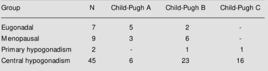
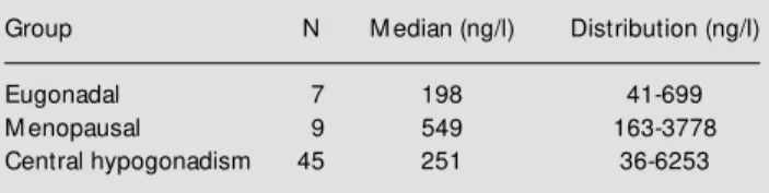
Mean serum albumin levels were 3.1 g/dl (normal values = 3.5-5.5; range 2.0-4.5). The severity of hepatic disease in each group, according to the CHILD score, is presented in Table 1; 22.9% of the patients were classified as Child-Pugh A, 50.8% as Child-Pugh B and 27% as Child-Pugh C. The analysis of a pos-sible correlation between gonadal function and Child-Pugh score was significant, sug-gesting a correlation between these variables. The concentration of the a-subunit ranged from 36 to 6253 ng/l (median: 273 ng/l). In the two patients with primary hypogonadism, the a-subunit levels were 1568 and 1005 ng/ l. The median and distribution of the a -subunit levels for the remaining hypogonadal, eugonadal and menopausal subjects are listed in Table 2. No statistically significant differ-ence in a-subunit levels was detected among these groups. The a-subunit levels of pa-tients with cirrhosis associated with alcohol (N = 28, median 222.5 ng/l) were not statis-tically different from the levels observed in the patients with cirrhosis of other etiologies (N = 35, median 313 ng/l).
The correlation between a-subunit and basal LH levels was significant both for the total sample (r = 0.48, P<0.01) and for cir-rhotic patients with central hypogonadism (r
Table 1 – Severity of hepatic disease and gonadal function in the cirrhotic patients w ho participated in the study.
Group N Child-Pugh A Child-Pugh B Child-Pugh C
Eugonadal 7 5 2
-M enopausal 9 3 6
-Primary hypogonadism 2 - 1 1
= 0.33, P = 0.02). A significant negative correlation between a-subunit levels and to-tal testosterone (r = -0.54, P<0.01) and free testosterone (r = -0.53, P<0.01) was ob-served in men with central hypogonadism. No association was observed between a -subunit levels or serum albumin levels and severity of hepatic disease according to the Child-Pugh score.
D iscussio n
The high prevalence of hypogonadism of central etiology in the population of cirrhotic patients evaluated here is related to the se-verity of liver disease, which triggers a hypo-thalamic disorder secondary to an adverse environment (21). This diagnosis, however, does not invalidate the presence of gonadal lesion in some patients from this group.
The production of the a-subunit is paral-lel to LH secretion during the different phases of life, increasing from prepuberty to adoles-cence (26). Thus, our finding of a positive correlation between a-subunit levels and LH levels agrees with data reported elsewhere (6,12,26). On the other hand, no statistically significant difference in a-subunit levels was detected between patients with central hy-pogonadism, eugonadism and menopausal women. Classically, a-subunit levels are el-evated in menopause (1-3,13), a fact that was observed here only as a tendency. Our findings are probably related to the small sample of menopausal women studied (9 patients), since their gonadotropin levels were as expected.
Studies on gonadotropin deficiency alone in hypogonadotropic hypogonadism have
reported low a-subunit values (12,14). Spratt and colleagues (12) studying 6 hypogonadal men with normal endogenous TSH secretion detected circulating a-subunit levels close to or below assay detection limits. Winters and Troen (14) studying 4 hypogonadal men observed low but usually detectable levels of
a-subunit. Several possibilities may justify the absence of this finding among the hypogonadal patients in the present series. The first is that our sample is not homoge-neous in terms of number of patients in each group and that there is dispersion of the results for hypogonadal patients, two of whom had the highest a-subunit value in the sample, including menopausal women. The second possibility is that, although a hypo-thalamic disorder exists both in gonadotro-pin deficiency alone and in hypogonadism associated with cirrhosis, the basic mecha-nism of the diseases is different: in the first condition endogenous GnRH secretion is undetectable, whereas in the second GnRH deficiency is transitory. This factor may be a determinant of the differential behavior of the a-subunit. Another explanation is that in hypogonadism associated with cirrhosis, basal LH levels are higher than those ob-served in individuals with gonadotropin de-ficiency alone, a possibility that was not investigated here. The possibilities of alter-ation in the hepatic clearance of the a -sub-unit or of a toxic factor associated with cirrhosis which may interfere with the pitu-itary gland dynamics should also be consid-ered.
Also important was our finding of a nega-tive correlation between the levels of a -subunit and testosterone, contrary to what would be expected for central hypogonadism. In conclusion, it seems clear that there is a parallelism between hepatic and gonadal dysfunction, and that in hypogonadism sec-ondary to cirrhosis of liver, a-subunit levels are correlated with LH levels but cannot be used as an additional diagnostic criterion of hypogonadism in these patients.
Table 2 – Serum a-subunit concentrations of cirrhotic patients. Group N M edian (ng/l) Distribution (ng/l)
Eugonadal 7 198 41-699
M enopausal 9 549 163-3778
Re fe re nce s
1. Kourides IA, Weintraub BD, Re RN, Ridgw ay EC & M aloof F (1978). Thyroid hormone, oestrogen, and glucocorticoid effects on tw o different pituitary glyco-protein hormone alpha subunit pools.
Clinical Endocrinology, 9: 535-542. 2. Kourides IA, Ridgw ay EC & M aloof F
(1979). Discordant responses of thyrotro-pin and its free a- and ß-subunits after thyrotropin-releasing hormone w ith incre-mental thyroid hormone replacement in primary hypothyroidism. Journal of Clini-cal Endocrinology and M etabolism, 49: 700-705.
3. Papapetrou PD (1982). Circulating a -sub-unit of glycoprotein hormones in normal subjects and its relationship to the LH and FSH serum levels. Acta Endocrinologica, 248: 1-2.
4. Oppenheim DS, Kana AR, Sangha JS & Klibanski A (1990). Prevalence of a -sub-unit hypersecretion in patients w ith pitu-itary tumors: clinically nonfunctioning and somatotroph adenomas. Journal of Clini-cal Endocrinology and M etabolism, 70: 859-864.
5. Ridgw ay EC, Klibanski A, Ladenson PW, Clemmons D, Beitins IZ, M cArthur JW, M artorana M A & Zervas NT (1981). Pure alpha-secreting pituitary adenomas. New England Journal ofM edicine, 304: 1254-1259.
6. M acfarlane IA, Beardw ell CG, Shalet SM , Darbyshire PJ, Hayw ard E & Sutton M L (1980). Glycoprotein hormone a-subunit secretion by pituitary adenomas: influ-ence of external irradiation. Clinical Endo-crinology, 13: 215-222.
7. Winters SJ & Troen P (1985). Pulsatile secretion of immunoreactive a-subunit in man. Journal of Clinical Endocrinology and M etabolism, 60: 344-348.
8. Winters SJ & Troen P (1990). Pituitary glycoprotein hormone a-subunit secretion after thyrotropin-releasing hormone stimu-lation in normal men and men w ith idio-pathic hypogonadotropic hypogonadism.
Journal of Clinical Endocrinology and M e-tabolism, 70: 544-547.
9. Hagen C & M cNeilly AS (1975). Changes in circulating levels of LH, FSH, LHß- and
a-subunit after gonadotropin-releasing hormone, and of TSH, LHß- and a-subunit after thyrotropin-releasing hormone. Jour-nal of Clinical Endocrinology and M etabo-lism, 41: 466-470.
10. Styne DM , Conte FA, Grumbach M M &
Kaplan SL (1980). Plasma glycoprotein hormone a-subunit in the syndrome of gonadal dysgenesis: the effect of estro-gen replacement in hypergonadotropic hy-pogonadism. Journal of Clinical Endocri-nology and M etabolism, 50: 1049-1052. 11. Hall JE, Whitcomb RW, Rivier JE, Vale
WW & Crow ley WF (1990). Differential regulation of luteinizing hormone, follicle-stimulating hormone, and free a-subunit secretion from the gonadotrope by gona-dotropin-releasing hormone (GnRH): evi-dence from the use of tw o GnRH antago-nists. Journal of Clinical Endocrinology and M etabolism, 70: 328-335.
12. Spratt DI, Chin WW, Ridgw ay EC & Crow ley WF (1986). Administration of low dose pulsatile gonadotropin-releasing hor-mone (GnRH) to GnRH-deficient men regulates free a-subunit secretion. Jour-nal of Clinical Endocrinology andM etabo-lism, 62: 102-108.
13. Kourides IA, Weintraub BD, Ridgw ay EC & M aloof F (1975). Pituitary secretion of free alpha and beta subunit of human thy-rotropin in patients w ith thyroid disorders.
Journal of Clinical Endocrinology and M e-tabolism, 40: 872-885.
14. Winters SJ & Troen P (1988). a-Subunit secretion in men w ith idiopathic hypogo-nadotropic hypogonadism. Journal of Clinical Endocrinology and M etabolism,
66: 338-342.
15. Whitcomb RW, O’dea LSL, Finkelstein JS, Heavern DM & Crow ley WF (1990). Utility of free a-subunit as an alternative neu-roendocrine marker of gonadotropin-re-leasing hormone (GnRH) stimulation of the gonadotroph in the human: evidence from normal and GnRH-deficient men.
Journal of Clinical Endocrinology andM e-tabolism, 70: 1654-1661.
16. Klibanski A, Jameson JL, Biller BM K, Crow ley WF, Zervas NT, Rivier J, Vale WW & Bikkal H (1989). Gonadotropin and
a-subunit responses to chronic gonado-tropin-releasing hormone analog adminis-tration in patients w ith glycoprotein hor-mone-secreting pituitary tumors. Journal of Clinical Endocrinology andM etabolism, 68: 81-86.
17. Smanik EJ, Barkoukis H, M ullen KD & M cCullough AJ (1993). Diseases of the liver. In: Schiff L & Schiff ER (Editors), The Liver and its Effect on Endocrine Function inHealth and Disease. JB Lippincott Com-pany, Philadelphia.
18. Bannister P, Handley T, Chapman C & Losow ski M S (1986). Hypogonadism in chronic liver disease: impaired release of luteinising hormone. British M edical Jour-nal,293: 1191-1193.
19. Van Thiel DH, Gavaler JS, Spero JA, Egler KM , Wight C, Sanghvi AT, Hasiba U & Lew is JH (1981). Patterns of hypotha-lamic-pituitary-gonadal dysfunction in men w ith liver disease due to differing etiolo-gies. Hepatology, 1: 39-46.
20. Bell H, Raknerud N, Falch JÁ & Haug E (1995). Inappropriately low levels of gona-dotropins in amenorrhoeic w omen w ith alcoholic and non-alcoholic cirrhosis. Eu-ropean Journal of Endocrinology, 132: 444-449.
21. Handelsman DJ, Strasser S, M cDonald JA, Conw ay AJ & M cCaughan GW (1995). Hypothalamic-pituitary-testicular function in end-stage non-alcoholic liver disease before and after liver transplantation. Clini-cal Endocrinology, 43: 331-337.
22. Guéchot J, Chazouillères O, Loria A, Hannovn L, Balladur P, Parc R, Giboudeau J & Poupon R (1994). Effect of liver trans-plantation on sex-hormone disorders in male patients w ith alcohol-induced or post-viral hepatitis advanced liver disease. Journal of Hepatology, 20: 426-430. 23. M adersbacher S, Ludvik G, Stulnig T,
Grünberger T & M aier U (1996). The im-pact of liver transplantation on endocrine status in men. Clinical Endocrinology, 44: 461-466.
24. Pugh RNH, M urray-Lyon IM , Daw son JL, Pietroni M C & William R (1973). Transec-tions of the oesophagus for bleeding va-rices. British Journal of Surgery, 60:
646-649.
25. Vieira JGH, Nishida SK, Lombardi M T, Abucham JZ & Kasamatsu TS (1995). M onoclonal antibodies specific for the free alpha subunit of glycoprotein hor-mones and their use in the development of a sensitive immunofluorometric assay. Brazilian Journal of M edical andBiological Research, 28: 633-636.