Cardiovascular Comorbidities and Obstructive Sleep
Apnea
Fátima Dumas Cintra, Dalva Poyares, Christian Guilleminault, Antonio Carlos Carvalho,
Sergio Tufik, Angelo A. V. de Paola
Universidade Federal de São Paulo e Stanford University - São Paulo, SP, Brazil - Stanford, CA , USA
Mailing Address: Fátima Dumas Cintra • Av. Marcos Penteado de Ulhoa Rodrigues, 1001 - 06543-330 – Santana de Parnaíba, SP - Brazil
E-mail: fatimacintra@interair.com.br Received on 12/08/05 • Accepted on 12/28/05
Obstructive sleep apnea-hypopnea syndrome (OSAHS)
is a prevalent condition in the general population. It is
associated with increased cardiovascular risk and often
goes unrecognized. Its diagnose requires a high degree of
clinical suspicion, particularly on the part of cardiologists,
and it may be confi rmed by polysomnography. Continuous
positive airway pressure (CPAP) therapy is highly effective,
since it improves sleep breathing pattern, promotes
restful sleep and thus enhances the quality of life of
these patients, in addition to attenuating or reversing
many cardiovascular complications related to OSAHS.
This paper addresses the pathophysiology and clinical
features of cardiovascular comorbidities associated with
the syndrome.
I
NTRODUCTION
Cardiovascular disease is one of the primary causes
of mortality worldwide
1. In the city of São Paulo,
cardiovascular diseases associated with atherosclerosis
are the leading cause of death, similar to developed
countries
2,3. A number of studies
4-6confi rm the role of
cigarette smoking, high LDL-cholesterol levels, low
HDL-cholesterol levels, diabetes mellitus, systemic arterial
hypertension, family history, obesity, physical inactivity,
central obesity, metabolic syndrome, and alcohol
consumption in the genesis of atherosclerosis and its
clinical complications.
In addition to these factors, recent evidence shows
increased cardiovascular mortality in patients with
obstructive sleep apnea-hypopnea syndrome (OSAHS)
7.
The role of this syndrome as a cardiovascular risk factor
deserves the careful attention of the cardiologist, because
this condition is often undiagnosed
8.
OSAHS is a disorder characterized by recurrent
complete or partial upper airway obstruction during
sleep, resulting in apneic episodes, oxyhemoglobin
desaturation, frequent arousals (fi gures 1 and 2) and
consequent daytime sleepiness. This syndrome is far
more common in men. Its incidence in middle-aged
men and women in the literature ranges from 1% and
5% and 1.2% and 2.5%, respectively
9-13, and may
increase with age
14. It is often associated with other
cardiovascular diseases; moreover, it is estimated that
40% of the patients with systemic hypertension have
OSAHS, undiagnosed and untreated
15,16.
Chart 1 shows some important defi nitions for
inter-preting and diagnosing this condition.
P
ATHOPHYSIOLOGY
Increased upper airway collapse during sleep is
associated with increased respiratory effort and change
in nasal and oral airfl ow, which may trigger hypoxemia
and hypercapnia
20. This process results in arousal with
resumption of breathing. Abnormal respiratory events and
arousals may alternate many times during the night
21.
During each episode of obstructive apnea/hypopnea,
the inspiratory effort against an occluded airway is
accompanied by negative pressure in the pleural space. As
apnea persists, hypoxemia and hypercapnia become more
marked, leading to pulmonary vasoconstriction and the
development of transient pulmonary hypertension. There
is however, a stimulation of the sympathetic nervous
system, causing systemic vasoconstriction and arterial
hypertension; in some cases, systolic blood pressure may
reach signifi cantly high nocturnal levels, even in subjects
with normal daytime blood pressure
Additionally, the hypoxia and subsequent reoxygenation
phenomenon, repeated many times during the night,
causes changes in reperfusion, with free radical
production
23, and oxidative stress is now considered a
major contributor to the cardiovascular consequences
observed in this group of patients
24. Association of
OSAHS with obesity
25, predominance in male and
postmenopausal women, as well as the systemic effects
triggered with its onset, strongly suggest that OSAHS is
a systemic disease, rather than a local abnormality
26,27.
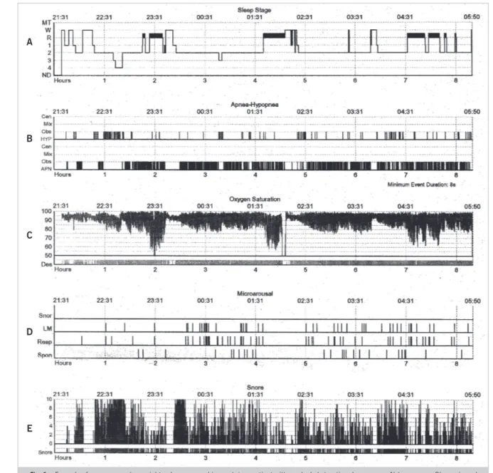
A
B
C
D
E
Fig. 1 – Example of a compressed overnight polysomnographic graph in a patient with marked obstructive sleep apnea. A) hypnogram; B) apneic and hypopneic events; C) frequent events with marked drops in oxyhemoglobin saturation; D) microarousals (sleep fragmentation); E) loud and frequent snoring
hypothesis for OSAHS is its correlation with higher
infl ammatory cytokine levels and insulin resistance.
Inflammatory cytokines, tumor necrosis factor-
α
(TNF-
α
), and interleukin-6 (IL-6) are involved in the
physiological regulation of sleep
28and are abnormally
elevated in apneic patients, when compared to normal
and obese subjects
29,30. However, the correlation between
OSAHS and insulin resistance seems to be independent
of obesity
31.
C
ARDIOVASCULAR
C
ONSEQUENCES
The main cardiovascular consequences are changes in
autonomic nervous system activity, arterial hypertension,
cardiac arrhythmias, coronary artery disease, stroke, and
congestive heart failure.
Changes in autonomic nervous system
activity
Heart rate and blood pressure changes observed
in obstructive sleep apnea are likely to be secondary
to autonomic nervous system activation during every
apneic event
32. This was demonstrated even in patients
with mild OSAHS
33,34. During these events, a progressive
increase in sympathetic activity occurs, reaching its peak
at apnea termination, followed by a marked decrease
during recovery
35,36.
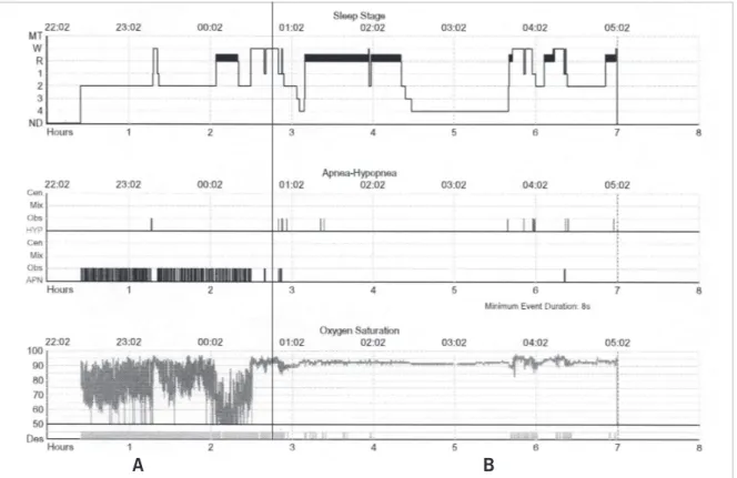
Fig. 2 – Example of a compressed overnight polysomnographic graph in a patient with marked obstructive sleep apnea followed by the introduction of nasal CPAP. A) observe the hypnogram, number of abnormal breathing events and intense oxyhemoglobin desaturation. B) after nasal CPAP introduction, observe the normalization of hypnogram with REM sleep rebound recording, abnormal breathing events, and oxyhemoglobin desaturation
A B
Chart 1 – Term Defi nition
17-19Term Defi nition
Polysomnography Polygraphic sleep recording, including electroencephalogram (EEG), electroocculogram (EOG), electromyogram (EMG), electrocardiogram (ECG), and respiratory parameters.
NREM sleep Characterized by electroencephalographic slow waves associated with specifi c graphic elements.
REM sleep Characterized by EEG desynchronization, rapid eye movements, muscle atonia, and dreams.
Arousals Transient awakening lasting more than three seconds.
Apnea Reduction of > 80% in oronasal airfl ow during more than ten seconds.
Hypopnea Reduction of > 20% in oronasal airfl ow during more than ten seconds, usually accompanied by either a decrease in oxyhemoglobin saturation or an arousal
Apnea-hypopnea index (AHI) Frequency of apneic or hypopneic episodes per hour of sleep; it is a measure of sleep apnea severity.
Oxygen desaturation Decrease of > 3% in oxyhemoglobin saturation, often caused by apnea or hypopnea.
Obstructive sleep apnea-hypopnea syndrome
AHI > fi ve per hour of sleep accompanied by symptoms such as snoring, restless sleep, nocturnal dyspnea, morning headaches, excessive daytime sleepiness, and arterial hypertension.
urinary norepinephrine concentrations are inversely
proportional to nadir nocturnal oxyhemoglobin saturation.
Furthermore, the ventilatory changes that determine
hypoxia and hypercapnia cause an increase in muscle
sympathetic nerve activity, which may persist for up
to twenty minutes following stimulus withdrawal
37.
The carotid sinus seems to play a role in the process
between exposure to intermittent nocturnal hypoxia and
development of sustained elevation in autonomic activation
levels, because barorefl ex regulation and sensitivity are
reduced in patients with OSAHS, compared to controls
38.
Current evidence suggests that the intermittent nocturnal
hypoxic stimulus of peripheral chemoreceptors increases
the sympathetic tone in these patients even during
wakefulness and optimal oxygen supply
39,40.
Hypertension
OSAHS is an independent risk factor for systemic
arterial hypertension (SAH)
42-47. SAH prevalence in
OSAHS patients ranges from 40% to 90%, and the
opposite, OSAHS prevalence in SAH patients, ranges
from 22% to 62%
43. Recent studies corroborate this high
prevalence between both conditions
48and show that, in
four years, subjects with apnea/hypopnea index (AHI)
greater than fi fteen per hour of sleep have a fi ve-fold risk
(45% likelihood) of developing SAH
43. A study with 44
patients with drug-resistant hypertension showed that
83% had OSAHS
49.OSAHS patients show great blood
pressure (BP) variability during the night, and they may
not experience the nocturnal dip observed in normal
patients, resulting in higher mean night-time BP values,
even when daytime BP is normal. This BP behavior may
be due to negative intrathoracic pressure with reduced
cardiac output and differential activation of baroreceptors,
hypoxia, hypercapnia, breathing-related arousal and
increased sympathetic activity
50.
Continuous positive airway
pressure (CPAP) has
proved to be effective in the treatment of patients with
OSAHS and SAH
51,52. In a series of eleven patients with
refractory SAH, CPAP reduced nocturnal blood pressure
53.
Furthermore, in twelve patients with severe OSAHS, the
use of CPAP decreased plasma norepinephrine and urinary
catecholamine metabolites
54.
Thus far, therefore, OSAHS should be considered in
the differential diagnosis of refractory systemic arterial
hypertension, and the use of CPAP may help in reducing
both daytime and night-time blood pressure
55,56.
Cardiac arrhythmias
Some authors have already studied the relationship
between OSAHS and cardiac arrhythmias
57,58; however,
there is no consensus in the medical literature concerning
results
57,58and prevalence of tachyarrhythmia and
bradyarrhythmia. This may be partly explained by the
unknown incidence of OSAHS in the general healthy
population and by the high incidence of hypertension and
cardiovascular diseases in OSAHS patients. However, it
is known that the use of CPAP in OSAHS patients may
reduce cardiac arrhythmias, just as artifi cial cardiac
stimulation may attenuate breathing disorders
59,60. Figure
3 shows an example of sinus pause associated with apneic
event in an OSAHS patient.
Atrial fi brillation (AF) deserves underscoring, because
it has been studied in the largest number of clinical
trials related to sleep-disordered breathing
61-66. Its
prevalence seems to increase in patients with OSAHS
and congestive heart failure (CHF) or recent myocardial
revascularization
61,62. As already stated, OSAHS causes
intermittent hypoxemia, sympathetic activation, and
abrupt changes in blood pressure, which may be related
to the development and recurrence of AF. In a prospective
study
of patients referred for electrical cardioversion
of atrial fi brillation/atrial fl utter, 82% of the untreated
or inadequately treated OSAHS patients experienced
recurrent AF, compared to 42% of the treated patients.
Moreover, in the untreated group, recurrence was even
higher among those who showed greater drop in oxygen
saturation during the apneic event. These data suggest
that appropriate treatment with CPAP may reduce AF
recurrence in OSAHS patients.
Ventricular ectopies were reported in up to 66% of
OSAHS patients. However, in a prospective study of 147
patients who underwent simultaneous polysomnography
and Holter monitoring, no increase in ventricular
arrhythmias was found
67. In view of discrepant reports
in the medical literature, it is diffi cult to establish a
direct relationship between the syndrome and ventricular
arrhythmias. Yet, analyzing the relationship between
ventricular ectopies and oxyhemoglobin desaturation
in patients with OSAHS
68, a marked increase is found
in ventricular ectopy frequency when oxygen saturation
drops below 60%, and the relationship between apneic
events and ventricular arrhythmias seems to exist solely in
patients with important desaturation during the night.
Patients with this syndrome experience ventricular
arrhythmias mainly during sleep, unlike patients with
normal sleep
68, and ventricular tachycardia is more
common in OSAHS patients (0-15%) than in the general
population (0-4%)
69. Bradyarrhythmias are strongly
associated with OSAHS. Guilleminault
et al
.
70have
found sinus pause (> 2.5 seconds), second-degree
atrioventricular block, and sinus bradycardia in 11%,
8%, and 7% of the patients, respectively. Koehler
et
al.
71analyzed factors involved in heart blocks in patients
with OSAHS and concluded that most cases occur during
REM sleep and periods with oxygen desaturation of at
least 4%.
Despite confl icting reports in the literature, oxygen
saturation drop during apneic episodes seems to be an
important factor triggering cardiac arrhythmias in patients
with sleep apnea-hypopnea syndrome, and its frequency
is associated with hypoxia severity.
Coronary artery disease
Despite the growing incidence of OSAHS and the
concomitant increase in cardiovascular mortality
9, most
studies face important limitations, because many risk
factors, such as obesity, masculine gender and age,
among others, are the same as those for hypertension
and coronary artery disease (CAD). Therefore, it seems
diffi cult to pinpoint the risk for CAD attributed to OSAHS;
however, convergent observations suggest that OSAHS is
an important factor associated with CAD.
In OSAHS, the increase in peripheral sympathetic
nerve activity during sleep to twice the normal values
persists during wakefulness
72and may contribute to
addition to autonomic nervous system involvement,
both the infl ammation and endothelial injury observed
in OSAHS are likely to participate in the mechanisms
involved in CAD
73.
In a well-conducted study evaluating the impact of
OSAHS therapy on long-term cardiovascular outcomes
(86.5 ± 39 months) in CAD patients, those appropriately
treated experienced a signifi cant reduction in cardiovascular
event risk, defined as cardiovascular death, acute
coronary syndrome, admission for heart failure, or need
of myocardial revascularization
74.
Nocturnal ST-segment changes consistent with
myocardial ischemia are common in patients with
OSAHS and CAD. Mooe et al
75evaluated the occurrence
of nocturnal myocardial ischemia and its relationship to
sleep-related breathing disorders. ST-segment depressions
occurred in 31% of the patients studied. Temporal
association between electrocardiographic fi ndings and
apneic events was found in 19% of the cases, more
frequently in men (p < 0.01) and in more severely
disordered breathing.
Stroke
Among stroke patients, sleep-disordered breathing
incidence, the obstructive form predominantly, may
exceed 50%
76. It remains quite unclear, however, whether
these events detected after stroke are a consequence of
the cerebrovascular event or a preexistent condition. As
with coronary artery disease, OSAHS and stroke also
share many risk factors, and it is equally diffi cult to prove
a cause-effect relationship.
Dziewas et al
77analyzed the frequency of
sleep-disordered breathing in groups of patients with fi rst and
recurrent ischemic stroke. Patients with recurrent stroke
showed higher mean apnea-hypopnea index (AHI) when
compared to patients with fi rst-ever stroke (26.6/h
vs.
15.1/h, p < 0.05) and most commonly had OSAHS. In
the multivariate analysis adjusted for clinical variables and
risk factors, sleep apnea was considered an independent
risk factor for stroke recurrence, and the authors advocate
the use of polysomnography in this group of patients for
risk stratifi cation.
OSAHS most probably contributes to recurrent stroke
through several mechanisms, such as systemic arterial
hypertension, increased platelet aggregation, blood
hypercoagulability, and endothelial dysfunction, among
others. Moreover, blood fl ow to the brain declines during
apnea, due to reduction in cardiac output, which may
predispose risk subjects to stroke, such as those with
atheromatous lesions in carotid and vertebral circulations.
This may be signifi cantly more important during REM
sleep, when brain oxygen demand is higher.
In addition, apnea may impair cognitive function in
patients with previous history of stroke, since it causes
excessive daytime sleepiness and poor concentration
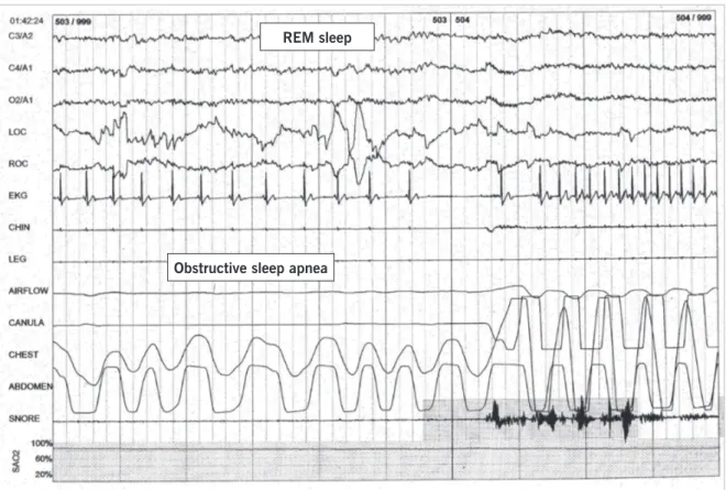
Fig. 3 – Example of a thirty-second epoch graphic containing an obstructive sleep apnea event with sinus pause followed by increment in heart rate
REM sleep
and memory
. Finally, randomized clinical trials
examining apnea therapy and neurological outcomes
are still lacking.
Congestive heart failure
Several studies also showed an important association
between congestive heart failure (CHF) and OSAHS. In
the Javaheri et al
79study, 81 men with CHF underwent
polysomnography, and 11% were found to have
obstructive sleep apnea syndrome. In the Sleep Heart
Health study
80, apneic patients with an AHI > 11 per
hour showed a 2.38 risk of developing CHF, regardless
of other established risk factors, exceeding those found
for other cardiovascular complications associated with
OSAHS, such as hypertension, CAD, and stroke.
It is also believed that CHF contributes to the
development of OSAHS for two reasons, namely,
decreased upper airway muscle tone during the resting
phase of the periodic breathing typical of CHF and fl uid
accumulation in the soft tissues of the cervical region,
contributing to the tendency of upper airway collapse.
Echocardiographic studies showed both systolic
81and diastolic
82dysfunction with increasing AHI. Possible
mechanisms include hypoxia effects (determining
ischemia and impaired contractility), myocyte injury
(due to higher levels of circulating catecholamines), and
intrathoracic pressure swings that accompany apneic
episodes (changing relaxation and left ventricular
end-systolic and end-diastolic volumes).
Finally, the diagnosis of sleep-disordered breathing
in CHF patients may provide valuable prognostic
information and a potential therapeutic option for this
group of patients
83.
Inflammatory, endothelial, and
thrombotic changes in osahs
Decline or absence of physiological mechanisms
involved in vascular relaxation was demonstrated
in response to endothelium-dependent substances
(increased vasoconstrictor, endothelin, and reduced nitric
acid and prostacyclin availability)
84,85. Another evidence
of endothelial damage was documented by enhanced
expression of adhesion molecules, leading to increased
adherence of monocytes to endothelial cells
86. OSAHS
may also trigger recurrent oxidative stress
87, probably
owing to greater production of oxygen-reactive species
by monocytes and granulocytes. These fi ndings reinforce
previous results regarding increased production of
superoxide radicals from polymorphonuclear neutrophils
in patients with OSAHS
88and low levels of nitric oxide
89.
Plasma homocystein levels were also reported to be
elevated in this syndrome
90, as a result of endothelial
dysfunction combined with excessive production of free
radicals. Serium haptoglobin and amyloid A protein were
also reported to be higher
91,92.
The relationship between infl ammatory process and
cardiovascular diseases has been repeatedly postulated
93.
OSAHS patients are chronically exposed to recurrent
hypoxia and sleep fragmentation, showing increased
C -reactive protein and interleukin-6 levels, when
compared to controls
94. It is also likely that C-reactive
protein levels in these patients are higher because of
associated obesity
95. All these endothelial, blood clotting,
and infl ammatory changes may contribute somewhat to
higher cardiovascular risk and atherogenesis in OSAHS
patients. Finally, CPAP therapy may improve these
changes; however, discontinuation of therapy leads to
the reappearance of abnormalities
96-103.
R
EFERENCES
1. Murray CJL, Lopez AD. The global burden of disease: a comprehensive assessment of mortality and disability from disease, injuries and risk factors in 1990 and projected to 2020. USA. Harvard School of Health; 1996.
2. Lolio CA, Laurenti R. Evolução da mortalidade por doença isquêmica do coração no município de São Pauo, 1970 a 1981. Arq Bras Cardiol. 1986; 46: 153.
3. Uemura K, Pisa Z. Recent trends in cardiovascular disease mortality in 27 industrialized countries. Wld Hlth Stat Quart. 1971; 38: 1617-25.
4. Kannel WB, D’Agostino RB, Wilson PW, Belanger AJ, Gagnon DR. Diabetes, fibrinogen, and risk of cardiovascular disease: the Framingham experience. Am Heart J. 1990; 120(3): 672-6.
5. Kannel WB. Lessons from curbing the coronary artery disease epidemic for confronting the impending epidemic of heart failure. Med Clin North Am. 2004; 88: 1129-33.
6. Avezum A, Piegas LS, Pereira JC. Fatores de risco associados com infarto agudo do miocárdio na região metropolitana de São Paulo. Uma região desenvolvida em um país em desenvolvimento. Arq Bras Cardiol. 2005; 84: 206-13.
7. Marin JM, Carrizo SJ, Vicente E, Agusti AGN. Long-term cardiovascular
outcomes in men with obstructive sleep apnea-hypopnoea with or without treatment with continuous positive airway pressure: an observational study. Lancet. 2005; 365: 1046-53.
8. Kramer NR, Cook TE, Carlisle CC, Corwin RW, Millman RP. The role of the primary care physician in recognizing obstructive sleep apnea. Arch Intern Med. 1999; 159: 965-8.
9. Young T, Palta M, Dempsey J, Skatrud J, Weber S, Badr S. The occurrence of sleep-disordered breathing among middle-age adults. N Engl Med. 1993; 328: 1230-35.
10. Olson LG, King MT, Hensley MJ, Saunders NA. A community study of snoring and sleep-disordered breathing: Prevalence. Am J Respir Crit Care Med. 1995; 152: 711-6.
11. Lavie P. Sleep apnea in industrial workers. In Guilleminault C, Lugaresi E, editors. Sleep-wake disorders: natural history, epidemiology and long-term evolution. Philadelphia: Lippincott Raven Press; 1983: 127-35.
12. Stradling JR, Crosby JH. Predictors and prevalence of obstructive sleep apnoea and snoring in 1001 middle aged men. Thorax. 1991; 46: 85-90.
an epidemiologic survey of middle-aged women. Chest. 1993; 103: 1147-51.
14. Bixler EO, Kales A, Soldatos CR, Vela-Bueno A, Jacoby JA, Scarone S. Sleep apneic activity in a normal population. Res Commun Chem Pathol Pharmacol. 1982; 36: 141-52.
15. Strohl KP, Redline S. Recognition of obstructive sleep apnea. Am J Respir Crit Care Med. 1996; 154: 279-89.
16. Silverberg DS, Oksenberg A. Are sleep-related breathing disorders important contributing factors to the production of essential hypertension? Curr Hypertens Rep. 2001; 3: 209-15.
17. EEG arousals: scoring rules and examples. A preliminary report from Sleep Disorders Atlas Task Force of the American Sleep Disorders Association. Sleep. 1992; 15: 173-84.
18. Rechtschaffen A, Kales A. A manual of standardized terminology, technique and scoring system for sleep stages of human sleep, Los Angeles Brain Information Service, Brain Information Institute, Los Angeles, CA; 1968.
19. Sleep-Related Breathing Disorders in Adults: Recommendations for syndrome defi nition and measurement techniques in clinical research. The report of an American Academy of Sleep Medicine Task Force. Sleep. 1999; 22: 667-89.
20. Remmers JE, deGroot WJ, Sauerland EK, Anch AM. Pathogenesis of upper airway occlusion during sleep. J Appl Physiol. 1978; 44(6): 931-8.
21. Wiegand L, Zwillich CW. Obstructive sleep apnea. Dis Mon. 1994; 40(4): 197-252.
22. Strohl KP, Novak RD, Singer W, Cahan C, Boehm KD, Denko CW, Hoffstem VS. Insulin levels, blood pressure and sleep apnea. Sleep. 1994; 17(7): 614-8.
23. McCord JM. Oxygen-derived free radicals in postischemic tissue injury. N Engl J Med. 1985; 312(3): 159-63.
24. Findley LJ, Boykin M, Fallon T, Belardinelli L. Plasma adenosine and hypoxemia in patients with sleep apnea. J Appl Physiol. 1988; 64(2): 556-61.
25. Guilleminault C. State of the art. Sleep and control of breathing. Chest. 1978; 73(2): 293, 297-9.
26. Vgontzas AN, Bixler EO, Chrousos GP. Sleep apnea is a manifestation of the metabolic syndrome. Sleep Med Rev. 2005;9(3):211-24.
27. Yun AJ, Lee PY, Bazar KA. Autonomic dysregulation as a basis of cardiovascular, endocrine, and infl ammatory disturbances associated with obstructive sleep apnea and other conditions of chronic hypoxia, hypercapnia, and acidosis. Med Hypotheses. 2004; 62(6): 852-6.
28. Opp MR, Kapas L, Toth LA. Cytokine involvement in the regulation of sleep. Proc Soc Exp Biol Med. 1992; 201(1): 16-27.
29. Vgontzas AN, Papanicolaou DA, Bixler EO, Kales A, Tyson K, Chrousos GP. Elevation of plasma cytokines in disorders of excessive daytime sleepiness: role of sleep disturbance and obesity. J Clin Endocrinol Metab. 1997; 82(5): 1313-6.
30. Fried SK, Bunkin DA, Greenberg AS. Omental and subcutaneous adipose tissues of obese subjects release interleukin-6: depot difference and regulation by glucocorticoid. J Clin Endocrinol Metab. 1998; 83(3): 847-50.
31. Vgontzas AN, Bixler EO, Chrousos GP. Metabolic disturbances in obesity versus sleep apnoea: the importance of visceral obesity and insulin resistance. J Intern Med. 2003; 254(1): 32-44.
32. Coy TV, Dimsdale JE, Ancoli-Israel S, Clausen J. Sleep apnoea and sympathetic nervous system activity. J Sleep Res. 1996; 5: 42-50.
33. Guilleminault C, Poyares D, Rosa A, Huang YS. Heart rate variability, sympathetic and vagal balance and EEG arousals in upper airway resistance and mild obstructive sleep apnea syndromes. Sleep Med.
2005; 6(5): 451-7.
34. Poyares D, Guilleminault C, Rosa A, Ohayon M, Koester U. Arousal, EEG spectral power and pulse transit time in UARS and mild OSAS subjects. Clin Neurophysiol. 2002; 113(10): 1598-606.
35. Smith ML, Neidermaier ON, Hardy SM, Decker MJ, Strohl KP. Role of hypoxemia in sleep apnea-induced sympathoexcitation. J Auton Nerv Syst. 1996; 56: 184-90.
36. Morgan BJ, Carbtree DC, Palta M, Skatrud J. Combine hypoxia and hypercapnia evokes long-lasting sympathetic activation in humans. J Appl Physiol. 1995; 79(1): 205-13.
37. Parati G, Di Rienzo M, Bonsignore MR, Insalaco G, Marrone O, Castiglioni P, et al. Autonomic cardiac regulation in obstructive sleep apnea syndrome: evidence from spontaneous barorefl ex analyss during sleep. J Hypertension. 1997; 15: 1621-6.
38. Jo JA, Blasi A, Valladares E, Juarez R, Baydur A, Khoo MC. Model-based assessment of autonomic control in obstructive sleep apnea syndrome during sleep. Am J Respir Crit Care Med. 2003; 167(2):128-36.
39. Arabi Y, Morgan BJ, Goodman B, Puleo DS, Xie A, Skatrud JB. Daytime blood pressure elevation after nocturnal hypoxia. J Appl Physiol. 1999; 87(2): 689-98.
40. Xie A, Skatrud JB, Puleo DS, Morgan BJ. Exposure to hypoxia produces long-lasting sympathetic activation in humans. J Appl Physiol. 2001; 91(4): 1555-62.
41. Belozeroff V, Berry RB, Khoo MC. Model-based assessment of autonomic control in obstructive sleep apnea syndrome. Sleep. 2003; 26(1): 65-73.
42. Nieto FJ, Young TB, Lind BK, Shahar E, Samet JM, Redline S, et al. Association of sleep-disordered breathing, sleep apnea, and hypertension in a large community-based study. Sleep Heart Health Study. JAMA. 2000; 283(14): 1829-36.
43. Peppard PE, Young T, Palta M, Skatrud J. Prospective study of the association between sleep-disordered breathing and hypertension. N Engl J Med. 2000; 342(19): 1378-84.
44. Hedner J. Regulation of systemic vasculature in obstructive sleep apnea syndrome. Sleep. 2000; 23(Suppl 4): S132-5.
45. Lavie P, Herer P, Hoffstein V. Obstructive sleep apnoea syndrome as a risk factor for hypertension: population study. BMJ. 2000; 320(7233): 479-82.
46. Sin DD, Fitzgerald F, Parker JD, Newton GE, Logan AG, Floras JS, et al. Relationship of systolic BP to obstructive sleep apnea in patients with heart failure. Chest. 2003; 123(5): 1536-43.
47. Silverberg DS, Oksenberg A, Iaina A. Sleep related breathing disorders are common contributing factors to the production of essential hypertension but are neglected, underdiagnosed, and undertreated. Am J Hypertens. 1997; 10(12 Pt 1): 1319-25.
48. Shahar E, Whitney CW, Redline S, Lee ET, Newman AB, Javier Nieto F, O’Connor GT, Boland LL, Schwartz JE, Samet JM. Sleep-disordered breathing and cardiovascular disease – Cross-sectional results of the Sleep Heart Study. Am J Respir Crit Care Med. 2001; 163: 19-25.
49. Logan AG, Perlikowski SM, Mente A, Tisler A, Tkacova R, Niroumand M, et al. High prevalence of unrecognized sleep apnoea in drug-resistant hypertension. J Hypertension. 2001; 19(12): 2271-7.
50. Fletcher EC. The relationship between systemic hypertension and obstructive sleep apnea: facts and theory. Am J Med. 1995; 98(2): 118-28.
51. Hla KM, Skatrtud JB, Finn L, Palta M, Young T. The effect of correction of sleep-disordered breathing on BP in untreated hypertension. Chest. 2002; 122(4): 1111-2.
patientes with obstructive sleep apnea syndrome. Am J Hypertens. 2002; 15(3): 251-7.
53. Logan AG, Tkacova R, Perlikowski SM, Leung RS, Tisler A, Floras JS, et al. Refractory hypertension and sleep apnoea: effect of CPAP on blood pressure and barorefl ex. Eur Respir J. 2003; 21(2): 241-7.
54. Hedner J, Darpo B, Ejnell H, Carlson J, Caidahl K. Reduction in sympathetic activity after long-term CPAP treatment in sleep apnoea cardiovascular implications. Eur Respir J 1995; 8: 222-9.
55. Somers VK, Dyken ME, Clary MP, Abboud FM. Sympathetic neural mechanisms in obstructive sleep apnea. J Clin Invest. 1995; 96(4): 1897-904.
56. Pepperell JC, Davies RJ, Stradling JR. Systemic hypertension and obstructive sleep apnoea. Sleep Med Rev. 2002; 6(3): 157-73.
57. Miller WP. Cardiac arrhythmias and conduction disturbances in the sleep apnea syndrome. Prevalence and signifi cance. Am J Med. 1982; 73: 317-21.
58. Guilleminault C, Connoly SJ, Winkle RA. Cardiac arrhythmias and conduction disturbances during sleep in 400 patients with sleep apnea syndrome. Am J Cardiol. 1983; 52: 490-4.
59. Javaheri S. Effects of continuous positive airway pressure on sleep apnea and ventricular irritability in patients with heart failure. Circulation. 2000; 101: 392-7.
60. Garrigue S, Bordier P, Jais P, Shah DC, Hocini M, Raherison C, et al. Benefi t of atrial pacing in sleep apnea syndrome. N Engl J Med. 2002; 346: 404-12.
61. Mooe T, Gullsby S, Rabben T, Eriksson P. Sleep-disordered breathing: a novel predictor of atrial fi brillation after coronary artery bypass surgery. Coron Artery Dis. 1996; 7: 475-8.
62. Kanagala R, Murali NS, Friedman PA, Ammash NM, Gersh BJ, Ballman KV, et al. Obstructive Sleep Apnea and recurrence of atrial fi brillation. Circulation. 2003; 107: 2589-94
63. Schulz R, Eisele HJ, Seeger W. Nocturnal atrial fi brillation in a patient with obstructive sleep apnoea. Thorax. 2005; 60(2): 174.
64. Gami AS, Pressman G, Caples SM, Kanagala R, Gard JJ, Davison DE, et al. Association of atrial fi brillation and obstructive sleep apnea. Circulation. 2004; 110(4): 364-7.
65. Porthan KM, Melin JH, Kupila JT, Venho KK, Partinen MM. Prevalence of sleep apnea syndrome in lone atrial fi brillation: a case-control study. Chest. 2004; 125(3): 879-85.
66. Flemons WW, Remmers JE, Gillis AM. Sleep apnea and cardiac arrhythmias. Is there a relationship? Am Rev Respir Dis. 1993; 148: 618-21.
67. Roche F, Xuong ANT, Court-Fortune I, Costes F, Pichot V, Duverney D, et al. Relationship among severity of sleep apnea syndrome, cardiac arrhythmias, and autonomic imbalance. PACE. 2003; 26: 669-77.
68. Shepard JW Jr, Garrison MW, Grither DA, Dolan GF. Relationship of ventricular ectopy to oxyhemoglobin desaturation in patients with obstructive sleep apnea. Chest. 1985; 88: 335-440.
69. Cutler MJ, Hamdan AL, Hamdan MH, Ramaswamy K, Smith ML. Sleep apnea: from nose to the heart. J Am Board Farm Pract. 2002; 15: 128-41.
70. Guilleminault C, Connolly SJ, Winkle RA. Cardiac arrhythmia and conduction disturbances during sleep in 400 patients with sleep apnea syndrome. Am J Cardiol. 1983; 52: 490-4.
71. Koehler U, Fus E, Grimm W, Pankow W, Schafer H, Stammnitz A, et al. Heart block in patients with obstructive sleep apnoea: pathogenetic factors and effects of treatment. Eur Respr J. 1998; 11: 434-9.
72. Carlson JT, Hedner J, Elam M, Ejnell H, Sellgren J, Wallin BG. Augmented resting sympathetic activity in awake patients with obstructive sleep apnea. Chest. 1993; 103: 1763-8.
73. Libby P, Ridker PM, Maresi A. Inflammation and atherosclerosis. Circulation. 2002; 105: 1135-43.
74. Milleron O, Pilliere R, Foucher A, de Roquefeuil F, Aegerter P, Jondeau G, et al. Benefi ts of obstructive sleep apnoea treatment in coronary artery disease: a long-term follow-up study. Eur Heart J. 2004; 25(9): 728-34.
75. Mooe T, Franklin KA, Wiklund U, Rabben T, Holmstrom K. Sleep- disordered breathing and myocardial ischemia in patients with coronary artery disease. Chest. 2000; 117(6): 1597-602.
76. Bassetti CL. Sleep and stroke. Semin Neurol. 2005; 52(1): 19-32.
77. Dziewas R, Humpert M, Hopmann B, Kloska S, Ludemann P, Ritter M, et al. Increased prevalence of sleep apnea in patients with recurring ischemic stroke compared with fi rst stroke victims. J Neurol. 2005; 252 (11): 1394-8; Epub 2005 Jul 20.
78. Engleman H, Joffe D. Neuropsychological function in obstructive sleep apnoea. Sleep Med Rev. 1999; 3(1): 59-78.
79. Javaheri S, Parker TJ, Liming JD, et al. Sleep apnea in 81 ambulatory male patients with stable heart failure: types and their prevalences, consequences, and presentations. Circulation. 1998; 97: 2154-9.
80. Shahar E, Whitney CW, Redline S, Lee ET, Newman AB, Javier Nieto F, et al. Sleep-disordered breathing and cardiovascular disease: cross-sectional results of the Sleep Heart Health Study. Am J Respir Crit Care Med. 2001; 163(1): 19-25.
81. Laaban JP, Pascal-Sebaoun S, Bloch E, Orvoen-Frija E, Oppert JM, Huchon G. Left ventricular systolic dysfunction in patients with obstructive sleep apnea syndrome. Chest. 2002; 122: 1133-8.
82. Fung JW, Li TS, Choy DK, Yip GW, Ko FW, Sanderson JE, et al. Severe obstructive sleep apnea is associated with left ventricular diastolic dysfunction. Chest. 2002; 121: 422-9.
83. Wilcox I, McNamara SG, Wessendorf T, Willson GN, Piper AJ, Sullivan CE. Prognosis and sleep disordered breathing in heart failure. Thorax. 1998; 53(3): S33-6.
84. Fletcher EC. Sympathetic over activity in the etiology of hypertension of obstruvtive sleep apnea. Sleep. 2003; 26(1): 15-9.
85. Pepin Jl, Levy P. Pathophysiology of cardiovascular risk in sleep apnea syndrom (SAS). Rev Neurol. 2002; 158: 785-97.
86. Dyugovskaya L, Lavie P, Lavie L. Increased adhesion molecules expression and production of reactive oxygen species in leukocytes of sleep apnea patients. Am J Respir Crit Care Med. 2002; 165(7): 934-9.
87. Lavie L. Obstructive sleep apnoea syndrome - an oxidative stress disorder. Sleep Med Rev. 2003; 7(1): 35-51.
88. Schulz R, Mahmoudi S, Hattar K, Sibelius U, Olschewski H, Mayer K, et al. Enhanced release of superoxide from polymorphonuclear neutrophils in obstructive sleep apnea. Impact of continuous airway pressure therapy. Am J Respir Crit Care Med. 2000; 162: 566-8.
89. Ip MS, Lam B, Chan LY, Zheng L, Tsang KW, Fung PC, et al. Circulation nitric oxide is suppressed in obstructive sleep apnea and is reversed by nasal continuous positive airway pressure. Am J Respir Crit Care Med. 2000; 162: 2166-71.
90. Lavie L, Perelman A, Lavie P. Plasma homicysteine levels in obstructive sleep apnea patients. Chest. 2001; 120: 900-8.
91. Lavie L, Lotan R, Hochberg I, Herer P, Lavie P, Levy AP. Haptoglobin polymorphism is a risk factor for cardiovascular disease in patients with obstructive sleep apnea syndrome. Sleep. 2003; 26(5): 592-5.
92. Svatikova A, Wolk R, Shamsuzzaman AS, Kara T, Olson EJ, Somers VK. Serum amyloid a in obstructive sleep apnea. Circulation. 2003; 108(12): 1451-4.
94. Yokoe T, Minoguchi K, Matsuo H, Oda N, Minoguchi H, Yoshino G, et al. Elevated levels of C-reactive protein and interleukin-6 in patients with obstructive sleep apnea syndrome are decreased by nasal continuous positive airway pressure. Circulation. 2003; 107(8):1129-34.
95. Guilleminault C, Kirisoglu C, Ohayon MM. C-reactive protein and sleep-disordered breathing. Sleep. 2004; 15; 27(8): 1507-11.
96. Kato M, Roberts-Thomson P, Phillips BG, Haynes WG, Winnicki M, Accurso V, et al. Impairment of endothelium-dependent vasodilation of resistance vessels in patients with obstructive sleep apnea. Circulation. 2000; 21; 102(21): 2607-10.
97. Kraiczi H, Caidahl K, Samuelsson A, Peker Y, Hedner J. Impairment of vascular endothelial function and left ventricular fi lling: association with the severity of apnea-induced hypoxemia during sleep. Chest. 2001; 119(4): 1085-91
98. Teramoto S, Kume H, Yamamoto H, Ishii T, Miyashita A, Matsuse T, et al. Effects of oxygen administration on the circulating vascular endothelial growth factor (VEGF) levels in patients with obstructive sleep apnea syndrome. Intern Med. 2003; 42(8): 681-5.
99. Shamsuzzaman AS, Gersh BJ, Somers VK. Obstructive sleep apnea: implications for cardiac and vascular disease.JAMA. 2003; 290(14): 1906-14.
100. Nieto FJ, Herrington DM, Redline S, Benjamin EJ, Robbins JA. Sleep apnea and markers of vascular endothelial function in a large community sample of older adults. Am J Respir Crit Care Med. 2004; 169(3): 354-60.
101. Dyugovskaya L, Lavie P, Lavie L. Phenotypic and functional characterization of blood gammadelta T cells in sleep apnea. Am J Respir Crit Care Med. 2003; 168(2): 242-9.
102. Schulz R, Hummel C, Heinemann S, Seeger W, Grimminger F. Serum levels of vascular endothelial growth factor are elevated in patients with obstructive sleep apnea and severe nighttime hypoxia. Am J Respir Crit Care Med. 2002; 165(1): 67-70.