Iranian Journal of Basic Medical Sciences
ijbms.mums.ac.ir
Effect of acetylcholine receptors on the pain‐related electrical
activities in the hippocampal CA region of morphine‐
addicted rats
Guan Zeng Li , Zhe Hui Liu , XinYa Wei , Pan Zhao , Chun Xiao Yang *, Man Ying Xu
Department of Neurology, Liaocheng People’s Hospital, Dongchang Xi Road, Liaocheng , ChinaDepartment of Cadre Health Protection, Liaocheng People’s Hospital, Dongchang Xi Road, Liaocheng , China
Department of Neurology, Second Affiliated Hospital, Harbin Medical University, Xuefu Road, Nangang District, Harbin , Heilongjiang, China
Laboratory of Neural Algesia Electrophysiology, Department of Physiology, Harbin Medical University, Xuefu Road, Nangang District, Harbin , Heilongjiang, China
A R T I C L E I N F O A B S T R A C T Article type:
Original article Objective(s):responses of pain excited neurons PEN and pain inhibited neurons PIN in hippocampal CA region To determine the effect of acetylcholine ACh , pilocarpine, and atropine on pain evoked of morphine addicted rats.
Materials and Methods:Female Wistar rats, weighing between ‐ g were used in this study. Morphine addicted rats were generated by subcutaneous injection of increasing concentrations of morphine hydrochloride for six days. Trains of electrical impulses applied to the sciatic nerve were used as noxious stimulation and the evoked electrical activities of PEN or PIN in hippocampal CA area were recorded using extracellular electrophysiological recording techniques in hippocampal slices. The effect of acetylcholine receptor stimulation byACh, the muscarinic agonist pilocarpine, and the muscarinic antagonist atropine on the pain evoked responses of pain related electrical activities was analyzed in hippocampal CA area of morphine addicted rats.
Results:Intra‐CA microinjection of ACh μg/ μl or pilocarpine μg/ μl decreased the discharge frequency and prolonged the firing latency of PEN, but increased the discharge frequency and shortened the firing inhibitory duration ID of PIN. The intra‐CA administration of atropine . μg/ μl produced opposite effect. The peak activity of cholinergic modulators was to min later in morphine addicted rats compared to peak activity previously observed in normal rats.
Conclusion: ACh dependent modulation of noxious stimulation exists in hippocampal CA area of morphine addicted rats. Morphine treatment may shift the sensitivity of pain related neurons towards a delayed response to muscarinergic neurotransmission in hippocampal CA region.
Article history: Received: Nov , Accepted: Apr , Keywords: Hippocampal CA Morphine addiction Muscarinergic
Acetylcholine receptors Pain
►Please cite this article as:
Li GZ, Liu ZH, Wei XY, Zhao P, Yang CX, Xu MY. Effect of acetylcholine receptors on the pain‐related electrical activities in the hippocampal CA region of morphine‐addicted rats. Iran J Basic Med Sci ; : ‐ .
Introduction
Opioids are commonly prescribed for chronic cancer pain management . Opioids are also known to be extremely addictive. The addictive features of morphine lead to its inappropriate use which creates significant medical and socioeconomic problems worldwide. Opioids are co‐stored with glutamate in several hippocampal pathways, including the lateral perforant path LPP input to granule cells in the dentate gyrus, the LPP input to CA pyramidal cells, and
the mossy fiber projection to CA pyramidal cells .
Several studies have shown that chronic exposure to morphine or heroin leads to the impairment of
hippocampal long‐term potentiation LTP and to
the impairment of acquisition or retention of
spatial memory.
Acetylcholine Ach is a major excitatory neurotransmitter in the central nervous system CNS
of vertebrates and invertebrates . More recently,
a study on the neurobiology of pain has revealed that the brain’s cholinergic system, particularly muscarinic acetylcholine receptors mAChRs , may be
involved in the modulation of pain . This action of
ACh is mediated by mAChRs . In addition, a vast
amount of literature has appeared describing the antinociceptive action of both cholinesterase
*Corresponding author: Chun Xiao Yang. Department of Neurology, Second Affiliated Hospital, Harbin Medical University, Xuefu Road, Nangang District,
inhibitors and cholinomimetic drugs . Various mAChRs have also been shown to be potential mediators of pain related neuroplasticity. Intrathecal administration of cholinergic muscarinic agonists or acetylcholinesterase inhibitors produces analgesia in
both animals and humans . Moreover, the
analgesic effect of morphine may be mediated by
components of the cholinergic system .
Therefore, it can be postulated that morphine or heroin may affect nociceptive neurotransmission in the hippocampus. However, the precise role of ACh in the reinforcement and addiction of abusive drugs is not well understood.
The hippocampus is a region of the mammalian brain that has been extensively studied due to its role in many forms of learning, memory, and pain response. Electrophysiological, pharmacological, behavioral, and clinical data indicate that the hippocampal formation is an integral component of the limbic system and, as such, plays an important role in the affective and motivational aspects of pain
perception . The hippocampus receives
cholinergic projections from the medial septal nucleus and Broca’s diagonal band, which terminate
in the CA , CA , and dentate gyrus regions .
Some scholars report that both morphine and AChR agonists can modulate hippocampal neuronal
excitability . Moreover, previous studies have
shown that the CA and dentate gyrus of the
hippocampus are involved in nociception , , .
However, the interplay of cholinergic and opioid effects for pain evoked responses of PEN and PIN, particularly in the hippocampal CA , is not well studied. Therefore, in this study, we have evaluated the effects of ACh, the muscarinic agonist pilocarpine, and the muscarinic antagonist atropine on pain related electrical activities in the hippocampal CA region of morphine addicted rats.
Materials
and
Methods
Experimentalanimals
Female Wistar rats Animal Centre of the Second Affiliated Hospital, Harbin Medical University,
Certificate No. ‐ ‐ weighing between ‐ g
were used in this study. Experiments complied with the Guide for the Care and Use of Laboratory Animals published by the US National Institutes of Health
NIH publication number ‐ ; revised, .
Morphine‐dependent rats were produced by subcutaneous injection of morphine hydrochloride for days at increasing concentrations Table as
previously described . Morphine hydrochloride
was injected subcutaneously three times per day according to a gradually increasing dose scheme, in which the morphine dose increased from mg/kg to mg/kg over five days to produce morphine addicted rats. Morphine‐dependent rats were
randomly divided into four groups: control group n= , neurons of rats were recorded : intra‐ CA administration of µl saline; ACh group
n= , neurons of rats were recorded : intra‐ CA administration of ACh μg/ µl ; atropine group n= , neurons of rats were recorded : intra‐CA administration of atropine . μg/ μl ; and pilocarpine group n= , neurons of rats were recorded : intra‐CA administration of
pilocarpine μg/ μl . All injections were
completed within min via a microliter syringe.
Neurosurgeryandelectrophysiologicalstudies
Rats were anesthetized with % urethane IP, g/kg . The right sciatic nerve was isolated and two skull windows were opened and covered with liquid paraffin. The head of the rat was fixed on a stereotaxic frame SN‐ , Narishige, Japan . After ‐ min, the rat was paralyzed with tubocurarine chloride IP, mg/kg and artificial ventilation was maintained. Single‐unit recordings were performed with a glass microelectrode . ‐ . μm, DC resistance – MΩ filled with KCl mol/l . According to the B coordinate system of
Pellegrino’s Atlas , the glass microelectrode was
inserted by a micromanipulator SM‐ , Narishige, Japan into the hippocampal CA A: − . ‐ − . mm; R or L: . ‐ . mm, H: . ‐ . mm to record the discharges of the CA neurons. Another glass microelectrode filled with drugs ACh, atropine, and pilocarpine was inserted by a micromanipulator SM‐
, Narishige, Japan into the hippocampal CA A: − . mm, R: . mm, or L: . mm, H: . mm for administration of drugs. The electrical activity was
amplified by a microelectrical amplifier MEZ‐ ,
Nihon Kohden, Japan and recorded by an output
isolator SS‐ J‐type, Narishige, Japan in combination
with a tape recorder ST‐CH X type, Panasonic,
Japan . Electrical activity was simultaneously monitored with an oscilloscope VC‐ , Nihon Kohden, Japan . As the neural discharges were recorded, the electrical stimulation of the sciatic nerve was performed through a double stainless steel electrode
delay: , interval: msesc,duration: . msec, and
train: ; SEN‐ ,NihonKohden,Japan asnoxious
Table1.Dose mg/kg and time of morphine injection
Dose mg/kg of morphine injection
: : :
stimulation. Articular movement and hair touching were set as the non‐noxious stimulation to identify the pain reactive neurons. The discharge of each neuron was monitored continuously for min, and typical discharges were recorded three times every two min.
Histological identification of the hippocampal CA3
region
At the end of the experiment in order to identify the tip position, pontamine sky blue in the microelectrode
was diffused out with a negative direct current mA,
min into the hippocampal CA A: − . ‐ − . mm; R or L: . ‐ . mm, and H: . ‐ . mm Figure .
Definitionofneurons
Recording PEN and PIN activity is widely considered as an appropriate electrophysiological
read‐out of pain , . PENs are defined as the
neurons that responded by increasing the discharge
frequency to noxious stimulus , while PINs are
defined as the neurons that responded by decreasing
the discharge frequency to noxious stimulus .
PEN showed very few spontaneous discharges before noxious stimulation. In contrast, spontaneous discharges were common in PIN, which were readily inhibited by the application of noxious stimulation.
Observationindex
The net increased value NIV, in Hz refers to the difference in the PEN or PIN between the average frequency of evoked discharges after noxious stimulation and the average frequency of the discharges within sec before noxious stimulation. The latency s refers to the latency time from the noxious stimulation to the appearance of the PEN discharges. Inhibitory duration ID, in sec refers to the latency time from the noxious stimulation to the appearance of the PIN discharges.
Statisticalanalysis
Data were scanned to a computer with Powerlab/ sec ADInstruments, Australia and analyzed with Chart v . software ADInstruments, Australia . All data were expressed as mean±SEM and were analyzed using SPSS
Figure 1. Location of the microelectrode arrow in the
hippocampal CA area of rat. Scale bar, . mm
. software SPSS, USA . Statistical differences were evaluated by repeated measures ANOVA, while the post hoc test was used to compare the differences between
the two groups. P< . was considered statistically
significant.
Results
Effects of saline on the electrical activities in
hippocampalCA3neuronsofmorphineaddictedrats
In the control group, the noxious stimulation increased the discharge frequency of PENs while decreasing the frequency of PINs. The average NIV of PENs was . ± . Hz, and the latency was . ± . sec. The NIV of PENs and the latency showed no significant differences ‐ min after the intra‐CA administration of saline compared to values before injection Figure A . The average NIV of PINs was – . ± . Hz and the average ID of
PINs was . ± . sec. Zero to min after the
intra‐CA administration of saline, the NIV of PINs and the ID showed no significant differences compared to values before injection Figure A . Administration of saline as a control treatment produced no significant change in the electrical activity of PENs and PINs Figure A and Figure A .
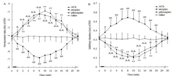
InfluenceofACh ontheelectricalactivities ofthe
CA3neuronsinmorphine‐addictedrats
In the ACh group, the average NIV of PENs was . ± . Hz, and the latency was . ± . sec. Immediately after the intra‐CA administration of ACh, the NIV of PENs began to decline and the latency began to prolong Figure B . These effects reached a peak at min after administration of ACh,
with the NIV decreasing to . ± . Hz F-statistic=
. , P< . and the latency being prolonged
to . ± . sec F= . , P< . . Between
the time points of ‐ min after administration of
ACh, the NIV F= . , P< . and latency F=
. , P< . of PENs showed significant
changes compared with those before administration. Between the time points of ‐ min after administration, the NIV, and between the time points of ‐ min after administration, latency showed
significant changes P< . or P< . compared
with those of the saline control group Figure . At min after ACh administration, the NIV and latency of PENs gradually returned to the values observed before treatment.
The average NIV of PINs was − . ± . Hz, and the average ID of PINs was . ± . sec. Immediately after injection of ACh, the average NIV began to increase and ID began to shorten Figure B . These changes reached a peak value at min after injection; with the average NIV at − . ± .
Hz F= . , P< . and the ID decreased to
. ± . sec F= . , P< . . The average NIV
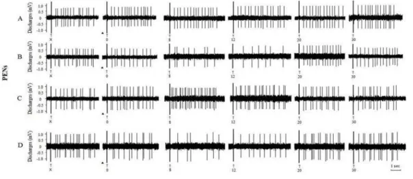
Figure2. Effects of intra‐CA injection of different substances on the evoked discharges of PENs in the CA of morphine‐addicted rats Morphine‐dependent rats were injected with A saline µl , B ACh μg/ µl , C atropine . μg/ μl ; and D pilocarpine μg/ μl ↑, Stimulus artifact; ▲, injection substance; X, before injection; , , , , , time after injection min
compared to values before injection between the time points of ‐ min after injection. Between ‐
min after injection, the ID F= . , P= . of
PINs showed significant differences compared to values before injection. Between the time points of ‐
min after administration, the NIV, and between the time points of ‐ min after administration, ID
showed significant changes P< . or P< .
compared with those of the control group Figure .
Effectsofatropineontheelectricalactivitiesofthe
CA3neuronsinmorphineaddictedrats
In the atropine group, at min after the intra‐CA administration of atropine, the average NIV of PENs significantly increased from . ± . Hz to . ± .
Hz F= . , P< . and the latency decreased
from . ± . sec to . ± . sec F= . ,
P< . Figure C . During ‐ min after the
injection of atropine, the NIV F = . , P= . and
the latency F= . , P< . of PENs showed
obvious changes compared to those before administration. Between the time points of ‐ min after administration, the NIV, and between the time points ‐ min after administration, latency showed
significant changes P< . or P< . compared with
those of the control group Figure .
The average NIV of PINs significantly decreased min after the intra‐CA administration of atropine from
− . ± . Hz to − . ± . Hz F= . ,
P< . and the ID was prolonged from . ± .
sec to . ± . sec F= . , P< . compared
to values before injection Figure C . At the time points
of ‐ min after the injection, the NIV F= . , P=
. and ID F= . , P= . of PINs showed
significant changes compared to values before administration. Between the time points of ‐ min after administration, the NIV, and between the time points of ‐ min after administration, ID showed
significant changes P< . or P< . compared to the
saline control group Figure .
Figure3. Effects of intra‐CA injection of different substances on the evoked discharges of PINs in the CA of morphine‐addicted rats
Figure4.Influence of intra‐CA injection of different substances on the NIV A and latency B of PEN in the CA of morphine‐addicted rats. ▃, injection of substance; X, before injection; , ,. . ., , time after injection min ; values are presented as means±SEM. *P< . ,
**P< . , #P< . , ##P< . , P< . , P< . , when compared with the saline control group
Influenceofpilocarpineontheelectricalactivities
oftheCA3neuronsinmorphineaddictedrats
In the pilocarpine group, the average NIV of PENs was . ± . Hz and the latency was . ± . sec before the injection of pilocarpine. After the intra‐ CA administration of pilocarpine, the NIV of PEN began to decrease and the latency began to prolong Figure D . These effects peaked at min after administration of pilocarpine. The average NIV
decreased to . ± . Hz F= . , P< . ,
and the latency was prolonged to . ± . sec
F= . , P< . compared to values before
injection. The NIV F= . , P= . and the
latency F= . , P= . showed significant
differences during the time points of ‐ min after the injection. Between the time points of ‐ min after administration, the NIV and latency showed
significant changes P< . or P< . compared
with those of control group Figure .
After the intra‐CA injection of pilocarpine, the NIV of PINs began to increase and ID began to shorten Figure D . These effects also reached a peak value at min after the injection, when the average NIV of PINs significantly increased from ‐ . ± . to ‐ . ± .
Hz F= . , P< . , and the average ID
decreased from . ± . to . ± . sec F= . ,
P< . compared to values before injection. The
time points of ‐ min after the injection showed
significant changes in the average NIV F= . ,
P= . and ID F= . , P= . of PINs compared
with measurements obtained prior to injection. Between the time points of ‐ min after administration, the NIV, and between the time points of ‐ min after administration, ID showed significant
changes P< . or P< . compared with the control
group Figure . About min after the
administration, the NIV and ID of PINs returned to the values observed before treatment Figure .
Figure5. Influence of intra‐CA injection of different substances on the NIV A and ID B of PIN in the CA of morphine‐addicted rats
▃, injection of substance; X, before injection; , ,. . ., , time after injection min ; values are presented as means±SEM. *P< . , **P< . ,
Discussion
In order to investigate the interplay of cholinergic and opioid effects for pain evoked responses in the CNS, we studied the effects of ACh on the electrical activities of PENs and PINs in hippocampal CA of morphine‐addicted rats. Our study revealed that, in this animal model for morphine dependence, ACh
and pilocarpine inhibited the electric activities of
evoked discharges of PENs, but potentiated discharges of PINs. On the other hand, atropine potentiated the electric activities of evoked discharges of PENs and inhibited those of PINs. These results demonstrate that intra‐CA administration of ACh or pilocarpine can produce
antinociceptive effects, whereas intra‐CA
administration of atropine can facilitate nociceptive effects in morphine‐dependent rats.
These results reveal that cholinergic neurons in the hippocampal CA region of morphine‐dependent rats are involved in the response to noxious stimuli, and these results are similar to the previously
studied effects in control rats . In the previous
study in normal rats, cholinergic substances evoked
a peak in electrical activity at to min after
injection. In morphine‐dependent rats, we observed a peak activity at to min after injection; to min later than in normal rats. Although further studies are needed comparing the effects in normal rats versus morphine‐dependent rats, the rather
large differences to the previous results indicate
that morphine treatment delays ACh‐dependent electrical activities of PENs and PINs in hippocampal CA . This may be a sign of delayed sensitivity of pain related neurons towards ACh in the hippocampal CA region after morphine treatment.
Early pharmacological experiments have shown that the microinjection of ACh or carbachol into specific brainstem nuclei can produce antinociceptive effects that can be reversed by muscarinic receptor
antagonists . Previous studies, including three
studies performed in our laboratory , ,
provided evidence that the hippocampal formation is involved in pain modulation and that nociceptive stimuli modify the electrical activity of the
hippocampus . Moreover, ACh can mediate some
antinociceptive effects, which are produced by other types of receptors or drugs. For instance, the antinociceptive effect of crotoxin is attributed to activation of muscarinic receptors, by action of the
toxin on central cholinergic neurons . Another
study showed that atropine blocked the antinociceptive effect of the crotoxin, further confirming that central muscarinic receptors mediate the antinociceptive effect
of the toxin . Our present results, demonstrating
that atropine potentiated the electric activities of evoked discharges of PENs and inhibited those of PINs, are consistent with the notion that ACh participates in antinociception in the CNS. On the other hand, centrally
administered opioids increase the ACh concentration in cerebrospinal fluid and in spinal cord dorsal horn microdialysates. In this case, analgesia resulting from central opioid injection is partially reversed by spinal
injection of mAChR antagonists . Therefore, it
appears that ACh participates in analgesic effects after systemic or central administration of opioids.
The hippocampus receives cholinergic innervation from the medial septum‐diagonal band complex. Every hippocampal region is connected by extensive fiber networks. The hippocampal CA area receives synaptic input from the granule cells of the dentate gyrus. The pyramidal cells in the CA area send their axon collaterals into the strata radiatum and strata oriens of the hippocampal CA area. Aside from these major fiber connections, the dendritic shafts of various types of interneurons can build connections with the spines of principal cells. Cholinergic suppression of synaptic
transmission has been reported in theCA region .
When synaptic transmission in the CA region was suppressed, the transmission of noxious stimulus was likely to be inhibited. Our previous study indicates that the hippocampal CA area is involved in the
modulation of nociception ; the effect in the
hippocampal CA of morphinistic rats was similar to
the effects in normal rats .
Compared to the results of our previous study
performed in normal rats , the peaks of the pain
evoked responses in morphine‐dependent rats are shifted to later response times. These results are similar to the previously studied effects in the hippocampal
CA area . The results indicate that the sensitivity
of pain related neurons in the CA and CA areas to noxious stimulation is attenuated in morphine addicted rats. Thus, morphine addiction seems to attenuate the sensitivity of pain related neurons to the noxious information processed in several hippocampal areas. Altered synaptic transmission and plasticity in brain areas involved in reward and learning are thought to underlie the long‐lasting effects of addictive drugs. Data
by Kahn etal showed a strong reduction of cellular
proliferation together with an increase of glutamate decarboxylase‐ mRNA transcription in the dentate gyrus‐CA region of the adult rat hippocampus after
repeated morphine treatment . It is likely that
reducing cell proliferation and neurogenesis and altering neuronal phenotypes, have an impact on ACh‐ dependent antinociceptive neurotransmission in the hippocampal CA . However, further investigation is required to determine the precise mechanisms underlying the changes in antinociceptive effects in morphine addicted rats.
Conclusion
rats; and the hippocampal CA area is involved in the ACh‐dependent modulation of noxious stimulation in morphine addicted rats. In comparison to ACh effects in
normal rats , morphine addicted rats show delayed
responses of pain related neurons to ACh in the hippocampal CA , which is likely related to adaptive changes of synaptic transmission and plasticity. Further comparative studies on the underlying mechanisms will deepen our knowledge about pain modulation by ACh and morphine and the effects of morphine dependence on pain.
Acknowledgment
This work was supported by the Union Foundation of Doctoral Students and New Teachers of National Ministry of Education in China Project
No. and the National Natural
Science Foundation of China Grant No. .
Conflicts
of
interest
The authors declare no conflicts of interest.
References
. Strumpf M, Willweber‐Strumpf A, Zenz M. Opioids:
modern concepts of pain management. Med Klin
Munich ; : ‐ .
. Bramham CR, Sarvey JM. Endogenous activation of
m and d‐ opioid receptors is required for long‐term potentiation induction in the lateral perforant path:
dependence on GABAergic inhibition. J Neurosci ;
: ‐ .
. Bao G, Kang L, Li H, Li Y, Pu L, Xia P, et al.
Morphine and heroin differentially modulate invivo
hippocampal LTP in opiate dependent rat.
Neuropsychopharmacology ; : ‐ .
. Li Z, Wu CF, Pei G, Xu NJ. Reversal of morphine‐
induced memory impairment in mice by withdrawal in Morris water maze: possible involvement of
cholinergic system. Pharmacol Biochem Behav ;
: ‐ .
. MiladiGorji H, Rashidy‐Pour A, Fathollahi Y.
Effects of morphine dependence on the performance of rats in reference and working versions of the
water maze. Physiol Behav ; : ‐ .
. Yang XF, Xiao Y, Xu MY. Both endogenous and
exogenous ACh plays antinociceptive role in the
hippocampus CA of rats. J Neural Transm ;
: ‐ .
. Mojtahedin A, Tamaddonfard E, Zanbouri A. Role of
central muscarinic cholinergic receptors in the formalin‐induced pain in rats. Indian J Pharmacol
; : ‐ .
. Li B, Duysen EG, Volpicelli‐Daley LA, Levey AI,
Lockridge O. Regulation of muscarinic acetylcholine receptor function in acetylcholinesterase knockout
mice. Pharmacol Biochem Behav ; : ‐ .
. Hartvig P, Gillberg PG, Gordh T Jr, Post C.
Cholinergic mechanisms in pain and analgesia. Trends
Pharmacol Sci ; : ‐ .
.Chen SR, Wess J, Pan HL. Functional activity of the
M and M receptor subtypes in the spinal cord studied with muscarinic acetylcholine receptor
knockout mice. J Pharmacol Exp Ther ; : ‐
.
.Taguchi K, Kato M, Kikuta J, Abe K, Chikuma T,
Utsunomiya I, etal. The effects of morphine‐induced
increases in extracellular acetylcholine levels in the rostral ventrolateral medulla of rat. J Pharmacol Exp
Ther ; : ‐ .
.McKenna JE, Melzack R. Blocking NMDA receptors
in the hippocampal dentate gyrus with AP produces
analgesia in the formalin pain test. Exp Neurol ;
: ‐ .
.Buño W, Cabezas C, Fernández de Sevilla D.
Presynaptic muscarinic control of glutamatergic
synaptic transmission. J Mol Neurosci ; : ‐
.
.Bramham CR, Sarvey JM. Endogenous activation of
mu and delta‐lopioid recptors is required for long‐ term potentiation induction in the lateral perforantpath: dependence on GABAergic inhibition. J
Neurosci ; : ‐ .
.Jiao RS, Yang CX, Zhang Y, Xu MY, Yang XF.
Cholinergic mechanism involved in the nociceptive modulation of dentate gyrus. Biochem Biophys Res
Commun ; : ‐ .
.Xiao Y, Yang XF, Xu MY. Effect of acetylcholine on
pain‐related electric activities in hippocampal CA area of normal and morphinistic rats. Neurosci Bull
; : ‐ .
.Zhao CY, Yan LX, Lu N, Zhang JY, Xu MY. Making
the model quickly for morphinomania in rats. J
Harbin Med University ; : ‐ .
.Pellegrino LJ, Pellegrino AS, Cushmanl AJ. A
stereotaxic atlas of the rat brain. nd ed. New York:
Plenum Press; .p. ‐ .
.Zhang Y, Yang CX, Xu XZ, Jiao RS, Jin HB, Lv YH,
et al. Morphine dependence changes the role of
droperidol on pain‐related electric activities in caudate
nucleus. Biochem Biophys Res Commun ;
: ‐ .
.Shi TF, Yang CX, Yang DX, Jiao RS, Zhang GW, Gao
HR,etal. MK‐ changes the role of glutamic acid on
modulation of algesia in nucleus accumbens. Biochem
Biophys Res Commun ; : ‐ .
.Zhang XT. The integration of thalamus in the
process of acupuncture analgesia. Sci China ;
: ‐ .
.Sun MZ, Chen LS, Gu HL, Cheng J, Yue LS. Effect of
acupuncture on unit discharge in nucleus parafascicularis of rat thalamus. Sheng li Xue Bao
; : ‐ .
.Li GZ, Liang QC, Jin YH, Yang CX, Zhang GW, Gao
HR, etal. The effect of acetylcholine on pain‐related
electric activities in the hippocampal CA of rats. J
Neural Transm ; : ‐ .
.Yaksh TL, Dirksen R, Harty GJ. Antinociceptive
effects of intrathecally injected cholinomimetic drugs
in the rat and cat. Eur J Pharmacol ; : ‐ .
.Khanna S, Zheng F. Morphine reversed formalin‐
induced CA pyramidal cell suppression via an effect on septohippocampal neural processing. Neurosci
; : ‐ .
.Zhang HL, Han R, Chen ZX, Chen BW, Gu ZL, Reid
PF, et al. Opiate and acetylcholine‐independent
analgesic actions of crotoxin isolated from crotalus
.Nogueira‐Neto FS, Amorim RL, Brigatte P, Picolo
G, Jr Ferreira WA, Gutierrez VP, etal. The analgesic
effect of crotoxin on neuropathic pain is mediated by central muscarinic receptors and ‐lipoxygenase‐
derived mediators. Pharmacol Biochem Behav ;
: ‐ .
.Xu ZM, Tong CY, Pan HL, Cerda SE, Eisenach JC.
Intravenous morphine increases release of nitric oxide from spinal cord by an a‐adrenergic and cholinergic
mechanism. J Neurophysiol ; : ‐ .
.Kremin T, Hasselmo ME. Cholinergic suppression of
glutamatergic synaptic transmission in hippocampal region CA exhibits laminar selectivity: Implication for
hippocampal network dynamics. Neurosci ;
: ‐ .
.Kahn L, Alonso G, Normand E, Manzoni OJ. Repeated
morphine treatment alters polysialylated neural cell adhesion molecule, glutamate decarboxylase‐ expression and cell proliferation in the adult rat