Time Course Study of Blood Pressure in
Term and Preterm Infants Immediately
after Birth
Gerhard Pichler1,2,3, Po-Yin Cheung1,2, Corinna Binder3, Megan O’Reilly1,2, Bernhard Schwaberger3, Khalid Aziz1,2, Berndt Urlesberger3,
Georg M. Schmo¨lzer1,2,3*
1.Centre for the Studies of Asphyxia and Resuscitation, Neonatal Research Unit, Royal Alexandra Hospital, Edmonton, Canada,2.Department of Pediatrics, University of Alberta, Edmonton, Canada,3.Division of Neonatology, Department of Pediatrics, Medical University, Graz, Austria
Abstract
Objective:To describe temporal changes in systolic, diastolic, and mean blood pressure (SBP, DBP, and MBP, respectively) in term and preterm infants immediately after birth.
Methods:Prospective observational two-center study. In term infants SBP, DBP, and MBP were assessed non-invasively every minute for the first 15 minutes, and in preterm infants every minute for the first 15 minutes, as well as at 20, 25, 30, 45, and 60 minutes after birth. Regression analyses were performed by gender and respiratory support in all neonates; and by mode of delivery, cord clamping time, and development of ultrasound-detected brain injury in preterm neonates. Results: Term infants (n554) had a mean (SD) birth weight of 3298 (442) g and gestational age of 38 (1) weeks, and preterm infants (n594) weighed 1340 (672) g and were 30 (3) weeks gestation. Term infants’ SBP, DBP and MBP within the first 15 minutes after birth were independent of gender or respiratory support. Linear mixed regression analysis showed that preterm infants, who were female, born vaginally, had delayed cord clamping and did not require positive pressure ventilation nor develop periventricular injury or ventriculomegaly, had significantly higher SBP, DBP, and MBP at some measurement points within the first hour after birth.
Conclusions:We present novel reference ranges of BP immediately after birth in a cohort of term and preterm neonates. They may aid in optimization of
cardiovascular support during early transition at all gestations.
OPEN ACCESS
Citation:Pichler G, Cheung P-Y, Binder C, O’Reilly M, Schwaberger B, et al. (2014) Time Course Study of Blood Pressure in Term and Preterm Infants Immediately after Birth. PLoS ONE 9(12): e114504. doi:10.1371/journal.pone. 0114504
Editor:Li-Min Huang, National Taiwan University Hospital, Taiwan
Received:April 20, 2014
Accepted:November 10, 2014
Published:December 16, 2014
Copyright:ß2014 Pichler et al. This is an open-access article distributed under the terms of the
Creative Commons Attribution License, which permits unrestricted use, distribution, and repro-duction in any medium, provided the original author and source are credited.
Data Availability:The authors confirm that all data underlying the findings are fully available without restriction. All relevant data are within the paper and its Supporting Information file.
Funding:GMS is a recipient of the Heart and Stroke Foundation/University of Alberta Professorship of Neonatal Resuscitation, the Heart and Stroke Foundation Canada Scholarship, the Banting Postdoctoral Fellowship, Canadian Institutes of Health Research, and the Alberta Innovates - Health Solutions Clinical Fellowship. MOR is supported by a Molly Towell Fellowship. The funders had no role in study design, data collection and analysis, decision to publish, or preparation of the manuscript.
Introduction
Throughout fetal life pulmonary vascular resistance remains high and the majority of right ventricular output bypasses the lungs through the ductus arteriosus into the systemic circulation [1–3]. Following the elimination of the low-resistance placenta and the reduction of pulmonary vascular resistance with the first breaths after birth, there is a transition from fetal to neonatal circulation and major hemodynamic changes occur. Pulmonary vascular resistance drops, systemic vascular resistance (SVR) increases, systemic blood flow is directed through the lungs, and within minutes to hours ductal shunt reverses, with complete physiologic closure of the ductus arteriosus within 48–72 hours after birth in healthy, term infants [1–5]. Blood pressure (BP) is a composite of cardiac function and peripheral circulation as determined by cardiac output (CO) and SVR, respectively. BP is usually monitored continuously in critically ill neonates using an indwelling arterial catheter [6] However, invasive BP monitoring is not available immediately after birth (or at all in healthy infants). Consequently, non-invasive BP monitoring is commonly used for cardiovascular assessment. There are no available reference values for BP in the first few minutes after birth.
This study aims to define reference ranges for systolic BP (SBP), diastolic BP (DBP) and mean BP (MBP) in the first hour after birth in preterm infants and in the first 15 minutes in term infants and to formulate percentile charts for use after delivery. We further examine differences in the temporal patterns of BP with respect to gender, mode of delivery, delayed cord clamping (DCC), gestational age, level of respiratory support, and development of severe cerebral ultrasound abnormalities.
Methods
This two-center prospective observational study was carried out at the Royal Alexandra Hospital, Edmonton, Canada and the Department of Pediatrics, Medical University Graz, Austria, both tertiary perinatal centers with approxi-mately 6000 and 3000 births per year and admitting approxiapproxi-mately 350 and 100 infants with a birth weight of ,1500 g to the neonatal nursery annually,
were stabilized for the first hour in the delivery room before admission to the Neonatal Intensive Care Unit.
Monitoring systems and data collection
IntelliVue MP50 monitors (Philips Healthcare, Philips Electronics Ltd., Markham, ON, Canada) were used to continuously measure heart rate, oxygen saturation, and blood pressure (BP). BP was measured using a non-invasive BP cuff of appropriate size (#1, #2 or #3) on the left upper arm. Cuff size diameter was chosen according to the circumference of the infants left arm. The left upper arm was chosen to avoid interference with the pulse oximetery measurements at the infant’s right hand or wrist. In term infants BP measurements were started immediately after birth and performed at each minute until the infants were 15 minutes of age and brought to the father or mother for kangaroo-care. In preterm infants BP measurements were performed at each minute for the first 15 minutes, then at 20, 25, 30, 45, and 60 minutes after birth. In the Edmonton site, DCC was routinely performed for 60 seconds after delivery unless the obstetric team noted contraindications (e.g. antepartum hemorrhage, fetal/neonatal bradycardia or apnea) and clamped earlier. All parameters were stored continuously in a multichannel system ‘‘alpha-trace digital MM’’ (B.E.S.T. Medical Systems, Vienna, Austria) for subsequent analysis.
Statistical analysis
Demographics of study infants were recorded. Centiles (10th, 25th, 50th, 75th, and 90th) for SBP, DBP and MBP were calculated by using the LMS-method described by Cole and Green and were fitted by using LMSchartmaker Version 2.54, a program to calculate age-related reference percentiles using the LMS method (Pan H, Cole TJ, http://www.healthforallchildren.co.uk/; 2011, Institute of Child Health, London, England). Subgroup analyses for term infants (gender, mode of delivery) and preterm infants were performed according to gender, gestational age (23+0–27+6 vs. 28+0–31+6 vs. 32+0–36+6), mode of delivery, DCC for 60 seconds versus earlier cord clamping ,60 seconds (ECC), level of respiratory support applied with T-piece mask (continuous positive airway pressure (CPAP) vs. positive pressure ventilation (PPV)), cardiovascular support, and ultrasound detected preterm brain injury defined as intraventricular haemorrhage, parench-ymal lesions and/or ventriculomegaly seen on cerebral ultrasound (performed on day 4¡1, 8¡1 and 14¡1), which were defined prior the start of study. The data are presented as mean ¡standard deviation (¡SD) for normally distributed continuous variables and median (interquartile range, IQR) when the distribution was skewed. Data were compared using Student’st-test and Mann-Whitney U test for parametric and nonparametric continuous variables, respectively, and x2 for
groups at each minute. p-values are 2-sided and p,0.05 was considered
statistically significant. Subgroup analyses were considered in an explorative sense; therefore, no multiple testing corrections were performed. Statistical analyses were performed with Stata (Intercooled 10, Statacorp, Texas, USA).
Results
Fifty-four term (Graz only) and 94 preterm infants (Edmonton n564, Graz n530,) were studied (Fig. 1) and their demographics are shown in Table 1. During the study period a total of 200 and 50 preterm infants were born in Edmonton and Graz, respectively. SBP, DBP, and MBP for term and preterm infants are presented in Tables 2–4.
Term infants
All term infants were delivered by cesarean section, none had DCC of 60 sec, and 10 (19%) of 54 required respiratory support (8/10 with CPAP, 2/10 with PPV; 1/ 10 required intubation and mechanical ventilation within the first 15 minutes and was admitted to the Neonatal Intensive Care Unit). The remaining term infants were given to the mothers for kangaroo care. The first BP measurements were obtained in the third minute after birth, after the baby was brought to the resuscitation table and the BP cuff was applied. There was no significant difference in SBP, DBP and MBP between infants who required respiratory support and those who did not. In addition, no gender differences were observed. Data from all term infants were therefore analyzed together. Median values of SBP, DBP, and MBP for all term infants are presented in Fig. 2.
Preterm infants
Of 94 preterm infants, 65 (74%) were born via cesarean section, 56 (60%) received DCC for 60 seconds, and all received respiratory support in the delivery room (54 (57%) PPV and 40 (43%) CPAP). Of the 94 preterm infants 26 infants were 23+0– 27+6 weeks, 33 infants were 28+0–31+6 and 35 infants were 32+0–36+6. Overall values of BP in preterm infants are presented in Tables 2–4.Fig. 3 shows the median values of SBP, DBP, and MBP for all preterm infants. None of the preterm infants required inotropes support in the delivery room, however, 18/94 preterm infants (15,28 weeks, two infants 28–32, and one infant.32 weeks) received cardiovascular support after admission to the neonatal intensive care unit.
Gender differences
minutes after birth. In female and male infants, SBP at 25 minutes was 51¡10 vs. 46¡9 mmHg (p50.03), DBP was 29¡9 vs. 25¡7 mmHg (p50.05), and MBP was 37¡9 vs. 33¡8 mmHg (p50.04), respectively.
Gestational age
Twenty-six of 94 (28%) infants were born,28 weeks, 33 (35%) infants were born between 28+0–31+6 weeks, and 35 (37%)$32 weeks gestation. Significantly more infants born ,32 weeks received antenatal steroids 51/59 vs. 18/35 (p50000.1),
Fig. 1. Flow diagram of eligible neonates.
doi:10.1371/journal.pone.0114504.g001
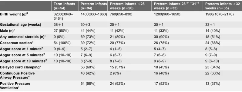
Table 1.Clinical data of study infants.
Term infants (n554)
Preterm infants (n594)
Preterm infants,28 weeks (n526)
Preterm infants 28+0231+6 weeks (n533)
Preterm infants.32 weeks (n535)
Birth weight (g)# 3230(3040–
3464)
1400(830–1860) 760(650–830) 1280(960–1650) 1980(1670–2170)
Gestational age (weeks) 38¡1 30¡3 25¡1 30¡1 33¡1
Male (n)* 27 (50%) 41 (44%) 11 (42%) 11 (33%) 14 (40%)
Any antenatal steroids (n)* 0 (0%) 69 (73%) 21 (80%) 30 (90%) 18 (51%)
Caesarean section* 54 (100%) 39 (72%) 20 (77%) 26 (78%) 24 (68%)
Apgar score at 1 minute# 9 (9–9) 5 (2–7) 4 (1–5) 5 (4–7) 8 (5–8)
Apgar score at 5 minutes# 10 (10–10) 7 (6–9) 6 (5–7) 7 (6–8) 9 (7–9)
Apgar score at 10 minutes# 10 (10–10) 8 (7–9) 8 (7–8) 9 (8–9) 9 (8–10)
Delayed cord clamping* 56 (60%) 15 (57%) 18 (45%) 23 (34%)
Continuous Positive Airway Pressure*
40 (42%) 2 (8%) 16 (48%) 22 (63%)
Positive Pressure Ventilation*
54 (58%) 24 (92%) 17 (52%) 13 (37%)
Data are presented as mean¡SD, unless indicated *n (%) or#median (IQR).
respectively. There were no significant differences in mode of delivery and DCC between infants born ,28, between 28+0–31+6 weeks and $32 weeks gestation. Centiles of SBP, DBP, and MBP for preterm infants born,28 weeks are presented in Fig. 4, for infants born 28+0–31+6 weeks in Fig. 5 and, for infants born $32 weeks gestation in Fig. 6. Analysis showed significantly higher SBP (at 1, 2, 3, 4, 10, 11, 12, 14, 20, 25, 30, and 60 minutes), MBP (at 1, 2, 3, 11, 12, 14, 20, 25, 30, 60 minutes) and DBP (at 1, 20, 30, 60 minutes) in preterm infants $32 weeks gestation compared to ,28 weeks infants, all p,0.05. Infants 28+0–31+6 weeks
had significant higher SBP at 1, 2, 5, 14, 20, and 25 minutes, MBP at 1 and 20 minutes, and DBP at 1 minute compared to ,28 weeks infants (p,0.05). Comparison of infants 28+0–31+6 weeks vs..32 weeks showed significant higher SBP at 1, 30 and 60 minutes, MBP at 1, 30 and 60 minutes, and DBP at 30 and 60 minutes in infants with .32 weeks (p,0.005).
Mode of delivery
There were no significant differences in gestational age, birth weight, DCC or antenatal steroids between vaginal and cesarean delivery. Regression model showed BP was significantly higher at 7 (SBP), 8 (SBP and MBP), 9 minutes
Table 2.Systolic blood pressure measurements of term and preterm infants.
Time after birth Term Infants (n554) Preterm Infants (n594)
1 min 59 (53–66)
2 min 53 (47–64)
3 min 62 (60–66) 54 (47–59)
4 min 54 (37–64) 53 (47–62)
5 min 63 (55–73) 52 (47–59)
6 min 74 (61–80) 56 (50–63)
7 min 65 (59–71) 54 (48–58)
8 min 74 (66–78) 52 (48–63)
9 min 72 (63–73) 54 (49–63)
10 min 62 (58–68) 54 (49–62)
11 min 65 (55–75) 54 (47–59)
12 min 67 (56–72) 51 (48–60)
13 min 61 (60–72) 53 (46–61)
14 min 66 (62–72) 52 (48–60)
15 min 63 (59–69) 51 (47–57)
20 min 49 (43–54)
25 min 50 (42–54)
30 min 47 (42–53)
45 min 47 (42–53)
60 min 46 (40–48)
Data are presented as median (IQR).
(MBP), and 45 minutes (SBP, DBP and MBP) after vaginal birth compared to caesarean section. SBP of vaginally born infants was 52¡11 mmHg at 45 minutes, compared to 46¡6 mmHg of those born by cesarean section, p50.009. DBP was also significantly higher at 45 min in vaginally born infants than that of infants born by caesarian section (32¡13 vs. 25¡7 mmHg, p50.01, respectively). MBP was also significantly higher at 45 minutes in vaginally delivered infants with 39¡13 mmHg compared to 32¡6 mmHg of infants born by caesarean section (p50.008). However, even though there is a statistically significant difference between the premature infants receiving DCC versus ECC, it is important to note that they were assisted at two different centers, with an unknown-and possibly differential-selection criteria and with different standards of care, which is a limitation of the study.
Delayed cord clamping
DCC for 60 seconds was only performed in one center (Edmonton) with a total of 56 (60%) preterm infants (14 infants were ,28 weeks, 21 infants were 28–32 weeks, and 20 infants were .32 weeks). There were no significant differences in gestational age, birth weight, mode of delivery, gender or antenatal steroids
Table 3.Diastolic blood pressure measurements of term and preterm infants.
Time after birth Term Infants (n-54) Preterm Infants (n594)
1 min 37 (25–40)
2 min 35 (25–40)
3 min 42 (35–45) 30 (27–36)
4 min 29 (24–35) 32 (28–37)
5 min 38 (33–46) 32 (27–35)
6 min 38 (24–58) 33 (27–34)
7 min 37 (26–42) 32 (27–35)
8 min 36 (32–42) 31 (26–36)
9 min 34 (28–37) 33 (29–36)
10 min 35 (28–38) 29 (26–36)
11 min 35 (24–36) 30 (26–36)
12 min 35 (30–45) 28 (25–39)
13 min 31 (24–40) 30 (23–35)
14 min 39 (28–45) 28 (24–33)
15 min 34 (29–40) 29 (24–35)
20 min 28 (24–34)
25 min 28 (22–33)
30 min 29 (23–33)
45 min 27 (21–32)
60 min 24 (21–27)
Data are presented as median (IQR).
between infants receiving DCC and ECC. At 60 minutes after birth MBP and DBP was significantly higher in infants receiving DCC for 60 seconds with
33¡6 mmHg and 26¡6 mmHg vs. 28¡4 mmHg and 22¡5 mmHg, p50.01, and p50.04, respectively. No significant differences in SBP were observed within the first hour.
Respiratory support
There were no significant differences in mode of delivery, DCC, gender, or antenatal steroids between groups who required different levels of respiratory support. However, infants receiving PPV had significantly lower gestational age and birth weight compared to infants receiving CPAP 28.5¡3.4 weeks vs. 31.3¡2.5 weeks (p,0.001) and 1169¡667 grams vs. 1567¡625 grams
(p50.041), respectively. Although, regression analysis showed significant higher DBP and MBP (p50.0001 and p50.001) in preterm infants receiving CPAP at 60 minutes after birth compared to PPV. T-test comparison had similar SBP, DBP and MBP values throughout the first 60 minutes after birth.
Table 4.Mean blood pressure measurements of term and preterm infants.
Time after birth Term Infants (n554) Preterm Infants (n594)
1 min 47 (35–49)
2 min 41 (33–49)
3 min 49 (42–52) 38 (35–46)
4 min 39 (27–41) 40 (36–44)
5 min 46 (37–54) 39 (35–45)
6 min 43 (41–65) 39 (35–44)
7 min 49 (38–51) 39 (34–45)
8 min 49 (45–52) 38 (35–45)
9 min 46 (40–52) 40 (36–45)
10 min 44 (39–48) 38 (33–49)
11 min 40 (36–44) 38 (34–44)
12 min 46 (41–49) 38 (34–46)
13 min 45 (36–49) 37 (34–43)
14 min 52 (40–54) 36 (31–41)
15 min 44 (41–49) 36 (32–41)
20 min 36 (31–40)
25 min 36 (30–40)
30 min 35 (30–40)
45 min 33 (29–39)
60 min 30 (28–33)
Data are presented as median (IQR).
Fig. 2. Median SBP, DBP, and MBP in mm Hg over the first 15 minutes after birth in term infants.
doi:10.1371/journal.pone.0114504.g002
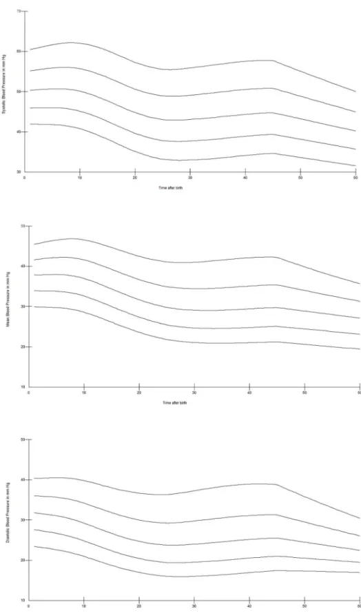
Fig. 3. Median SBP, DBP, and MBP in mm Hg over the first 60 minutes after birth in preterm infants.
Fig. 4. SBP (a), DBP (b), and MBP (c) centile over the first 60 minutes after birth in preterm infants 23+0
– 27+6
Ultrasound-detected brain injury
All preterm infants had cranial ultrasound prior to discharge and 13/94 (14%) had intraventricular hemorrhage grade 3 or 4 or a periventricular ischemic lesion (n54). Multiple regression analysis did not show any difference, however t-test comparison showed a significant lower SBP, and MBP at 25, 30 and 60 minutes of age and DBP 25 and 60 minutes in infants who subsequently developed
intraventricular hemorrhage during their hospital admission. SBP at 25, 30 and 60 minutes in the infants developing intraventricular hemorrhage was
40¡11 mmHg, 41¡3 mmHg, and 38¡6 mmHg compared to 50¡9 mmHg, 49¡7 mmHg, and 46¡6 mmHg, p50.0005, p50.0008, and p50.001 respec-tively. MBP was 28¡11 mmHg, 30¡4 mmHg, and 26¡3 mmHg compared to 36¡7 mmHg, 36¡6 mmHg, and 32¡5 mmHg, p50.003, p50.01, and
p50.002, respectively. DBP was 22¡11 mmHg and 21¡2 mmHg compared to 25¡6 mmHg and 29¡5 mmHg, p50.02 and p50.02, respectively.
Discussion
To our knowledge this is the first study describing SBP, DBP, and MBP reference values in term and preterm infants immediately after birth. Overall, SBP, DBP, and MBP remained similar within the first 15 minutes after birth in term infants and decreased throughout the first 60 minutes after birth in preterm infants. Preterm infants, who were born vaginally, were female, had DCC for 60 seconds, and required CPAP had significantly improved SBP, DBP, and MBP values at some measurement points. Furthermore, preterm infants developing severe brain injury had significantly lower SBP, DBP, and MBP at 25, 30, and 60 minutes after birth.
Although, our results demonstrate that a low BP immediately after birth might requires cardiovascular support, however, the clinical condition of the infant has to be taken into account and this should be investigated in future studies. In addition, we report on physiological changes at birth and their clinical impact has to be defined in randomized studies.
Term infants
Only term infants after elective caesarian section were included and therefore mode of delivery comparison was not possible, which is a limitation of the study. Infants, were not excluded from analyses, who needed cardio circulatory support after the first hour. Exclusion of neonates, in whom we don’t know yet if the blood pressure in the first hour after birth is normal or not, would have caused a bias in overall analyses of data. In addition, ethic approval was only granted for
Fig. 5. SBP (a), DBP (b), and MBP (c) centile over the first 60 minutes after birth in preterm infants 28+0–
31+6weeks gestation.
Fig. 6. SBP (a), DBP (b), and MBP (c) centile over the first 60 minutes after birth in preterm infants 32+0
elective caesarian section and therefore we were unable to recruit infants born after vaginal delivery. Furthermore, we only measured for the first 15 minutes after birth as term infants were returned to their parents for kangaroo-care at that time point. Therefore we don’t know if BP changes afterwards.
Preterm infants
We observed a steady decrease in SBP, DBP, and MBP throughout the first hour after birth. At first glimpse, this contradicts several echocardiography studies reporting a steady increase in stroke volume and CO in term infants within 1 hour after birth [4,6–8]. However observations of BP, being a composite of both CO and SVR, may be explained by ductal flow and/or blood vessel relaxation and therefore blood redistribution.
Mode of delivery
We observed a significant higher SBP, DBP, and MBP at 45 minutes after vaginal birth compared to caesarian section delivery. Our observations are similar to published data on peripheral oxygen saturation (from 1 until 5 minutes) and heart rate (from 2 to 10 minutes), which reported higher values after vaginal delivery; in comparison published data on cerebral oxygenation was similar regardless of mode of delivery during neonatal transition[9–13]. Observational studies in vaginally born term infants reported an increased left stroke volume over the first 15 minutes, whereas right stroke volume was unchanged [2]. In addition, Walther et al and Agata et alreported that CO peaked between 30 minutes to 2 hours after birth with a steady decreased to reach a steady state thereafter [4,7].
Delayed Cord Clamping
DCC is believed to improve cardiovascular function and stability during the immediate transition to neonatal life after birth. DCC for 60 seconds can increase an infant’s blood volume by approximately 16 mL/kg and reduce rates of intraventricular hemorrhage in premature infants [14–16]. Some of the benefits of DCC are due to the circulatory transition initiated by air breathing [17], as well as due to improved circulating blood volume, including brain blood flow resulting in increased blood flow in the superior vena cava. In the current study MBP and DBP, but not SBP, was significantly increased 60 minutes after birth in the DCC group compared to the ECC group. Nevertheless the present comparison of DCC and ECC must be interpreted with caution, although DCC was routinely
exposed to DCC trended to have a higher right ventricular stroke volume, however no differences in shortening fraction compared to ECC was identified [18]. In a small group of healthy term infants DCC of 4 minutes resulted in improved venous return and left ventricular function on day 3 of age compared to immediately clamped infants [19]. These improvements are most likely
redistribution from the capillary blood toward alternative vascular beds,
preventing a relative ischemia and hypovolemia and have the potential to improve neonatal adaptation. However,Bhatt et alrecently described that ventilation prior to umbilical cord occlusion improved cardiovascular function and stability during the immediate transition to neonatal life after birth in preterm lambs [17]. This indicates that lung aeration mitigated most of the adverse cardiovascular
responses to cord clamping [17]. In comparison, early clamping causes a
significant decrease in pulmonary vascular resistance, reduced blood volume and has a profound influence on cardiovascular function after birth [17,20].
Respiratory Support
Lung aeration at birth results in an 8–10 times increase in pulmonary blood flow, which is essential for both pulmonary gas exchange and maintenance of left ventricular output [3,21,22]. A recent study by Bhatt et al demonstrated that infants receiving ventilation before DCC had significantly improved hemody-namics [17]. In addition, a study byBinder et al reported no differences in BP-values within the first 10 minutes after birth [22], which is similar to our findings. One study that examined hemodynamic changes after surfactant administration in the delivery room reported an increase in right ventricular output but decrease in left ventricular output after surfactant administration [23].
In the current study we had similar BP values for infants requiring CPAP or PPV. Complicated effects of intrathoracic pressure and cardiopulmonary interaction in the transitional circulation may explain the lack of significant difference between groups who required different levels of respiratory support.
Preterm brain injury
Limitations
Observational studies such as this are limited by patient selection, however, the bias was mitigated to some extent by our larger patient numbers, more than one delivering institution, and prospective inclusion of potential confounders, such as gender and mode of delivery. Measurement of BP may vary considerably with the underlying methodology and the BP cuff size, which potentially are confounder in measurement of peripheral BP. However, these methodologies for ascertaining BP are in common use in intensive care units around the world, giving our reference ranges clinical generalizability. In addition, the non-invasive BP recording has its limitations and correlates poorly with invasive BP [24]. A more recent study comparing invasive and non-invasive found a good agreement between
oscillometric and invasive reading with the exception blood pressure at the lower limit [25]. These findings emphasize the necessity of careful evaluation of low blood pressure in preterm neonates. We did not perform echocardiography or peripheral blood flow measurements, which would have added substantial information to the cardiovascular changes during neonatal transition after birth and is a limitation to the current study. Echocardiography might have identified changes in CO, whereas measurements of peripheral blood flow using near-infrared spectroscopy or laser doppler flow study [26] might have revealed changes in systemic vascular resistance, regional blood flow, or oxygen extraction. Newer point-of-care technologies will hopefully address these gaps in future. It will be interesting to determine why BP continuously decreases during the first minutes after birth.
Conclusions
Non-invasive measurements of blood pressure are feasible during immediate neonatal transition and provide important information on the cardiovascular status of the newborn. These are the first reference ranges of BP in a cohort of term and preterm neonates. We have formulated centile charts that may be used to optimization cardiovascular support during immediate transition in both term and preterm infants.
Acknowledgments
We would like to thank the parents and infants agreeing to be part of the study. We would like to thank the Resuscitation-Stabilization-Triage team at the Royal Alexandra Hospital and at the Medical University Graz for helping and
supporting the study.
Author Contributions
Conceived and designed the experiments: GP PYC GMS. Performed the
MOR BS KA BU GMS. Contributed reagents/materials/analysis tools: GP PYC CB MOR BS KA BU GMS. Wrote the paper: GP PYC CB MOR BS KA BU GMS.
References
1. Polglase GR, Hooper SB, Gill AW, Allison BJ, McLean CJ, et al. (2009) Cardiovascular and pulmonary consequences of airway recruitment in preterm lambs. J Appl Physiol 106: 1347–1355.
2. Noori S, Wlodaver A, Gottipati V, McCoy M, Schultz D, et al.(2012) Transitional changes in cardiac and cerebral hemodynamics in term neonates at birth. J Pediatr 160: 943–948.
3. Rudolph AM(1977) Fetal and neonatal pulmonary circulation. Am Rev Respir Dis 115: 11.
4. Walther FJ, Benders MJ, Leighton JO (1993) Early changes in the neonatal circulatory transition. J Pediatr 123: 625–632.
5. Urlesberger B, Brandner A, Pocivalnik M, Koestenberger M, Morris N, et al.(2013) A Left-to-Right Shunt via the Ductus Arteriosus Is Associated with Increased Regional Cerebral Oxygen Saturation during Neonatal Transition. Neonatology 103: 259–263.
6. Noori S, Drabu B, Soleymani S, Seri I(2012) Continuous non-invasive cardiac output measurements in the neonate by electrical velocimetry: a comparison with echocardiography. Arch Dis Child - Fetal Neonatal Ed;97F340–F343.
7. Agata Y, Hiraishi S, Misawa H, Han JH, Oguchi K, et al.(1995) Hemodynamic adaptations at birth and neonates delivered vaginally and by Cesarean section. Biol Neonate 68: 404–411.
8. Agata Y, Hiraishi S, Oguchi K, Misawa H, Horiguchi Y, et al.(1991) Changes in left ventricular output from fetal to early neonatal life. J Pediatr 119: 441–445.
9. Agata Y, Hiraishi S, Misawa H, Hirota H, Nowatari M, et al.(1994) Regional blood flow distribution and left ventricular output during early neonatal life: a quantitative ultrasonographic assessment. Pediatr Res 36: 805–810.
10. Urlesberger B, Kratky E, Rehak T, Pocivalnik M, Avian A, et al.(2011) Regional oxygen saturation of the brain during birth transition of term infants: comparison between elective cesarean and vaginal deliveries. J Pediatr 159: 404–408.
11. Dawson JA, Kamlin COF, Wong C, te Pas AB, Vento M, et al.(2010) Changes in heart rate in the first minutes after birth. Arch Dis Child - Fetal Neonatal Ed 95: F177–81.
12. Rabi Y, Yee W, Chen SY, Singhal N(2006) Oxygen saturation trends immediately after birth. J Pediatr 148: 590–594.
13. Kamlin COF, O’Donnell CPF, Davis PG, Morley CJ (2006) Oxygen saturation in healthy infants immediately after birth. J Pediatr 148: 585–589.
14. Mercer JS (2006) Delayed Cord Clamping in Very Preterm Infants Reduces the Incidence of Intraventricular Hemorrhage and Late-Onset Sepsis: A Randomized, Controlled Trial. Pediatrics 117: 1235–1242.
15. Yao AC, Moinian M, Lind J(1969) Distribution of blood between infant and placenta after birth. Lancet 2: 871–873.
16. Yao AC, Hirvensalo M, Lind J(1968) Placental transfusion-rate and uterine contraction. Lancet 1: 380– 383.
17. Bhatt S, Alison BJ, Wallace EM, Crossley KJ, Gill AW, et al.(2013) Delaying cord clamping until ventilation onset improves cardiovascular function at birth in preterm lambs. J Physiol 591: 211–26.
18. Sommers R, Stonestreet BS, Oh W, Laptook A, Yanowitz TD, et al.(2012) Hemodynamic Effects of Delayed Cord Clamping in Premature Infants. Pediatrics 129: e667–e672.
19. Zaramella P, Freato F, Quaresima V, Secchieri S, Milan A, et al. (2008) Early versus late cord clamping: Effects on peripheral blood flow and cardiac function in term infants. Early Hum Dev 84: 195– 200.
21. Polglase GR, Hooper SB(2006) Role of intra-luminal pressure in regulating PBF in the fetus and after birth. Current Pediatric Reviews 2: 287–299.
22. Binder C, Urlesberger B, Avian A, Pocivalnik M, Mu¨ller W, et al. (2013) Cerebral and peripheral regional oxygen saturation during postnatal transition in preterm neonates. J Pediatr 163: 394–9.
23. Sehgal A, Mak W, Dunn M, Kelly E, Whyte H, et al.(2010) Haemodynamic changes after delivery room surfactant administration to very low birth weight infants. Arch Dis Child Fetal Neonatal Ed 95: F345–51.
24. Northern Neonatal Nursing Initiative(1999) Systolic blood pressure in babies of less than 32 weeks gestation in the first year of life. Arch Dis Child Fetal Neonatal Ed 80: F38–F42.
25. Takci S, Yigit S, Korkmaz A, Yurdako¨ k M(2012) Comparison between oscillometric and invasive blood pressure measurements in critically ill premature infants. Acta Paediatr 101: 132–5.