CLINICAL SCIENCE
Intraocular pressure in very low birth weight preterm
infants and its association with postconceptional age
Rodrigo L. Lindenmeyer,ILucas Farias,I Taı´s Mendonc¸a,IJoa˜o Borges Fortes Filho,IRenato S. Procianoy,II Rita C. SilveiraII
IMedical School, Federal University of Rio Grande do Sul, Hospital de Clı´nicas de Porto Alegre, Department of Ophthalmology, Porto Alegre/RS, Brazil.
IIMedical School, Federal University of Rio Grande do Sul, Department of Pediatrics, Hospital de Clı´nicas de Porto Alegre, Newborn Section, Porto Alegre/RS, Brazil.
OBJECTIVE: To evaluate intraocular pressure in very low birth weight preterm infants and correlate it with postconceptional age.
METHODS:The intraocular pressure in a prospective cohort of very low birth weight premature infants (defined as a birth weight#1,500 g and gestational age#32 weeks) admitted to Hospital de Clı´nicas de Porto Alegre, Brazil was evaluated weekly. The evaluated outcome was the variation in the intraocular pressure following changes in the postconceptional age (defined as the gestational age at birth plus the age in weeks at the time of examination) in the weeks following preterm birth. Mixed-effects models were used for the statistical analysis to determine the intraocular pressure variation according to postconceptional age, and means and 10th and 90thpercentiles were calculated for the intraocular pressure values.
RESULTS: Fifty preterm infants with a mean gestational age of 29.7¡1.6 weeks and a mean birth weight of 1,127.7¡222.7 g were evaluated. The mean intraocular pressure for the entire cohort considering both eyes was 14.9¡4.5 mmHg, and 13.5% of all recorded intraocular pressure values were greater than 20 mmHg. The analysis revealed a mean reduction in the intraocular pressure of 0.29 mmHg for each increase in postconceptional age (p= 0.047; 95% CI:20.58 to20.0035). The mean intraocular pressure (P10–P90) decreased from 16.3 mmHg (10.52– 22.16) at 26.3 weeks to 13.1 mmHg (7.28–18.92) at 37.6 weeks of postconceptional age.
CONCLUSIONS:The mean intraocular pressure in very low birth weight preterm infants was 14.9¡4.5 mmHg. This value decreased 0.29 mmHg per week as the postconceptional age increased.
KEYWORDS: Prematurity; Very Low Birth Weight Preterm Infants; Intraocular Pressure; Tonometry.
Lindenmeyer RL, Farias L, Mendonc¸a T, Fortes Filho JB, Procianoy RS, Silveira RC. Intraocular pressure in very low birth weight preterm infants and its association with postconceptional age. Clinics. 2012;67(11):1241-1245.
Received for publication onApril 6, 2012;First review completed onJune 15, 2012;Accepted for publication onJuly 10, 2012
E-mail: [email protected]
Tel.: 55 51 9969 8081
INTRODUCTION
The survival rate for very low birth weight (VLBW) preterm infants has increased since the 1950s as a result of numerous advances in general perinatal care. The first studies detailing the intraocular pressure (IOP) in this population were published in the 1950s by Dolcet (1) and Brockhurst (2), who found mean IOPs of 35 mmHg and 24.5 mmHg, respectively; both values were considered much higher than those expected for normal adults. Thirty years later, Musarella & Morin (3) re-examined the IOP in VBW preterm infants and obtained isolated IOP values that could be correlated with corneal diameter. Similarly, Tucker et al. (4) identified the correlations between the IOP, corneal
diameter and the axial length of the eye and the postconcep-tional age (PCA) and birth weight. In 1999, Ricci (5) eva-luated the IOP of 20 preterm infants in a longitudinal study with five visits and concluded that the mean IOP following birth decreased progressively.
Currently, more clinical information is available regarding the IOP variation among VLBW preterm infants. In 2008, Ng et al. (6) reported that the IOP decreases as the PCA increases in preterm infants.
Our study aimed to evaluate the behavior of the IOP in VLBW preterm infants, as well as its association with PCAs up to 37.6 weeks.
METHODS
Study design
This longitudinal study included VLBW preterm infants (VLBW was defined as a birth weight [BW]#1,500 g; preterm was defined as a gestational age [GA]#32 weeks) admitted to the Hospital de Clı´nicas de Porto Alegre (HCPA) between 2008 and 2010.
Copyrightß2012CLINICS– This is an Open Access article distributed under
the terms of the Creative Commons Attribution Non-Commercial License (http:// creativecommons.org/licenses/by-nc/3.0/) which permits unrestricted non-commercial use, distribution, and reproduction in any medium, provided the original work is properly cited.
Setting
The study was conducted in the neonatal intensive care unit (NICU) of HCPA, Porto Alegre, Brazil. HCPA is a tertiary university hospital in an urban area with a popula-tion of approximately 3 million.
Patients
The study included VLBW preterm infants who were admitted to the NICU. Exclusion criteria were ocular or systemic malformations, genetic anomalies, death before 14 days of life, and infants with Stage III or IV intraventricular hemorrhage. The gestational age of each infant was deter-mined based on obstetric history and early obstetric ultra-sound and confirmed by clinical examination of the newborn infant. The PCA was defined the GA of the infant plus the number of weeks since birth. All the infants were examined while hospitalized in the NICU. Routine screenings for retinopathy of prematurity (ROP) did not occur on the same day that the IOP measurements took place.
Outcome evaluated IOP.
Studied variables
The gestational age and BW were evaluated for the entire cohort.
Intraocular pressure
Ophthalmological evaluations included IOP measure-ments at four weekly evaluations. Because of possible cardiovascular and respiratory complications, all the evalua-tions were performed when the patient was clinically stable. Subsequent evaluations were performed, preferably at weekly intervals, and the PCA was adjusted at each exa-mination date. All IOP measurements were obtained with the infant lying supine and, when necessary, in the incubator. After the application of anesthetic eye drops (0.5% prox-ymetacaine hydrochloride) in both eyes, a neonatal Barraquer eyelid speculum was placed on the left eye (LE). The left eye IOP was measured, and the procedure was repeated for the right eye (RE). Three sequential IOP measurements, with 5% confidence for each eye, were made using a previously calibrated tonometer (Tonopen XLTM, Mentor O & O Inc. Santa Barbara, CA, USA). The IOP measurement recorded for each evaluation was the mean value of these three measure-ments. The measurements were taken when the infant was
quiet and still to avoid a Valsalva-like effect. All the IOP measurements were recorded by the same individual (R.L.L.).
Statistical analysis
To detect a difference of 2.3 mmHg (¡2.0) with 90% study
power and a significance level ofp,0.05, 16 patients (32 eyes) were required. Neonatal data were evaluated using descrip-tive statistics and are reported as absolute values and percentages. Mixed-effects models were used to analyze the association between IOP and PCA. Percentiles (P10 and P90) were used to describe the range of normality. For the comparison of IOP and PCA, the level of significance was set at 5% for a two-tailed sample distribution.
Ethical issues
This study and its informed consent form were approved by the Ethics and Research Committee of HCPA (n
˚
07-667).RESULTS
Between November 2008 and June 2010, 50 VLBW preterm infants who met the inclusion criteria were examined. The mean GA at birth was 29.7¡1.6 weeks (range: 26 to 32 weeks) and the mean BW was 1,127.2¡222.7 grams (range: 710 to
1,500 g) for the entire cohort. All of the included patients were also evaluated for ROP, and none of the infants developed any stage of ROP.
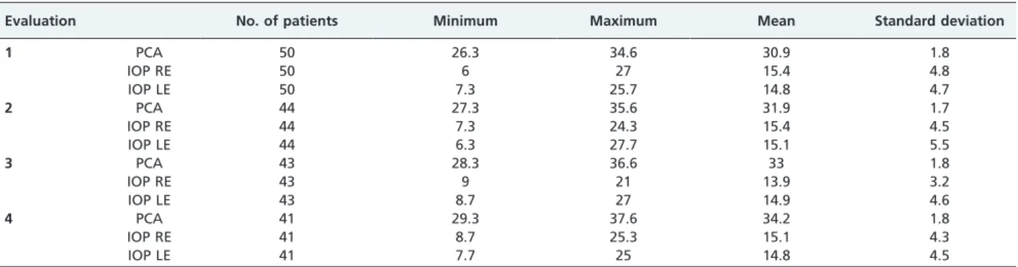
The four planned evaluations to measure the IOP were performed in 41 patients. Two patients underwent three evaluations, one patient underwent two evaluations and six patients underwent only one IOP measurement. The evalua-tions were missed because of prolonged clinical instability or death. The mean time between birth and the first IOP examination was 8.1¡5.4 days. A total of 356 (89%) IOP measurements, out a total of 400 that were initially planned, were performed (Table 1).
The mean IOP in both eyes for all measurements was 14.9¡4.5 (range: 6 to 27.7) mmHg. The mean RE IOP was
15¡4.3 (range: 6 to 27), and the LE IOP was 14.9¡4.8 (range: 6.3 to 27.7) mmHg. The IOP values were greater than 20 mmHg in 13.5% of the measurements. The mean IOP values at timepoints 1, 2, 3 and 4 are shown in Table 1. Table 2 shows the mean IOP values at each PCA week.
An analysis using mixed-effects models revealed a trend towards a reduced RE and LE IOP as a function of PCA. In the REs, the reduction was 0.29 mmHg for each one week
Table 1 -Descriptive analysis of the evaluations.
Evaluation No. of patients Minimum Maximum Mean Standard deviation
1 PCA 50 26.3 34.6 30.9 1.8
IOP RE 50 6 27 15.4 4.8
IOP LE 50 7.3 25.7 14.8 4.7
2 PCA 44 27.3 35.6 31.9 1.7
IOP RE 44 7.3 24.3 15.4 4.5
IOP LE 44 6.3 27.7 15.1 5.5
3 PCA 43 28.3 36.6 33 1.8
IOP RE 43 9 21 13.9 3.2
IOP LE 43 8.7 27 14.9 4.6
4 PCA 41 29.3 37.6 34.2 1.8
IOP RE 41 8.7 25.3 15.1 4.3
IOP LE 41 7.7 25 14.8 4.5
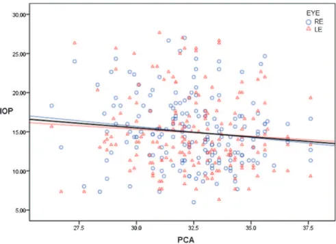
increase in PCA (p= 0.072; 95% CI,20.6 to+0.026). In the LEs, the reduction was 0.15 mmHg for each one week increase in PCA (p= 0.42; 95% CI,20.5 to+0.21). When both eyes were analyzed together, the mean reduction was 0.29 mmHg for each one week increase in PCA (p= 0.047; 95% CI,20.58 to20.0035). Figure 1 shows the separate IOP measurement values and their association with PCA. Lines indicate the reduction in each eye and in both eyes. Based on the correlation formula that we determined (y= 23.97 – 0.29x), the mean IOP varied according to the PCA from 16.4 mmHg at 26.3 weeks to 13.1 mmHg at 37.6 weeks. Moreover, the variation between the 10th and the 90th
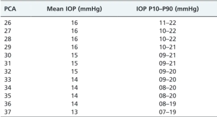
percentiles ranged from 10.5 to 22.2 mmHg at P10 and from 7.3 to 18.9 mmHg at P90 (Figure 2).
DISCUSSION
This study determined the IOP of VLBW preterm infants using longitudinal measurements over four weeks. We found that IOP of VLBW preterm infants, analyzed longitudinally, was 14.9¡4.5 mmHg over a mean of four observations taken
four consecutive weeks after birth. Using mixed-effects models, we observed a significant reduction in the IOP (approximately 0.29 mmHg for each week of PCA). Our
study evaluated the behavior of the IOP in VLBW preterm infants and its association with PCAs of up to 37.6 weeks because we expected that preterm infants would have the same IOP as term infants after that timepoint. Our findings reflect the IOP values for prematurity for IOP examinations performed before 37.6 weeks of PCA. To the best of our knowledge, this value has not previously been described.
A study by Ng et al. (6) found an IOP reduction of 0.11 mmHg (p,0.001) for each one week increase in the PCA. In their study, six IOP measurements were taken in infants with PCAs that ranged from 26.1 to 46.4 weeks. While our study found a greater reduction in IOP (0.29 mmHg; p= 0.047), our subjects had a narrower PCA range (26.3 to 37.6 weeks). This difference suggests that the IOP reduction reported in the Ng et al. study may have been greater because the infants were evaluated after 40 weeks PCA, a point at which the IOP might not undergo any additional changes. By contrast, our study determined the IOP in infants younger than 37.6 weeks PCA; therefore, our measurements were limited to infants who were actually preterm. Our results may better reflect the behavior of the IOP in the weeks following premature birth.
A previous study by Ricci (5) found that a reduced IOP was associated with increases in the PCA and suggested this was caused by the maturation of the aqueous drainage system. Improvements in the aqueous flow coincide with the complete formation of the aqueous drainage system. Despite the differences in methods and statistical analyses, our results agree with those of Ricci (5) and Ng et al. (6). However, Musarella and Morin (3) did not find a direct correlation between the IOP and PCA; they found that the IOP decreased according to the increase in infant weight and that the IOP might be associated with physical development and maturity.
Historically, most studies of IOP in preterm infants used only one isolated IOP measurement. The longitudinal studies that found a negative correlation between IOP and PCA were conducted by Ricci in 1998 and by Ng et al. in 2008. Ricci evaluated the IOP in 40 eyes of 20 premature infants with GAs ranging from 26 to 32 weeks (mean: 29.5¡1.5 weeks) and used repeated measures ANOVA for Table 2 -Intraocular pressure (P10–P90) according to
postconceptional age.
PCA Mean IOP (mmHg) IOP P10–P90 (mmHg)
26 16 11–22
27 16 10–22
28 16 10–22
29 16 10–21
30 15 09–21
31 15 09–21
32 15 09–20
33 14 09–20
34 14 08–20
35 14 08–20
36 14 08–19
37 13 07–19
PCA: postconceptional age in weeks; IOP: intraocular pressure.
the statistical analysis of the IOP, although the IOP measurements were not controlled for differences between the various PCAs (5). By contrast, Ng et al. used mixed-effects models to analyze IOP measurements in 104 preterm infants with a median (interquartile range) GA of 29.8 weeks (range: 28.7–30.9 weeks) and a median BW of 1,208 g (range: 1,049–1,370 g) to adjust the IOP values to the corresponding PCAs (6). In our study, the mean GA was 29.7¡1.6 weeks
(range: 26–32 weeks), and the mean BW was 1,127.2¡
222.7 g (range: 710–1,500 g) for the entire cohort.
Few studies have conducted similar investigations, and an adequate method for studying this topic has rarely been adopted. The best method for this type of study seems to be longitudinal IOP measurements taken while the infant is hospitalized to gain weight or to treat the many comorbidities that may affect them after preterm birth. However, because infants are born at different GAs and often have unstable clinical conditions in the first weeks of life, it is difficult to follow a protocol that meets all the requirements for the use of repeated measures ANOVA to analyze the data. Therefore, we chose to use mixed-effects models for statistical analysis because it is more flexible for handling data and because it can be used to evaluate both eyes at the same time.
To measure the IOP in newborn infants, we used a protocol based on experiences in previous studies. Anesthetic eye drops were mandatory to make the examination less uncomfortable for the infant. Furthermore, the use of general anesthesia would not be ethically correct and might affect evaluations by reducing the IOP. A Barraquer eyelid speculum was used because of the small size of the eye and adnexa. There are no previous studies related to the Barraquer eyelid speculum as a source of bias in IOP measurements in preterm infants. However, a single study (7) suggests that the use of the Alfonso eyelid speculum may falsely elevate IOP by 4 mmHg in children 6 to 252 months of age (mean: 70 months). Therefore, the use of the Barraquer speculum could be one limitation of our study.
We used a Tonopen XPTM tonometer because we have found that it has a good correlation with Goldman/Perkins tonometers. Our study found that the mean IOP in VLBW preterm infants is 14.9 mmHg and that the IOP decreases as the infant develops (by 0.29 mmHg per PCA week). Our study found a greater reduction, 0.29 mmHg (p= 0.047), over a narrow range of PCAs (26.3 to 37.6 weeks). Overall, our study provides information regarding the behavior of the IOP in VLBW premature infants. Further studies on this subject are warranted to improve our understanding of the IOP in this particular group of patients.
ACKNOWLEDGMENTS
The authors certify that the protocol for the research project has been approved by a suitably constituted ethics committee in the institution within which the work was undertaken and that the research protocol conforms to the provisions of the Declaration of Helsinki in 1995 (as revised in Edinburgh, 2000). The authors declare no financial support or relationships that may pose a conflict of interest.
AUTHOR CONTRIBUTIONS
Silveira RC planned, conducted and revised all steps of the study and approved the final manuscript. Lindenmeyer RL was the main investigator and planned, conducted and collected all of the IOP measurements and approved the final manuscript. Farias L organized the protocol and data and approved the final manuscript. Mendonc¸a T organized the protocol and data and approved the final manuscript. Fortes Filho JB was also a main investigator and planned, conducted and revised all steps of the study and approved the final manuscript. Procianoy RS planned, conducted and revised all steps of the study and approved the final manuscript.
REFERENCES
1. Dolcet L. Tension ocular del recien nacido. Arch Soc Oftal Hispano-am. 1952;12(9):1057-63.
2. Brockhurst RJ. The intraocular pressure of premature infants. Am J Ophthalmol. 1955;39(6):808-811.
3. Musarella MA, Morin JD. Anterior segment and intraocular pressure measurements of the un anesthetized premature infant. Metab Pediatr Syst Ophthalmol. 1985;8(2–3):53-60.
4. Tucker SM, Enzenauer RW, Levin AV, Morin JD, Hellmann J. Corneal diameter, axial length, and intraocular pressure in premature infants. Ophthalmology. 1992;99(8):1296-00.
5. Ricci B. Intraocular pressure in premature babies in the first month of life. J AAPOS. 1999;3(2):125-7.
6. Ng PC, Tam BS, Lee CH, Wong SP, Lam HS, Kwok AK, et al. A longitudinal study to establish the normative value and to evaluate
perinatal factors affecting intraocular pressure in preterm infants. Invest Ophthalmol Vis Sci. 2008;49(1):87-92, http://dx.doi.org/10.1167/iovs. 07-0954.