Muscles of
Cebus libidinosus
(Rylands
et al.
2000):
Manipulatory Behavior and Tool Use
Tales Alexandre Aversi-Ferreira1,2*, Rafael Souto Maior3, Frederico O. Carneiro-e-Silva4,
Roqueline A. G. M. F. Aversi-Ferreira1,4, Maria Clotilde Tavares3, Hisao Nishijo2, Carlos Tomaz3
1Department and Faculty of Nursing, Neurosciences and Primates Behavior Center (NECOP), Federal University of Goia´s, Catala˜o, Brazil,2Department of Physiology, School of Medicine and Pharmaceutical Sciences, System Emotional Science, University of Toyama, Toyama, Japan,3Department of Physiology, Institute of Biology, Laboratory of Neuroscience and Behavior, University of Brasilia, Brasilia, Brazil,4Department of Animal Anatomy, Faculty of Veterinary Medicine, Federal University of Uberlaˆndia, Uberlaˆndia, Brazil
Abstract
The present study describes the flexor and extensor muscles inCebus libidinosus’forearm and compares them with those from humans, chimpanzees and baboons. The data is presented in quantitative anatomical indices for similarity. The capuchin forearm muscles showed important similarities with chimpanzees and humans, particularly those that act on thumb motion and allow certain degree of independence from other hand structures, even though their configuration does not enable a true opposable thumb. The characteristics of Cebus’ forearm muscles corroborate the evolutionary convergence towards an adaptive behavior (tool use) betweenCebusgenus and apes.
Citation:Aversi-Ferreira TA, Maior RS, Carneiro-e-Silva FO, Aversi-Ferreira RAGMF, Tavares MC, et al. (2011) Comparative Anatomical Analyses of the Forearm Muscles ofCebus libidinosus(Rylandset al.2000): Manipulatory Behavior and Tool Use. PLoS ONE 6(7): e22165. doi:10.1371/journal.pone.0022165
Editor:Sharon Gursky-Doyen, Texas A&M University, United States of America
ReceivedApril 7, 2011;AcceptedJune 16, 2011;PublishedJuly 15, 2011
Copyright:ß2011 Aversi-Ferreira et al. This is an open-access article distributed under the terms of the Creative Commons Attribution License, which permits unrestricted use, distribution, and reproduction in any medium, provided the original author and source are credited.
Funding:This work was supported by Core Research for Evolutional Science and Technology, Japan Science and Technology Agency, Japan, Japan Society for the Promotion of Science Asian Core Program, and the Ministry of Education, Science, Sports and Culture, Grant-in-Aid for Scientific Research (A) (22240051); and by National Council of Technology and Development - Brazil. The funders had no role in study design, data collection and analysis, decision to publish, or preparation of the manuscript.
Competing Interests:The authors have declared that no competing interests exist. * E-mail: [email protected]
Introduction
In the last two decades, several behavioral studies have focused on the capuchin’s ability to use tools. In its strictest sense, tool use is only found in a handful of old world monkeys (OWM) and apes. The only exceptions among new world monkeys (NWM) are the capuchins, which have been reported to use tools both in the captivity and in the wild [1,2,3]. Such studies have reported that the Cebus is capable to handle rocks to open coconuts, to use toothpicks to push food out of a pipe or to extract molasses through the orifices of a box [4,5,6]. Recently wild capuchins were observed to fish for termites using twigs, an activity until then only seen in chimpanzees [7]. Such complex behaviors are dependent on versatile grasping ability [8,9]. Accordingly, Cebushave been reported to display a wide array of grasping strategies and manipulative, comparable to chimpanzees and humans [10,11].
Dexterous hand ability, and consequently tool use, is associated with the development of primate intelligence and culture [12,13]. This adaptive behavior therefore denotes an important evolution-ary convergence, especially between capuchins and chimpanzees. Capuchin tool use seems also dependent on other neurological, cognitive and morphological convergences [10,14]. In this sense, capuchins stand as an important model for testing hypotheses regarding the evolution of primate cognition.
Comparative anatomical analysis of primates may yield important knowledge regarding behavior and phylogeny. More specifically,
the hand. Although a few studies have focused on comparative behavioral assessment of capuchin tool use [8,9], the literature on their forearm myology is scarce. Early studies have indicated that precision grips were untenable to capuchins due to lack of saddle joint in the hand and therefore tool use ability was not related to thumb mobility [15,16]. Further behavioral studies, however, have reported that this genus can adduct the thumb towards the index finger, favoring the flexing of the interphalangeal rather than the metacarpophalangeal joint, coined ‘lateral opposability’ [8]. Howev-er, there are still no anatomical confirmations of these findings.
In the present study, the flexors muscles of the forearm in the Cebus libidinosus[17] monkey were investigated. Origin, insertion, arterial branching and innervation of each muscle were charac-terized to provide an anatomical understanding of the manual skills observed inCebus. The anatomical observations here were then compared to the analogous muscles found in humans [18] and chimpanzees and baboons [19]. The degree of anatomical similarity among the forearm muscles in these species was compared using the Comparative Anatomy Index (CAI) [20].
Materials and Methods
Samples
anatomical collection of the Neuroscience and Behavior of Primates Laboratory (NECOP) from the Federal University of Goias-Catala˜o-Goias. The remaining of them belonged to the Brazilian Institute of Environment and Renewable Natural Resources (IBAMA) archive and were donated to the University of Goia´s in the 1970’s. This work was approved by the Institutional Ethical Committee from the Federal University of Goia´s (CoEP-UFG 81/ 2008, authorization from the IBAMA number 15275).
Preparation of the animals for dissection
All procedures involving the animals were done in accordance to the guidelines of the Brazilian Society of Animal Experimen-tation (COBEA). After the trichotomy with a razor blade, the animals were incubated in water at room temperature for 10– 12 hours; and then received perfusion, by the femoral vein, 10% of formaldehyde with 5% of glycerin for fixation. The animals were conserved in 10% of formaldehyde, in covered opaque cubes,
to avoid the penetration of light and the evaporation of the formaldehyde.
To undertake the anatomical observations for the present work, after receiving the specimens at the anatomical collection of the Federal University of Goia´s each one was processed, by the first author, as follows: (1) it received an injection of latex 601-A (Dupont) stained with Wandalar red diluted in ammonium hydroxide in the abdominal aorta in order to facilitate the visualization of small arteries; (2) it was incubated in water at room temperature for 10– 12 hr; and then (3) it received a perfusion of 10% formaldehyde with 5% glycerin through the femoral vein for fixation. The monkeys were preserved in 10% formaldehyde in closed opaque boxes to avoid light penetration and formaldehyde evaporation.
Dissection and documentation
The dissection of the forearm was performed with emphasis on the flexors muscles of the forearm and registered with a digital
Table 1.Comparative analyses of the flexor superficial muscles forearm amongCebus libidinosus(C.l.), human (Homo), chimpanzee (Pan) and baboon (Papio).
Muscle Features Cebus libidinosus Homo Pan Papio
Flexor carpi ulnaris
Origin Medial epicondyle of humerus and olecranon
Highly similar toC.l. CAI = 0.0
Highly similar toC.l. CAI = 0.0
Highly similar to C.l.
CAI = 0.0 Insertion Pisiform bone
Innervation Ulnar nerve
Vascularization Ulnar artery
Palmaris longus
Origin Medial epicondyle
of humerus
Variable, may be absent Somewhat similar toC.l. CAI = 0.425
Highly similar toC.l. CAI = 0.0
Highly similar to C.l.
CAI = 0,0 Insertion Palmar aponeurosis
Innervation Median nerve
Vascularization Ulnar artery
Flexor carpi radialis
Origin Medial epicondyle
of humerus
Similar toC.l. Vascularized by radial artery
CAI = 0.125
Double insertion in metacarpal II and III Somewhat similar to C.l.
CAI = 0.250
Highly similar to C.l.
CAI = 0.0
Insertion Base of metacarpal II
Innervation Median nerve
Vascularization Ulnar artery
Flexor digitorum superficialis
Origin Humeral head – medial epicondyle of the humerus; Radial head – anterior surface of the radius
Three heads of origin – humeral, radial and ulnar. Somewhat similar toC.l. CAI = 0.375
Highly similar toHomo Somewhat similar to C.l.CAI = 0.375
Highly Similar to C.l.
CAI = 0.0
Insertion Middle phalanges of II to V fingers
Innervation Median nerve
Vascularization Ulnar artery and branches of the radial artery
Pronator teres
Origin Medial epicondyle of humerus Two heads of origin, humeral and ulnar Somewhat similar to C.l.CAI = 0.375
Highly similar toHomo Somewhat similar to C.l.CAI = 0.375
Highly similar to C.l.
CAI = 0.0
Insertion Postero-lateral portion of the radius
Innervation Median nerve
Vascularization Ulnar artery
Superficial group
Somewhat similar
GCAI = 0.26
SimilarGCAI = 0.20 Highly similar
GCAI = 0.0
camera. The first author of the present paper dissected both sides of the eight specimens ofCebus libidinosus. The nomenclature of the forelimb muscles follows, whenever it is possible, that used in human anatomy (The Federative Committee on Anatomical Terminology, 1998). When no such parallel was possible, they were referred to following the patterns of the international nomenclature from the Human Anatomic Nominal. The data collected were analyzed and compared with the patterns described for human, chimpanzee and baboon species.
Statistical analysis
Based on Aversi-Ferreira [20], we used a simple comparative non-parametric method for two different species associated on anatomical concepts of normality and variation as nominal variables. Relative frequency (RF) was defined as:RF = (N2nv)/ N; whereNis the total number of specimens of the sample andnvis the number of individuals presenting variation of the normal pattern.
When more than one parameter (location, nerve, blood vessel, origin and insertion of a muscle) was necessary, they were associated to a specific pondered value with respect to their degree of relevance in comparative analysis. Parameters with less variation were ascribed a higher value. Therefore, innervation, origin and insertion, and vascularization were ascribed the weighs 3, 2, 1, respectively.
The Pondered Average of Frequencies (PAF) was calculated using the RF values:
PAF~fðRF1:P1ÞzðRF2:P2ÞzðRF3:P3Þg=P1zP2zP3 whereRF1is the frequency of the muscle innervation andP1is 3;
RF2is the frequency of the muscle origin andP2is 2,RF3is the frequency of the muscle vascularization andP3is 1.
To consider the proximity between the structures studied, the difference in the relative frequency is calculated, or Comparative Anatomy Index (CAI) between samples from different species:
CAI~jPAFi{PAFiij; where indexes i and ii represent samples 1 and 2.
From the equations above, it follows that the close to zeroCAI
values represents greater similarity between samples it represents, whereas a CAI closer to 1.0 means higher divergence between samples. More specifically, CAI value of 0 indicates high similaritybetween the structures analyzed, from 0 to 0.200 as
similarstructures, from 0.200 to 0.650 assomewhat similar, from 0,650 and 1,000 asdissimilar.
For the purposes of the present work, theCebuswas primarily chosen as the reference for comparison against human, chimpan-zee and baboon morphology, although they were also analyzed among themselves.
For example, regarding the muscle flexor carpi radialis,RFwas 1 to all parameters inCebusspecimens andRF= 0 was set for the absence of any parameters in the other species.
Then,
PAFCe~fð1x3Þzð1x2Þzð1x2Þzð1x1Þg=8~1,000; PAFH~fð1x3Þzð1x2Þzð1x2Þzð0x1Þg=8~0,875; PAFCh~fð1x3Þzð1x2Þzð0x2Þzð1x1Þg=8~0,750; PAFB~fð1x3Þzð1x2Þzð1x2Þzð1x1Þg=8~1,000;
Where the indexes Ce, H, Ch and B represents respectively Cebus, humans, chimpanzees and baboons. In order, the first parameter is innervation, the second is origin of muscle, the third
Note that vascularization is the only difference betweenCebusand humans whereas insertion differs betweenCebusand chimpanzees and no differences were observed betweenCebusand baboons in this muscle.
From these values, the CAI is calculated,
CAICe-H~j1,000{0,875j~0:125; CAICe-Ch~j1,000{0,750j~0:250; CAICe-B~j1,000{1,000j~0:000:
The CAI calculated indicate that the muscle flexor carpi radialis ofCebusand baboons arehighly similar,Cebusand humans are
similar, and Cebus and chimpanzees are somewhat similar
structures, according to the parameters adopted here.
A special case is the palmaris longus muscle that is absent in 10% of human population [18] and that presents many variations. To calculate theFR, the same purposed pondered values were used, but they were adjusted by a 10% decrease to innervation and vascularization. Adjustment to origin and insertion were set at 50% decrease since Gray [18] observed that variations in this muscle occur mainly found at its origin and insertion.
Other important parameter to be calculated is the ‘Group CAI’ (GCAI) for structures, such as superficial and deep muscles in the forearm, which combines the summationRFof individual muscles summation average ofCAI for the species not used as reference (Homo,PanandPapio). To calculatedGCAIwe used the following equation: GCAICe{H X n i~1 RFCe n { X n i~1 RFH n ~
1z1z1z1z1
5 {
1z0:575z0:875z0:625z0:625 5 ~ 5 5{
3:7 5
~j1:00{0:74j~0:26
or, P n i~1 CAIH n ~
0:0z0:425z0:125z0:375z0:375
5 ~0:26
GCAICe{Ch X n i~1 RFCe n { X n i~1 RFCh n ~ 1z1z1z1z1 5 {
1z1z0:750z0:625z0:625 5 ~ 5 5{ 4 5
~j1:00{0:8j~0:20
or,
P
n
i~1 CAICh
GCAICe{B
X
n
i~1 RFCe
n { X
n
i~1 RFB
n
~
1z1z1z1z1
5 {
1z1z1z1z1 5
~
5 5{
5 5
~j1:00{1:00j~0:00
or,
P
n
i~1 CAIB
n ~
0:0z0:0z0:00z0:0z0:0
5 ~0:00
The GCAI calculated above shows that the group of superficial muscles ofCebus’s forearm is highly similar to baboon, similar to chimpanzees, and somewhat similar to humans.
Results and Discussion
Recently, we have reported a descriptive anatomical compar-ison of the extensor forearm muscles of Cebus libidinosus [21], Figure 1. Photograph of the anterior aspect of forearm right of
a Cebus libidinosus (C.l.). 1).brachioradialis muscle, 2) flexor carpi radialis muscle, 3) flexor digitorum superficialis muscle, 4) palmaris longus muscle, 5) flexor carpi ulnaris muscle and, 6) pronator teres muscle. (bar = 1,2 cm).
doi:10.1371/journal.pone.0022165.g001
Table 2.Comparative analysis of the flexor deep muscles forearm amongC.l., human (Homo), chimpanzee (Pan) and baboon (Papio).
Muscle Features Cebus libidinosus Homo Pan Papio
Pronator quadratus Origin Internal portion antero-lateral of the distal third of the ulna
Innervated by median nerve Somewhat similar toC.l. CAI = 0.375
Highly similar toC.l. CAI = 0.0
Highly similar toC.l. CAI = 0.0
Insertion Border antero-medial of the distal third of the radius
Innervation Ulnar nerve
Vascularization Ulnar artery
Flexor digitorum profundus
Origin Proximal portion of the anterior surface of the ulna
Highly similar toC.l. CAI = 0.0
Not included tendon to index finger. Similar toC.l. CAI = 0.0625
Tendons from radial head to I, II and III fingers; and from ulnar head to III, IV and V fingers; associated with flexor digitorium superficial. ToCAI
purposes, it was considered inexistent. Dissimilar fromC.l. CAI = 1.00 Insertion Base of the phalanges
Innervation Ulnar nerve
Vascularization Ulnar artery
Flexor pollicis longus
Origin Medial epicondyle of the antero-medial surface of the radius
Also originates from the adjacent part of the interosseous membrane. Similar toC.l.
CAI = 0.125
Highly similar toC.l. CAI = 0.0
Attached to belly of the flexor digitorum profundus muscle. Somewhat similar toC.l. CAI = 0.250
Insertion Distal phalange of the thumb and a tendon to index finger
Innervation Median nerve
Vascularization Ulnar artery
Deep muscles SimilarGCAI = 0.167 SimilarGCAI = 0.020 Somewhat similar
GCAI = 0.417
especially in comparison with new world monkeys. In the present study, we have expanded on those previous data by applying a non-parametric statistical test (Comparative Anatomy Index) [20] to compare the anatomy of the forearm flexor of muscles ofCebus libidinosuswith those of other primates that use tools (humans and chimpanzees) and to baboons, which has not be reported to show this behavior. We also further expanded the findings of the previous report by applying the same statistical test to forearm extensor muscles.
Table 1 summarizes the similarities and differences across the superficial muscles forearm fromCebus,Homo,PanandPapio.
According to Aversi-Ferreira and colleagues [22], the flexor carpi ulnaris muscle and palmaris longus muscle (Figure 1) are similar in all primates species analyzed here with regards to origin, insertion, innervation and vascularization. Flexor carpi radialis, flexor digitorum superficialis and pronator teres muscles, on the other hand, (Figure 1) are more similar betweenCebusandPapio. The insertion of the flexor carpi ulnaris muscle on pisiform in Cebusis evident because this bone is well developed in this species. In general, all superficial muscles in theCebus’forearm present similarities to the other primates considered here. However, considering some specific details (shown in table 1), more similarities, based on the statistical analysis used in the present work, are found between superficial muscles in theCebusandPapio. Table 2 indicates the similarities and differences among the forearm deep muscles from Cebus, Homo, Pan and Papio, with regards to origin, insertion, innervation and vascularization.
Pronator quadratus muscle (Figure 2) is identical in all non-human primates considered. In non-humans, however this muscle is
innervated by the median nerve [18], not the ulnar nerve as shown in non-human primates.
The features observed in table 2 indicate that, in general,Cebus and Pan show great similarity regarding the flexor digitorum profundus and flexor pollicis longus muscles (CAI = 0.0625 and CAI = 0.0, respectively; also Figure 3), closely followed by Homo (CAI = 0.0 and CAI = 0.125, respectively). Interestingly, these muscles are more distinct in baboons, especially the digitorum profundus (CAI = 1.0), which was not the case for any superficial flexor muscles. The flexor digitorum profundus and flexor pollicis longus muscles are involved in finger (including thumb) move-ment. This anatomical evidence is consistent with the Cebus’ manipulation skills.
The bellies of the flexor digitorum profundus and flexor pollicis longus muscles are clearly attached to each other, in contrast to humans where bellies are separated and individualized [18]. Indeed, these differences allow for the hand skills required by capuchins’ arboreal habits, such as grabbing and holding [23].
The descriptive analysis of the extensor muscles has been detailed in length elsewhere [21] (shown here in Figure 4). Here we provide a brief assessment of those findings under the light of CAI. In Table 3, we show the CAI and GCAI values for the extensor forearm muscles. High similarity (i.e. CAI = 0) betweenCebus,PanandPapio is evident in almost all extensor muscle, except for the deep dorsal sub-group. In this sub-group, theCebus’abductor pollicis longus and extensor pollicis brevis show a greater similarity to modern humans and chimpanzees, respectively. It is important to note that the extensor pollicis brevis is not completely differentiated as a distinct muscle from the abductor pollicis longus inCebusor in any other primate, except for humans and gibbons. The fleshy part, which constitutes this bundle, is deeply blended but it is differentiated into two separate tendons [21]. This configuration is highly similar to that ofPanand it is further differentiated inHomo, but not seen in Papio. Interestingly, the deep dorsal sub-group was pointed by [21] as the major point, in the forearm, which sets capuchins apart from the remaining new world monkeys.
Since the majority of forearm muscles acts on the hand and fingers, the present study described the anatomy of the capuchin forearm muscles and compared them with those of humans, chimpanzees and baboons. The superficial group of flexor muscles was more similar to those of baboons (GCAI = 0.00) than chimpanzees and humans (GCAI = 0.20 and GCAI = 0.26, respectively). On the other hand, the deep flexor muscles were more similar to those of chimpanzees (GCAI = 0.02). They were even found more similar to those of humans (GCAI = 0.167) than Figure 2. Photograph of the anterior aspect of forearm right of
C.l. The arrow is indicating the pronator quadratus muscle.
(bar = 0,5 cm).
doi:10.1371/journal.pone.0022165.g002
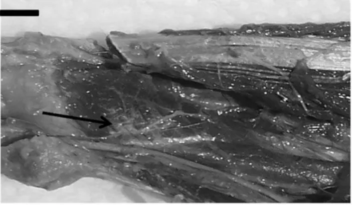
Figure 3. Photograph of the anterior aspect of forearm left of
C.l.The arrow is indicating the tendon of flexor pollicis longus. 1) flexor digitorum profundus muscle and 2) flexor pollicis longus muscle. (bar = 0,7 cm).
Figure 4. Photograph of the posterior aspect of right forearm of C.l. 1) tendon of the extensor pollicis brevis, 2) tendon of the abductor pollicis longus muscle, 3) tendon of the extensor pollicis longus muscle. (Bar = 1,2).
Table 3.Comparative analyses of the extensor muscles forearm amongC.l., human (Homo), chimpanzee (Pan) and baboon (Papio). (Based on Aversi-Ferreira et al., 2010).
Muscle Features Cebus libidinosus Homo Pan Papio
Radial Group Brachioradialis muscle
Origin Latero-distal portion of the humerus on supracondylar ridge
Highly similar to C.l. CAI = 0.0
Highly similar to C.l. CAI = 0.0.
Highly similar to C.l. CAI = 0.0
Insertion Styloid process of radius Innervation Radial nerve
Vascularization Radial artery Extensor carpi
radialis longus
Origin Latero-distal portion of the humerus on the supracondylar ridge
No attached bellies with others muscles. Somewhat similar to C.l. CAI = 0.22
Highly similar to C.l. CAI = 0.0
Highly similar to C.l. CAI = 0.0
Insertion Dorsal surface of the base of the second metacarpal on its radial side Innervation Radial nerve
Vascularization Radial artery Extensor carpi
radialis brevis
Origin Lateral epicondyle of the humerus Highly similar to C.l. CAI = 0.0
Highly similar to C.l. CAI = 0.0
Highly similar to C.l. CAI = 0.0
Insertion Dorsal surface of the base of the second metacarpal
Innervation Radial nerve Vascularization Radial artery
Supinator Origin Lateral epicondyle of the humerus Highly similar to C.l. CAI = 0.0
Highly similar to C.l. CAI = 0.0
Highly similar to C.l. CAI = 0.0
Insertion Medium portion of the radious Innervation Radial nerve
Vascularization Radial artery Superficial Dorsal
Group
Extensor digitorum communis
Origin Lateral epicondyle of the humerus Lesser variation regarding the distribution of tendons to fingers.
Somewhat similar to C.l. CAI = 0.22
Highly similar to C.l. CAI = 0.0
Highly similar to C.l. CAI = 0.0
Insertion Dorsal aponeurosis in the second to fifth proximal phalanges Innervation Radial nerve
Vascularization Radial artery Extensor digiti
quinti proprius
Origin Lateral epicondyle of the humerus Only one insertion tendon to little finger.
Somewhat similar to C.l. CAI = 0.22
Fleshy portion is well detached. Somewhat similar to C.l.
CAI = 0.22
Highly similar to C.l. CAI = 0.0
Insertion Dorsum of the IV and V fingers Innervation Radial nerve
Vascularization Radial artery Ulnar Group
Extensor carpi ulnaris
Origin Lateral epicondyle of the humerus Highly similar to C.l. CAI = 0.0
Highly similar to C.l. CAI = 0.0
Highly similar to C.l. CAI = 0.0
Insertion Metacarpal of the little finger Innervation Radial nerve
extensor muscles, where capuchins were overall more similar to chimpanzees and humans (GCAI = 0.063 and GCAI = 0.125, respectively) than to baboons (GCAI = 0.134).
Baboons are mainly terrestrial monkeys with no reported use of tools either in captivity or in the wild [24]. Capuchins and chimpanzees are both arboreal and terrestrial, and even show an occasional bipedalism [25,26]. Higher similarities in deep flexor muscles and extensor muscles between capuchin and chimpanzee, as opposed to baboon, suggest a possible link between lifestyle and forearm morphology. For instance, baboons do not show a clear separation among the extensor indicis propius, the extensor digiti quinti proprius and extensor pollicis brevis muscles, as seen in capuchin and chimpanzee. The only exception among extensor muscles is the extensor pollicis longus muscle. Also, the insertions of the extensor muscles in chimpanzees and capuchins are similar between both species but distinct from those in humans. They
skills, which is associated with arboreal habits [21]. Nevertheless, Aversi-Ferreira et al., [27] noted that the capuchin shoulder and arm muscles, which aid in locomotion with thoracic members, are more similar to baboons than chimpanzees.
Another important factor regarding complex tool use is thumb opposability. Contrary to apes and macaques, capuchins present only lateral opposability [8]. This concept incorporates thumb prehensive grips observed in this genus. This finding was later corroborated by [21] which pointed to 3 thumb related move-ments that distance Cebus from NWM, namely: the extensor pollicis longus inserts in digit 1 only, abductor pollicis longus’ anterior part is separated into 2 tendons, and extensor pollicis longus is not completely blended with extensor indicis. These features allow the uncoupling of the movements of the thumb from other digits. These are important differences that set capuchins away from closely related NWM. In the case of capuchin abductor
Muscle Features Cebus libidinosus Homo Pan Papio
Extensor pollicis longus
Origin Posterior surface in the medium third of the ulna and interosseous membrane
Single insertion in distal phalange of the thumb. Somewhat similar to C.l. CAI = 0.250
Describe as derived from of a common extensor muscle primitive. Somewhat similar to C.l.
CAI = 0.22
Similar to Pan. Somewhat similar to C.l.
CAI = 0.22
Insertion Bases of the proximal and distal phalanges of the thumb Innervation posterior interosseous nerve Vascularization posterior interosseous artery Extensor indicis
propius
Origin Posterior surface of the ulna and interosseous membrane
Tendon isolated to index finger.
Somewhat similar to C.l. CAI = 0.22
Highly similar to C.l. CAI = 0.0.
Highly similar to C.l. CAI = 0.0
Insertion Proximal phalanges of the II, III and IV fingers
Innervation Posterior interosseous nerve Vascularization Posterior interosseous artery Abductor pollicis
longus
Origin Posterior surface of the ulna, radius and interosseous membrane
Highly similar to C.l. CAI = 0.0
Double insertion into the trapezoid and base of the first metacarpal. Somewhat similar to C.l.
CAI = 0.250
Similar to Pan. Somewhat similar to C.l.
CAI = 0.250
Insertion Base of the first metacarpal Innervation Posterior interosseous nerve Vascularization Posterior interosseous artery Extensor pollicis
brevis
Origin Proximal third of the radius and interosseous membrane
Single insertion in distal phalange of the thumb, Somewhat similar to C.l. CAI = 0.250
Highly similar to C.l. CAI = 0.0
Absent. Dissimilar from C.l. CAI = 1.0
Insertion articular capsule of the trapezoid-metarcapal I articulation and the base of this last bone Innervation posterior interosseous nerve Vascularization posterior interosseous artery
Extensor group Similar GCAI = 0.125 Similar
GCAI = 0.063
Similar GCAI = 0.134
doi:10.1371/journal.pone.0022165.t003
chimpanzees and baboons (CAI = 0.0; CAI = 0.250; and CAI = 0.250, respectively). The conjunct rotation that occurs at the capuchin carpo-metacarpal joint, which also allow this relative opposability, is more similar to OWM than NWM [28]. These findings confirm the evolutionary convergence of hand and forearm anatomy between capuchins and OWM, particularly chimpanzees and humans. They also support the high proximity in grasping and manipulative tasks among these species [9,10,11]. Finally, the use of fine, independent hand movement and thumb opposability for a wide variety of grasping and manipulation in capuchins is further supported by abundant corticospinal termi-nations [29]. These termitermi-nations are very dense at the ventral horn of cervical segments of the spine, from where motorneurons originate to innervate hand muscles. Rilling and Insel [30] also suggested that the increased number of sensorimotor fibers in Cebus brain may contribute to the wide variety of grasping strategies and manipulation skills. Capuchins also show high level of encephalization indices, in some cases, rivaling those of chimpanzees [30,31,32]. The highly developed cognitive skills shown by Cebus [33,34] are also critical to solving tasks that requires tools.
Overall, capuchins’ muscle and neural organization as well as behavioral habits and lifestyle point to an evolutionary conver-gence with chimpanzees, and even humans, despite a phylogenetic
branching of around 30 million years ago [12]. The role phylogenetic constraints on the evolution of the forearm muscles of NWM cannot be underplayed: most of these muscles are remarkably similar across this very diverse group of primates [21]. The myological uncoupling of thumb movement and independent finger movements found capuchins, unique among NWM, is an important adaptive. The acquisition of such features may have allowed for the neurological and cognitive developments of tool use behavior.
In conclusion, the forearm anatomical data amassed from the present study as well as previous ones [21,22] support the behavioral grasping and manipulation abilities observed in this genus. Forearm muscle shape and differentiation in capuchins is in keeping with capuchin’s high encephalization indices and cognitive skills. These findings further corroborate the evolution-ary convergence towards an adaptive behavior (tool use) between Cebusgenus and apes.
Author Contributions
Conceived and designed the experiments: TA-F HN CT. Performed the experiments: TA-F RA FCeS. Analyzed the data: TA-F FCeS MT RM. Wrote the paper: TA-F RM.
References
1. Parker ST, Gibson KR (1977) Object manipulation, tool use and sensorimotor intelligence as feeding adaptations inCebusmonkeys and great apes. Journal of Human Evolution 6: 623–641.
2. Waga IC, Dacier AK, Pinha PS, Tavares MCH (2006) Spontaneous tool use by wild Capuchin monkeys (Cebus libidinosus) in the cerrado. Folia Primatologica 77: 337–344.
3. Ottoni EB, Izar P (2008) Capuchin monkey tool use: Overview and implications. Evolutionary Anthropology 17: 171–178.
4. Westergaard GC, Fragaszy DM (1987) The manufacture and use of tools by capuchin monkeys (Cebus apella). Journal of Comparative Psychology 101: 159–168.
5. Visalberghi E, Fragaszy DM, Savagerumbaugh S (1995) Performance in a tool-using task by common chimpanzees (Pan troglodytes), bonobos (Pan paniscus), an orangutan (Pongo pygmaeus), and capuchin monkeys (Cebsu apella). Journal of Comparative Psychology 109: 52–60.
6. Fragaszy D, Izar P, Visalberghi E, Ottoni EB, De Oliveira MG (2004) Wild capuchin monkeys (Cebus libidinosus) use anvils and stone pounding tools. American Journal of Primatology 64: 359–366.
7. Souto A, Bione CB, Bastos M, Bezerra BM, Fragaszy DM, et al. (2011) Critically endangered blonde capuchins fish for termites and use new techniques to accomplish the task. Biology Letters In press.
8. Christel MI, Fragaszy D (2000) Manual function inCebus apella. Digital mobility, preshaping, and endurance in repetitive grasping. International Journal of Primatology 21: 697–719.
9. Spinozzi G, Truppa V, Lagana T (2004) Grasping behavior in tufted capuchin monkeys (Cebus apella): Grip types and manual laterality for picking up a small food item. American Journal of Physical Anthropology 125: 30–41. 10. Pouydebat E, Gorce P, Coppens Y, Bels V (2009) Biomechanical study of
grasping according to the volume of the object: Human versus non-human primates. Journal of Biomechanics 42: 266–272.
11. Pouydebat E, Reghem E, Borel A, Gorce P (2011) Diversity of grip in adults and young humans and chimpanzees (Pan troglodytes). Behavioural Brain Research 218: 21–28.
12. Byrne RW (2000) Evolution of primate cognition. Cognitive Science 24: 543–570.
13. Roth G, Dicke U (2005) Evolution of the brain and intelligence. Trends in Cognitive Sciences 9: 250–257.
14. Perry S (2011) Social traditions and social learning in capuchin monkeys (Cebus). Philosophical Transactions of the Royal Society B-Biological Sciences 366: 988–996.
15. Napier JR (1962) The evolution of the hand. Scientific American 12: 49–55. 16. Napier JR (1980) Hands. Princeton: Princeton University Press.
17. Rylands AB, Schneider H, Langguth A, Mittermeier R, Groves CP, et al. (2000) An assessment of the diversity of new word primates. Neotropical Primates 8: 61–93.
18. Standring S (2008) Forearm, wrist and hand. Gray’s anatomy: The anatomical basis of clinical practice. London: Churchill Livingstone. pp 839–898. 19. Swindler DR, Wood CD (1973) Superior member. An Atlas of Primates Gross
Anatomy. Washington: University of Washington Press. pp 148–155. 20. Aversi-Ferreira TA (2009) A New Statistical Method for Comparative Anatomy.
International Journal of Morphology 27: 1052–1059.
21. Aversi-Ferreira TA, Diogo R, Potau JM, Bello G, Pastor JF, et al. (2010) Comparative Anatomical Study of the Forearm Extensor Muscles of Cebus libidinosus(Rylands et al., 2000; Primates, Cebidae), Modern Humans, and Other Primates, With Comments on Primate Evolution, Phylogeny, and Manipulatory Behavior. Anatomical Record-Advances in Integrative Anatomy and Evolu-tionary Biology 293: 2056–2070.
22. Aversi-Ferreira TA, Aversi-Ferreira RAGMF, Silva Z, Gouveˆa-e-Silva LF, Penha-Silva M (2005) Anatomical study of the deep muscles of the forearm of the Cebus apella (Linnaeus, 1766). Acta Scientiarum Biological Sciences 27: 297–301.
23. Aversi-Ferreira TA, Vieira LG, Pires RM, Silva Z, Penha-Silva N (2006) Comparative study of the superficial muscles of the forearm of theCebusand Homo. Bioscience Journal 22: 139–144.
24. Rose MD (1973) Quadrupedalism in primates. Primates 14: 337–357. 25. Fragaszy DM, Visalberghi E, Fedigan LM (2004) The Complete Capuchin: The
biology of the genusCebus. Cambridge: Cambridge University Press. 26. Stanford CB (2006) Arboreal bipedalism in wild chimpanzees: Implications for
the evolution of hominid posture and locomotion. American Journal of Physical Anthropology 129: 225–231.
27. Aversi-Ferreira TA, Pereira-de-Paula J, Prado YCL, LIma-e-Silva MS, Mata JR (2007) Anatomy of the shoulder and arm muscles ofCebus libidinosus. Brazilian Journal of Morphological Sciences 24: 3–14.
28. Rose MD (1992) Kinematics of the trapezium-1st metacarpal joint in extant anthropoids and miocene mominoids. Journal of Human Evolution 22: 255–266.
29. Bortoff GA, Strick PL (1993) Corticospinal terminations in 2 Newmonkeys -Further evidence that corticomotoneuronal conections provide part of the neural substrate for manual dexterity. Journal of Neuroscience 13: 5105–5118. 30. Rilling JK, Insel TR (1999) The primate neocortex in comparative perspective
using magnetic resonance imaging. Journal of Human Evolution 37: 191–223. 31. Stephan H, Andy OJ (1969) Quantitative comparative neuroanatomy of
primates - an attempt at a phylogenetic interpretation. Annals of the New York Academy of Sciences 167: 370–&.
32. Kudo H, Dunbar RIM (2001) Neocortex size and social network size in primates. Animal Behaviour 62: 711–722.
33. Tavares MCH, Tomaz C (2002) Working memory in capuchin monkeys (Cebus apella). Behavioural Brain Research 131: 131–137.