Transcriptional Auto-Regulation of RUNX1 P1
Promoter
Milka Martinez1, Marcela Hinojosa1, Daniel Trombly2, Violeta Morin1, Janet Stein2,3, Gary Stein2,3, Amjad Javed4, Soraya E. Gutierrez1*
1Departamento de Bioquimica y Biologia Molecular, Facultad de Ciencias Biologicas, Universidad de Concepcion, Concepcion, Chile,2Department of Cell and Developmental Biology, University of
Massachusetts Medical School, 55 Lake Avenue North, Worcester, 01655, Massachusetts, United States of America,3Department of Biochemistry and Vermont Cancer Center, University of Vermont College of Medicine, 89 Beaumont Avenue, Burlington 05405, Vermont, United States of America,4Department of Oral and Maxillofacial Surgery, School of Dentistry, University of Alabama at Birmingham, Alabama, United States of America
*sgutierr@udec.cl
Abstract
RUNX1 a member of the family of runt related transcription factors (RUNX), is essential for hematopoiesis. The expression of RUNX1 gene is controlled by two promoters; the distal P1 promoter and the proximal P2 promoter. Several isoforms of RUNX1 mRNA are gener-ated through the use of both promoters and alternative splicing. These isoforms not only dif-fers in their temporal expression pattern but also exhibit differences in tissue specificity. The RUNX1 isoforms derived from P2 are expressed in a variety of tissues, but expression of P1-derived isoform is restricted to cells of hematopoietic lineage. However, the control of hematopoietic-cell specific expression is poorly understood. Here we report regulation of P1-derived RUNX1 mRNA by RUNX1 protein.In silicoanalysis of P1 promoter revealed presence of two evolutionary conserved RUNX motifs, 0.6kb upstream of the transcription start site, and three RUNX motifs within 170bp of the 5’UTR. Transcriptional contribution of these RUNX motifs was studied in myeloid and T-cells. RUNX1 genomic fragment contain-ing all sites show very low basal activity in both cell types. Mutation or deletion of RUNX motifs in the UTR enhances basal activity of the RUNX1 promoter. Chromatin immunopre-cipitation revealed that RUNX1 protein is recruited to these sites. Overexpression of RUNX1 in non-hematopoietic cells results in a dose dependent activation of the RUNX1 P1 promoter. We also demonstrate that RUNX1 protein regulates transcription of endogenous RUNX1 mRNA in T-cell. Finally we show that SCL transcription factor is recruited to regions containing RUNX motifs in the promoter and the UTR and regulates activity of the RUNX1 P1 promoterin vitro. Thus, multiple lines of evidence show that RUNX1 protein regulates its own gene transcription.
a11111
OPEN ACCESS
Citation:Martinez M, Hinojosa M, Trombly D, Morin V, Stein J, Stein G, et al. (2016) Transcriptional Auto-Regulation of RUNX1 P1 Promoter. PLoS ONE 11(2): e0149119. doi:10.1371/journal.pone.0149119
Editor:Andre van Wijnen, University of Massachusetts Medical, UNITED STATES
Received:July 30, 2015
Accepted:January 27, 2016
Published:February 22, 2016
Copyright:© 2016 Martinez et al. This is an open access article distributed under the terms of the
Creative Commons Attribution License, which permits unrestricted use, distribution, and reproduction in any medium, provided the original author and source are credited.
Data Availability Statement:All relevant data are within the paper.
Funding:This work was supported by the SEG Fondo Nacional de Desarrollo Cientifico y Tecnologico grant number 1130697 and AJ National Institute of Health grant number R01AR062091.
Introduction
Runt-related transcription factor 1 (RUNX1) belongs to a family of three transcription factors. All three members share homology in a 128 amino acids region designated as runt homology domain (RHD), which mediates the binding to consensus core sequence5’ -PuACCPuCA-3’in the target DNA. RHD is also required for nuclear import, interaction with core binding factorβ(CBFβ) for an efficient binding to target DNA, and physical and functional interaction with several other proteins to regulate gene transcription [1,2]. Members of RUNX family are key regulators of lineage-specific gene expression and development of distinct organs [2,3]: RUNX1 is essential for definitive hematopoiesis during embryonic development [4–6],
RUNX2 is required for osteogenesis [7–9] and RUNX3 for development of gut and propriocep-tive neurons of the dorsal root ganglia [10–13]. Thus, despite the presence of evolutionary con-served RHD, RUNX family members exhibit distinct and non-redundant biological functions.
Global deletion of RUNX1 gene results in embryonic lethality at midgestation due to hem-orrhages in the central nervous system [4,5]. In adult mice, RUNX1 is required for develop-ment and maturation of thymocytes, T and B lymphocytes, as well as megakaryocytes [14–16]. Conditional deletion of RUNX1 gene in hematopoietic organs revealed that in early postnatal life RUNX1 is not essential for maturation of myeloid lineage cells or the maintenance of hematopoietic stem cells [14]. In contrast, in adult animals hematopoietic tissue specific loss of RUNX1 results in progressive splenomegaly, expansion of the myeloid compartment, cytope-nia in the peripheral blood and increased fraction of the immature cells in the bone marrow [16]. Thus, RUNX1 continue to play an important regulatory function in adult hematopoiesis and postnatal development.
In leukemia RUNX1 gene is one of the most frequent targets of mutations and chromo-somal rearrangements. In human, rearrangements of RUNX1 locus are associated with 30% of all acute leukemia [17–19]. Indeed, RUNX1 gene is involved in multiple leukemia associated chromosomal translocations (8;21) RUNX1-ETO, (16;21) RUNX1-MTG16, (3;21) RUN-X1-Evi1, (12;21) TEL-RUNX1, and (X;21) RUNX1-FOG2 [20,21]. The resultant fusion pro-teins are involved in leukemiogenesis with a wide range of pathological features. For example, t (8;21) RUNX1-ETO tends to occur in early adulthood and is characterized by enhanced granu-lopoiesis and inhibition of erythropoiesis. RUNX1-ETO is found in 12–15% of patients withde novoacute myeloid leukemia [22].
Dysregulation of RUNX1 gene also results in development of other hematological disorders such as Myelo Dysplastic Syndrome (MDS), Acute Lymphoblastic Leukemia (ALL) and Famil-ial Platelet Disorder (FPD). Somatic mutations in the RUNX1 gene is one of the major driving factors in the etiology of the MDS which is characterized by 20% blasts in the blood or bone marrow. FPD is characterized by haploid insufficiency mutation of RUNX1 gene with qualita-tive and quantitaqualita-tive defects in platelet. FPD patients show high frequency (20–50%) of acute myeloid leukemia development [23–25]. Thus, dominant inhibition of RUNX1 function is con-sidered a common, and necessary, alteration for the development of several hematological disorders.
lymphocytes present in thymus and spleen, [27–30]. Despite the importance of RUNX1 tran-scription factor in hematopoiesis, the regulatory mechanism and the factors involved in con-trollingRUNX1gene transcription remains poorly understood.
In addition to RUNX1, several nuclear regulators such as GATA1, PU.1 and SCL play important roles in the RUNX1 gene transcription during hematopoiesis. For instance, the SCL, a basic helix loop helix containing transcription factor, is predominantly expressed in hemato-poietic tissues [31]. SCL also interacts with RUNX1 protein to form regulatory complexes for gene transcription. Gene knockout studies have stablished critical requirement of SCL in regu-lating hematopoiesis specific gene expression and establishment of the vascular system [32–
35]. SCL null embryos show a complete absence of blood formation and display defects in yolk sac angiogenesis [35,36]. SCL exerts its activity in hematopoietic progenitors as part of a multi-protein complex. In erythroid cells SCL interacts with other transcription factors such as I47, Limb domain binding protein (LDB1), limb only domain protein (LMO 2), GATA1 and RUNX1 [37–39].
In this paper we have investigated the role of RUNX1 protein in transcriptional regulation of P1 promoter. Through the use of chromatin immunoprecipitation assays, we show in hematopoietic cells that RUNX1 protein is bound to multiple sites situated in P1 promoter and first exon. Site directed mutagenesis revealed that the promoter and UTR RUNX regulatory motifs show a differential response. RUNX motifs in the promoter enhance transcription, whereas RUNX motifs in the UTR inhibit gene transcription. In hematopoietic cells a positive feedback loop regulates RUNX1 gene transcription.
Methods
Cell culture
The human myeloid HL-60 cells and Jurkat T-cells were obtained from The European Collec-tion of Cell Cultures (ECACC), cultured in RPMI media supplemented with 10% fetal bovine serum (FBS), 2 mM L-glutamine, 100 U/mL penicillin, and 100 mg/mL streptomycin. Human cervical carcinoma, HeLa cells, which do not express RUNX1, were obtained from Stein’s lab, grown in DMEM supplemented with 10% FBS, 2 mM L-glutamine, 100 U/mL penicillin, and 100mg/mL streptomycin. Cells were maintained at 37°C in a humidified atmosphere with 95% air and 5% CO2.
Cloning of the RUNX1 P1 promoter and expression vectors
Human RUNX-P1 promoter was amplified using genomic DNA isolated from HL-60 cells. The*1.1kb genomic fragment spans 591bp of P1 promoter, 444bp of the UTR and 41bp of coding sequence. The P1 promoter and UTR fragment (P1+UTR) was amplified using forward and reverse primers indicated inTable 1. In addition, the -591 to +54bp of RUNX1 P1 pro-moter was amplified using forward and reverse primers described inTable 1.
carrying the mutation in RUNX site 3 was then amplified using the same forward and reverse primer as described above. To generate RUNX site 3,4 double mutant complementary oligonu-cleotides carrying mutations in both RUNX sites (seeTable 1) were synthesized. A similar two step strategy described for RUNX site 3 mutant was used to generate 1.1kb P1+UTR fragment carrying mutation in both RUNX sites 3 and 4. The single and double mutant PCR products were ligated into SmaI digested pGL3 Luc plasmid. To generate RUNX site 3–5 triple mutant, a complementary oligonucleotide carrying two base pair mutation in RUNX site 5 (seeTable 1) was synthesized and the same two step strategy was employed using site 3,4 mutant plasmid as template DNA. Incorporation of the substitution mutations in RUNX sites was confirmed by direct sequencing. The CMV-driven RUNX1 expression construct was described previously [40]. The expression vector carrying full length SCL cDNA was purchased from Origene (Rockville, MD, USA).
Table 1. List of Primers.
Promoter Cloning
P1 Forward 5’GTTGTCCATTTAGGGGGAATAAAA3’
Reverse 5’GGGTACGAAGGAAATGACTCAAATA3’
P1+UTR P1 Forward 5’GTTGTCCATTTAGGGGGAATAAAA3’
UTR Reverse 5’GCCCAAAGAAGTTTTCACACAA3’
Cloning of RUNX Mutant Promoters
Site 3 Product 1 (667 bp) Forward 5’GTTGTCCATTTAGGGGGAATAAAA3’
Reverse 5’TGGTTCTGTGtgTGTTTATGAGG3’
Site 3 Product 2 (449 bp) Forward 5’CTCATAAACAcaCACAGAACCAC3’
Reverse 5’GCCCAAAGAAGTTTTCACACAA3’
Site 3, 4 Product 1 (764 bp) Forward 5’CTAGCAAAATAGGCTGTCCC3’
Reverse 5’TACCCAACTTGtgTGTCTGTGtgTGTTTATGAGGCCC3’
Site 3, 4 Product 2 (535 bp) Forward 5’GGCCTCATAAACAcaCACAGACAcaCAAGTTGGGTAG3’
Reverse 5’GCCCAAAGAAGTTTTCACACAA3’
Site 5 Product 1 (859 bp) Forward 5’GTTGTCCATTTAGGGGGAATAAAA3’
Reverse 5’AAACCCTGTGtgTTGCATTCAG3’
Site 5 Product 2 (357 bp) Forward 5’CTGAATGCAAcaCACAGGGTTT3’
Reverse 5’GCCCAAAGAAGTTTTCACACAA3’
Primers for real time PCR
RUNX Site 1 Forward 5’GTTGTCCATTTAGGGGGAATAA3’
Reverse 5’TTGGTAACGTCTATCATGGCATA3’
RUNX Site 2 Forward 5’AATCAGTAGTTCCAAAAACCACAA3’
Reverse 5’CAGGCTGTGCAAGAAAATAGC3’
RUNX Site 3,4,5 Forward 5’GAAAACTTCTTTGGGCCTCAT3’
Reverse 5’CTGTGGGTTGGTGATGCTC3’
SCL Site Forward 5’TTTTCTTGCACAGCCTGGGGGAG3’
Reverse 5’GCCCAAAGAAGTTTTCACACAACCC3’
SCL Site +23 enhancer Forward 5’AACTGCCGGTTTATTTTTCG3’
Reverse 5’TCTCTGGGAAGCCTCTTGAC3’
Control 1 Forward 5’TACCTGTGAGTTGCCAGCCCGT3’
Reverse 5’GGCTACCCAACTTGTGGTTC3’
Control 2 Forward 5’TACCTGTGAGTTGCCAGCCCGT3’
Reverse 5’CAGGCTGTGCAAGAAAATAGC3’
RUNX1 hnRNA Forward 5’CGATGGCTTCAGACAGCATA3’
Reverse 5’GGTGAAACAAGCTGCCATTT3’
Transient transfections and reporter assays
HeLa cells cultured in 12-well plates were transiently transfected with NanoJuice™Transfection reagent (EMD Millipore, Pillerica, MA, USA), as per manufacturer’s instructions. Briefly, the cells were co-transfected with 200ng of promoter reporter, and increasing concentrations of RUNX1 and SCL expression vectors and a fix amount (5ng) ofRenillaluciferase plasmid
(Pro-mega Corp. Madison, WI, USA) used as internal control. The transfected DNA was maintained at a constant total amount of 1400ng by using pBluescript plasmid (Agilent Technologies Inc, Santa Clara, CA, USA) as a filler DNA.
Jurkat and HL-60 cells were electroporated with P1+UTR-Luc and P1-Luc to determine basal activity of RUNX1 promoter. Briefly, 1×10⁶cells were re-suspended in 100μl of Neon
transfection buffer and electroporated with 10μg of each promoter reporter plasmid and 50ng
ofRenillaluciferase plasmid. Electroporated cells were seeded in 6-well plates and harvested 18
hours later. Cell pellets were collected by centrifugation and lysed and suspended in 50μL of
passive lysis buffer (Promega Corp. Madison, WI, USA). Luciferase activity was determined in 20μl of cell lysates using the Dual-Luciferase Reporter Assay System (Promega Corp. Madison,
WI, USA). Promoter-luciferase activity was normalized withRenillaluciferase values obtained
in each sample.
Chromatin immunoprecipitation
Chromatin immunoprecipitation (ChIP) assays were performed as described earlier [41] with some modifications. Jurkat cells (10×106) were fixed with 1% formaldehyde at 37°C for 10 min. Crosslinking was stopped by using a final concentration of 0.125M glycine and cells were col-lected by centrifugation. Cells were washed with ice cold PBS, and re-suspended in 1mL of lysis buffer (25mM HEPES pH7.8, 1.5mM MgCl2, 10mM KCl, 0.1% (v/v) NP-40, 1X Cømplete, 25mM MG132) on ice for 10 min. Following dounce homogenization (20 strokes, pestle A), nuclei were collected by centrifugation at 750g for 5 min. Nuclear pellets were re-suspended in 1mL sonication buffer (50mM HEPES pH7.9, 140mM NaCl, 1mM EDTA, 1%(v/v) Triton X-100, 0.1%(v/v) Na-deoxycholate, 0.1%(v/v) SDS, 1X Cømplete, 25mM MG132) and sonicated on ice to an average DNA size of 200-300bp. The samples were centrifuged at 16,000 g for 15 min and precleared with A/G plus- agarose beads precoated with 2μg/mL sonicated salmon
sperm DNA, and 1mg/mL BSA. For immunoprecipitation 3 units (A260) of pre-cleared chro-matin was mixed with 2μg of RUNX1 polyclonal antibody (Active Motif, Carlsbad, CA), for 12
hours at 4°C. The immuno complexes were collected by binding to A/G plus- agarose beads for 1h at 4°C. The beads were washed twice with each of the following buffers: sonication buffer, sonication buffer containing 500mM NaCl, LiCl buffer (20mM Tris pH 8, 1mM EDTA, 250mM LiCl, 0.5% NP-40, 0.5% Na-deoxycholate) and 10mM Tris pH 8. The immunocomplexes were eluted in 50mM Tris pH 8, 1mM EDTA and 1% SDS at 65°C for 15 min. To reverse the cross-linking eluted chromatin was incubated with 200mM NaCl at 65°C for 12 hours. The immuno-precipitated DNA was purified by ethanol precipitation and 2μl of DNA was used for PCR
reactions. Occupancy of RUNX1 protein was determined on promoter and UTR by independent PCR reactions. The forward and reverse primers for site 1, site 2 and sites 3,4,5 are described in
Table 1. Primer sequences used to determine SCL occupancy in the RUNX1-P1 promoter and +23 enhancer located in intron 1 of the RUNX1 gene are described inTable 1. Data was analyzed with Prism 6 software, and statistical significance determined by student t-test.
RNA interference, antibodies and western blot analysis
mRNA (Dharmacon Inc, Lafayette, CO) were initially screened. Only the siRNA with greater than 50% knock down efficiency of RUNX1 protein, were used for subsequent experiment. Briefly, 1×10⁶Jurkat cells were electroporated with either a control or two different RUNX1 siRNA (siR1, siR2) for 18 hours. Cells were harvested for either ChIP or western blot analysis.
Blots were probed with polyclonal RUNX1 antibody (Cell Signaling Technology Inc, Bev-erly, MA) or SCL antibody (Santa Cruz Biotechnologies, Santa Cruz, CA). Blots were stripped and re-probed with polyclonal Actin, Lamin B antibody or mouse monoclonal GAPDH anti-body (Santa Cruz Biotechnologies, Santa Cruz, CA). Protein were detected using species matched HPR-conjugated secondary antibodies and chemiluminiscence imagining.
Real time reverse transcription
—
PCR
Jurkat cells were electroporated with siRNA as described above and RNA was extracted using RNeasy1Mini Kit (Qiagen Inc., Valencia, CA) according to the manufacturer’s protocol. The purified DNA-free hnRNA were used for synthesis of cDNA using SuperScript™First-Strand Synthesis System (Life Technologies, Grand Island, NY) as per manufacturer’s instructions.
The relative level of P1 derived RUNX1 hnRNA was determined with a forward primer located in exon 1 and a reverse primer located in intron 1 (seeTable 1). RT-PCR for RUNX1 hnRNA was used to detect transcriptional activity as a substitute for the nuclear run-on assay. The level of RUNX1-hnRNA was determined usingΔΔCt method with relative quantification to GAPDH used as an internal control.
Results
RUNX motifs regulate activity of the RUNX1 P1 promoter
To identify potential transcription factors that regulates expression of RUNX1 mRNA derived from P1 promoter we performedin silicoanalysis. Genomic sequences of the human
We next examined direct regulation of RUNX1 promoter by RUNX1 protein using co-transfection experiments in HeLa cells. We and others have reported that HeLa cells do not express endogenous RUNX transcription factors and thus serve as a cell system with zero back-ground. Our results show that wild type RUNX1 P1+5’UTR-Luc responds only modestly to increasing dosage of RUNX1 protein. In contrast, P1+5’UTR-Luc with mutated RUNX sites show a dose dependent 2-5-fold increase in promoter activity (Fig 3A). We further established that strong transcriptional response of mutant promoter is not due to any difference in amount of overexpressed RUNX1 protein. Western blot analysis from parallel plates showed compara-ble levels of RUNX1 protein in transfected cells (Fig 3B). These data suggest that RUNX motifs in the UTR and the promoter region respond differently to RUNX1 overexpression. To con-firm a positive response by RUNX motifs located in the promoter region, we performed co-expression studies with P1 promoter lacking the UTR region (Fig 3C). Interestingly, the P1 promoter containing two RUNX motifs, showed a similar RUNX1-dose dependent increase in Fig 1. RUNX motifs regulate basal activity of the RUNX1 P1 promoter.A) The sequences of 591bp P1 promoter and 445bp UTR along with 41bp of coding region of the RUNX1 gene are shown. Binding sites for RUNX and SCL transcription factors are indicated in bold letters. The 5’UTR sequences are represented in gray color and translational start site ATG is indicated in bold letters. Diagrammatic representation of the RUNX1 P1 promoter (B) with UTR sequences and (C) without the UTR sequences. Relative position of five RUNX binding motifs labeled as R1-R5 is indicated with dark gray boxes. The basal activity of RUNX1 P1+UTR and RUNX1 P1 promoter in hematopoietic (D) HL-60 and (E) Jurkat cells is shown. D) HL-60 cells were electroporated with 200ng of either P1+UTR-Luc or P1-Luc promoter plasmids. Parallel plate of cells were transfected with promoterless pGL3-Luc plasmid as a baseline control. Cells were harvested 18h later to determine luciferase activity. Data were normalized with Renilla luciferase values used as an internal control. The
normalized data from four independent experiments performed in triplicate is pooled and presented in bar graph with standard error of the mean. E) Jurkat cells were electroporated with 200ng of either P1+UTR-Luc or P1-Luc plasmids. Cells were harvested 18h later and promoter reporter activity determined as described above. Asterisks indicate statistically significant increase in basal promoter activity in HL-60 and Jurkat cells (**p<0.01,***p<0.001).
activity (Fig 3C and 3D). Taken together our results demonstrate that RUNX1 autoregulates transcriptional activity of P1 promoter through RUNX motifs.
RUNX1 directly binds to P1 promoter and regulates RUNX1 gene
transcription
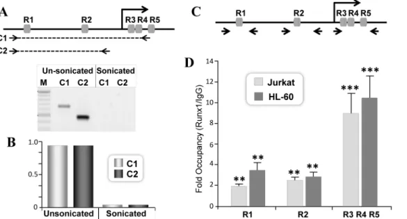
To investigate whether endogenous RUNX1 is recruited to the P1 promoter, we performed ChIP experiments in hematopoietic cells, in which RUNX1 protein is highly expressed. Chro-matin immunoprecipitated from Jurkat cells with RUNX1 antibody was quantified by qPCR. The two RUNX motifs in the P1 promoter (R1 and R2) are separated by 338bp. To evaluate independent occupancy of RUNX1 at each site, we initially optimized our sonication condi-tions (Fig 4A). Traditional PCR showed successful amplification of P1 promoter fragment encompassing R1 and R2 motifs from unsonicated DNA template but not from sonicated DNA (Fig 4A, lower panel). The effective separation of R1 and R2 motifs in the RUNX1 chro-matin was further confirmed by lack of any DNA amplification and detection in real time PCR (Fig 4B). Together these data demonstrate that our experimental conditions allow assessment of independent occupancy of RUNX1 on R1 and R2 motifs.
We find RUNX1 protein is recruited to both R1 and R2 motifs and exhibit significant enrichment over IgG ranging from two to three fold in Jurkat cells (Fig 4D). Binding of Fig 2. RUNX binding sites within 5’UTR inhibit RUNX1 P1 promoter activity.A) Schematic illustration of the wild type and RUNX sites mutated promoters. Mutant promoters were generated by introducing two base pair substitution mutations in one (mR3), two (mR3,4) or three RUNX sites (mR3,4,5) in the UTR region. Bases selected for mutation in each RUNX binding site are indicated in lower case (AACCACA!AcaCACA).
B) HL-60 cells were electroporated with 200ng of either wild type or mutated promoter reporters. Cells were harvested 18h later and equal amount of cell lysates were evaluated for luciferase activity using the dual luciferase reporter system. Normalized values from four independent experiments with three replicates each are presented in bar graphs with standard error of the mean. Asterisks represent statistically significant increase in basal activity of the RUNX1 mutant promoters compared to wild type promoter (***p<0.001).
RUNX1 protein to these motifs was further confirmed in HL-60 cells (Fig 4D). We also deter-mined RUNX1 protein recruitment at the three RUNX sites located in 5’UTR. Due to their close proximity, we could not assess their occupancy individually (Fig 4C). ChIP assay revealed RUNX1 occupancy of these sites in both Jurkat and HL-60 cells. The total enrichment of the UTR-RUNX sites (R3,R4,R5) is significantly higher relative to R1 and R2 sites (Fig 4D). How-ever, if adjusted per RUNX motif they are equivalent to those of RUNX sites within the pro-moter (Fig 4D). Occupancy of both promoter and UTR sites is consistent with involvement of these sites in transcriptional regulation shown in Figs1and2.
To determine transcriptional function of endogenous RUNX1 protein, we depleted RUNX1 protein from Jurkat cells. Several small interfering RNA targeting various regions of RUNX1 RNA were initially screened (data not shown). Two siRNA targeting different coding regions of the RUNX1 mRNA showed consistent knock down of RUNX1 protein by 54% and 56% respectively (Fig 5A). ChIP analysis from siRNA treated cells showed a decreased RUNX1 occupancy on all RUNX sites (Fig 5B). However the effect was more pronounced in cells treated with RUNX1 siRNA2 (Fig 5B). To understand consequences of decreased RUNX1 occupancy at these sites we determined RUNX1 gene transcription by evaluating the levels of Fig 3. Mutation or deletion of RUNX motifs in the UTR enhances RUNX1 mediated transcriptional activity of the P1 promoter.A) Non-hematopoietic HeLa cells were transiently co-transfected with a fix amount (200ng) of either wild type or RUNX mutant promoter-reporter plasmids and increasing concentration (100-1200ng) of RUNX1 expression vector. Cells were harvested 18h later and luciferase activity was determined by dual luciferase assay system. Values were normalized with Renilla luciferase activity used as an internal control. Pooled values from at least four independent experiments performed in triplicate are presented in the bar graph along with the standard error of the mean. Asterisks indicate statistical significance between wild type and mutant promoter (*p<0.1,**p<0.01. B) Parallel plates of HeLa cells were transiently co-transfected exactly as described in (A) and harvested 18h later. Cell were directly lysed and equal amount of proteins were resolved by SDS-PAGE. Blots were probed with polyclonal RUNX1 antibody, strip and re-probed with mouse monoclonal GAPDH antibody. C) HeLa cells were co-transfected with P1-Luc and increasing concentrations of RUNX1 expression plasmid using nanojuice transfection reagent. Cells were harvested 18h later for luciferase activity. Normalized data pooled from four independent experiments are presented in bar graph. Asterisks indicate statistically significant increase in promoter activity in response to RUNX1 overexpression as determined by student t-test (*p<0.1, p**<0.01). D) HeLa cells were co-transfected with 200ng of P1-Luc and increasing concentrations of RUNX1 expression vectors.
Cells were processed for western blot analysis 18h later and a representative blot is shown.
Fig 4. Endogenous RUNX1 protein is recruited to RUNX motifs located in the P1 promoter and the 5’UTR.A) Efficiency of chromatin shearing of the RUNX1 locus in Jurkat cells. Control (unsonicated) and sheared (sonicated) chromatin was subjected to PCR using either (A) traditional or (B) real time qPCR analysis. Primer pair amplifying RUNX1 chromatin loci encompassing R1-R4 (C1) or R1-R2 (C2) is indicated with dashed lines. The amplified 650bp C1 and 500bp C2 region of the RUNX1 gene were detected in control but not in sonicated chromatin. M denote 100bp ladder. B) Lack of intact DNA between the promoter and the UTR RUNX sites in sonicated chromatin was confirmed by real time qPCR. C) Arrowhead indicates relative position of primer pairs used to assess RUNX1 occupancy on promoter and UTR RUNX motifs D) Equivalent amount of chromatin (3U OD260) from Jurkat or HL-60 cells were used for ChIP assays. Chromatin was immunoprecipitated with 2μg of IgG or RUNX1 polyclonal antibody. Site specific occupancy was determined by real time
PCR. Mean values of IgG or RUNX1 immunoprecipitated DNA from three independent experiments with three replicates were obtained for each site. Data is expressed as fold changes over IgG in respective cells. Asterisks indicate statistically significant enrichment of DNA immunoprecipitated with RUNX1 antibody (**p<0.01,***p<0.001).
doi:10.1371/journal.pone.0149119.g004
Fig 5. Depletion of endogenous RUNX1 protein in hematopoietic cells decreases occupancy and activity of RUNX1 P1 promoter.A) Jurkat cells were electroporated with 80nM of either control siRNA (Cont) or two different siRNA directed against RUNX1 (siR1 and siR2). Cells were harvested 48h later and equal amount of proteins were resolved by SDS PAGE. Blots were probed with polyclonal antibody against RUNX1, stripped and re-probed with polyclonal antibody against Actin. A representative image of chemiluminescence signal is shown. B) Jurkat cells were electroporated with indicated siRNA for 48h and processed for chromatin immunoprecipitation. Equal amount of immunoprecipitated DNA was subjected to real time PCR amplification. Data was averaged from three independent experiments and is presented as fold over IgG for respective RUNX motifs. C) Jurkat cells electroporated with control or RUNX1 siRNA were pelleted 48h later to isolate RNA. Level of P1 promoter derived RUNX1 hnRNA was determined by qPCR using primer pair located in exon 1 and intron 1. Pooled data from three independent experiments with three replicate each is shown for non electroporeted (NE) and siRNA
electroporated Jurkat cells. Asterisks indicate statistically significant decrease in expression of RUNX1 hnRNA (**p<0.01,***p<0.001).
the RUNX1 heterogeneous nuclear RNA (hnRNA) present in the nucleus. The hnRNA reflects the primary transcript and is normally not degraded by siRNA treatment (Elferink & Reiners, 1996; Köhler & Roos, 2008). We find a significant decrease in P1 promoter derived
RUNX1-hnRNA from siRNA treated cells (Fig 5C). Decrease in RUNX1 primary transcript in hematopoietic cells is consistent with the promoter reporter data of a positive autoregulatory loop. Taken together these results indicate direct occupancy of RUNX1 is an integral compo-nent of RUNX1 mediated regulation of the P1 promoter.
SCL transcription factor activates RUNX1-P1 promoter
Several transcription factors are critical for development of hematopoietic cells. Here we assessed involvement of basic helix-loop-helix containing protein SCL that binds E box motif in the target DNA. The initial two hundred base pairs of the RUNX1-P1 promoter contain an evolutionary conserved E-box motif (Fig 1A). To assess SCL occupancy, ChIP assays were per-formed in hematopoietic cells. We find SCL is expressed in both Jurkat and HL-60 cells (Fig 6A). However, level of endogenous SCL protein is ten times higher in Jurkat cells. SCL occu-pancy of the RUNX1-P1 promoter was modest in Jurkat cells (Fig 6B). To assess if this is due to a poor efficiency of immunoprecipitation, we also evaluated SCL occupancy at the +23 RUNX1 enhancer region. We find a 9-fold enrichment of the RUNX1 enhancer region in immunoprecipitated DNA (Fig 6C). Our data is consistent with previous report showing SCL occupancy at the +23 enhancer in Jurkat cells [31]. Thus, the SCL motif in the RUNX1 pro-moter exhibit a poor occupancy compared to the enhancer region. Interestingly occupancy of SCL on the RUNX1-P1 promoter was significantly higher in HL-60 compared to Jurkat cells (Fig 6B).
These results are consistent with increased basal activity of the RUNX1 P1+UTR promoter in HL-60 cells (Fig 1D and 1E). The ability of SCL to regulate RUNX1 P1+UTR promoter was studied in HeLa cells that lack endogenous expression of SCL protein (Fig 6D). SCL overex-pression resulted in a 2-fold increased activity of the RUNX1 P1+UTR promoter (Fig 6E).
Like several hematopoietic transcription factors, SCL forms a molecular complex with RUNX1 protein. Therefore, we assessed interplay between RUNX1 and SCL transcription fac-tor for regulation of the RUNX1-P1 promoter. Interestingly, chromatin immunoprecipitation revealed that SCL protein is recruited to both the P1 promoter and the UTR region that do not contain SCL motif (Fig 7A). These data suggest that SCL is recruited to the RUNX1-P1 pro-moter by protein-DNA, and protein-protein interaction. We next assessed if RUNX1 and SCL can functionally interact to regulate RUNX1 promoter activity. HeLa cells transfected with RUNX1 or SCL showed activation of both the P1+UTR and P1 promoter (Fig 7B). To our sur-prise, RUNX1-P1 promoter activity did not change when RUNX1 and SCL were co-expressed (Fig 7B). Thus, co-expression of RUNX1 and SCL does not enhance transcriptional function of RUNX1 protein. In summary, our data show that RUNX1 and SCL proteins can regulate P1 promoter derived transcription of the RUNX1 gene.
Discussion
promoter. Taken together, our results demonstrate that in hematopoietic cells RUNX1 gene expression is regulated by a positive feedback mechanism.
The P1 and P2 promoters of the RUNX1 gene in both human and mice are separated by 160kb sequences. Earlier reports have established that a 531bp enhancer region is present in the intron 1 of the mouse RUNX1 gene. This enhancer region located 23.5kb after the RUNX1 translational start site, is highly conserved among different mammalian species. This RUNX1 enhancer can drive hematopoietic cell specific expression of a reporter genein vivo[45]. To
define contribution of the promoter sequences in transcription of the RUNX1 gene, we studied sequences of both the P1 promoter and the exon 1 of the RUNX1 gene. We find that a 0.6kb fragment of the P1 promoter and 0.6kb region of exon 1 exhibit a 20-fold higher basal activity in hematopoietic cells when compared with non-hematopoietic cells.
Interestingly, UTR of the RUNX1 gene contains potential repressor regions, as removal of these sequences enhances basal transcription of the RUNX1-P1 promoter. To identify factors contributing to repressor response, we performed anin silicoanalysis of UTR sequences. The
UTR region contains three RUNX sites in close proximity. These sites are evolutionary
Fig 6. SCL transcription factor activates RUNX1-P1 promoter.A) Equal amount of total protein from Jurkat and HL-60 cells were resolved by SDS-PAGE. Blots were probed with polyclonal SCL antibody, stripped and reprobed for tubulin antigen, used as a loading control. B) Jurkat and HL-60 cells were processed for chromatin immunoprecipitation. SCL occupancy of SCL motif in the RUNX1 P1 promoter was determined by qPCR. The positions of the SCL motif and the primer pair for ChIP assays are indicated in the diagram. Average data from three independent ChIP experiments, with three replicate each is shown as relative to IgG. C) Jurkat cells were processed for ChIP assays exactly as described in (B). Occupancy of SCL motif present in +23 RUNX1 enhancer was determined by qPCR. Average data from three independent ChIP experiments, with three replicate each is shown as relative to IgG. Statistical significance of SCL occupancy is indicated by asterisks (***p<0.001). D) HeLa cells were transiently co-transfected with RUNX1 promoter and indicated
amounts of SCL expression vector. Cells were harvested 18h later for western blot analysis. SCL protein was detected by probing blots with SCL antibody. GAPDH is shown as internal loading control in the same blots. E) HeLa cells were co-transfected with 200ng of the RUNX1 P1+UTR Luc and increasing concentration of SCL expression vector. Luciferase activity was determined 18h later and normalized with Renilla luciferase. Bars in graph represent mean values of three independent experiments each performed in triplicate.
conserved, suggesting a role in the transcriptional regulation of RUNX1 gene. Indeed, chroma-tin immunoprecipitation revealed that native RUNX1 protein in hematopoietic cells is recruited to these sites. Deletion or mutation of these RUNX motifs results in a 4 to 20-fold increase in activity of the RUNX1-P1 promoter. These data indicates that repressor activity of UTR sequences is due to recruitment of RUNX1 protein to the Runx sites. Interestingly, two evolutionary conserved RUNX motifs are also present within initial 570 bp of the P1 promoter. In hematopoietic cells, endogenous RUNX1 protein is directly recruited to these regulatory motifs. In contrast occupancy of the Runx motifs in the promoter by RUNX1 protein enhances transcriptional activity of the P1 promoter. The ability of RUNX1 protein to serve as transcrip-tional activator was further confirmed by overexpression and knockdown approaches. Deple-tion of RUNX1 protein by siRNA in hematopoietic cells, significantly reduces RUNX1 occupancy at these sites, as well as transcription of the RUNX1 gene. Thus, a positive feedback loop is required for expression of RUNX1 gene in hematopoietic cells.
Our over-expression results indicate that RUNX protein activates P1 promoter. However, RUNX motifs located in the promoter and UTR showed a differential response in the presence of RUNX1 protein. The two RUNX sites located in the P1 promoter showed an enhanced tran-scriptional response to RUNX1 protein. In sharp contrast, the three RUNX motifs located in UTR inhibits transcriptional activation by the RUNX1 protein. These findings are consistent with earlier reports demonstrating that based on promoter and cell context, RUNX proteins can act as either transcriptional activators or transcriptional repressors [46]. Most likely, this differential response by RUNX regulatory motifs is necessary to maintain tight levels of RUNX1 mRNA within hematopoietic tissues. The ability of RUNX proteins to form cell Fig 7. SCL is recruited to regions containing RUNX motifs in the promoter and the UTR.A) Chromatin from Jurkat and HL-60 cells were
immunoprecipitated with SCL antibody. DNA regions containing indicated RUNX motifs were amplified by qPCR. Mean values of SCL immunoprecipitated DNA from three independent experiments and three replicates were obtained for each site. Data is presented as fold changes over IgG in Jurkat and HL-60 cells. Statistically significant occupancy of SCL in promoter region containing RUNX motifs was observed only in Jurkat cells (*p<0.1) B) HeLa cells were
cotransfected with either P1+UTR-Luc or P1-Luc and 200ng of RUNX1 and SCL expression vectors. For co-expression 200ng of RUNX1 was transfected with increasing amounts of SCL expression vector. Luciferase activity was determined 18h later and values normalized with Renilla luciferase used as internal control. Normalized data pooled from three independent experiments each performed in triplicate, is presented with standard error of the mean. Statistically significant changes in RUNX1 promoter activity is indicated by asterisks (**p<0.01).
specific and combinatorial regulatory complexes allows either activation or repression of gene transcription [47–50]. For example, RUNX1 protein establishes activating or repressing com-plexes by physical interaction with other hematopoietic transcription factors such as GATA, SCL, C/EBP, SMAD coactivators and Groucho/TLE, mSin3A and NCoR corepressors [51–56]. Additional parameters, such as chromatin structure and interaction with chromatin modifying proteins also determines the functional response of RUNX1 protein [54]. RUNX1 binding to P1 promoter and the exon 1 region could permit the formation of a chromatin modifying regu-latory hub in hematopoietic cells. In fact, previous reports have shown that chromatin looping can bring together both promoter and enhancer regions and that RUNX1 binding to its cog-nate motif is the initial event in chromosomal looping [45,57]. Thus, RUNX1 gene transcrip-tion is controlled by differential response of RUNX motifs and formatranscrip-tion of regulatory complexes with other transcription factors and chromatin modifying proteins.
Several transcription factors including members of the SMAD family, GATA1 and PU.1 regulates RUNX1 gene expression in hematopoietic cells. During hematopoiesis, these tran-scription factors work cooperatively by either forming a physical and/or functional interaction with RUNX1 protein. In this paper we also studied involvement of SCL transcription factor in regulating P1 promoter derived RUNX1 gene transcription. A putative SCL binding motif is present in the RUNX1 P1 promoter. This SCL motif is located between RUNX sites present in the UTR and the P1 promoter. In hematopoietic cells the endogenous SCL protein is recruited to this site. However, SCL occupancy in the promoter site is rather modest when compared with the SCL motif located in the +23 enhancer of RUNX1 gene. Overexpression of SCL pro-tein in non-hematopoietic cells enhances transcriptional activity of the RUNX1-P1 promoter. Interestingly, SCL protein is recruited to both the UTR and the P1 promoter regions. Our find-ings are consistent with previous reports that showed physical interaction between SCL and RUNX1 protein [53]. However, co expression of SCL and RUNX1 did not cause synergistic activation of the RUNX1-P1 promoter. Thus, occupancy of SCL at the RUNX1-P1 promoter and enhancer regions may support chromatin conformation that is conducive for RUNX1 gene transcription [45].
In summary, our findings demonstrate that in hematopoietic cells RUNX1 protein is recruited to its own promoter to regulate RUNX1 gene transcription in a positive feedback loop. This auto-regulation is also noted for other members of the RUNX gene family [58,59]. Thus, auto-regulatory process maybe a key mechanism for sensing and maintaining RUNX1 protein threshold necessary for specification and differentiation of hematopoietic cells.
Author Contributions
Conceived and designed the experiments: MM MH SEG. Performed the experiments: MM MH DT. Analyzed the data: MM AJ SEG. Contributed reagents/materials/analysis tools: VM JS GS. Wrote the paper: AJ SEG.
References
1. Tahirov TH, Inoue-Bungo T, Morii H, Fujikawa A, Sasaki M, Kimura K, et al. Structural analyses of DNA recognition by the AML1/Runx-1 Runt domain and its allosteric control by CBFbeta. Cell. 2001; 104 (5):755–67. PMID:11257229
2. Lian JB, Javed A, Zaidi SK, Lengner C, Montecino M, van Wijnen AJ, et al. Regulatory controls for oste-oblast growth and differentiation: role of Runx/Cbfa/AML factors. Crit Rev Eukaryot Gene Expr. 2004; 14(1–2):1–41. PMID:15104525
3. Coffman JA. Runx transcription factors and the developmental balance between cell proliferation and differentiation. Cell Biol Int. 2003; 27(4):315–24. PMID:12788047
5. Okuda T, van Deursen J, Hiebert SW, Grosveld G, Downing JR. AML1, the target of multiple chromo-somal translocations in human leukemia, is essential for normal fetal liver hematopoiesis. Cell. 1996; 84:321–30. PMID:8565077
6. North T, Gu TL, Stacy T, Wang Q, Howard L, Binder M, et al. Cbfa2 is required for the formation of intra-aortic hematopoietic clusters. Development. 1999; 126(11):2563–75. PMID:10226014
7. Choi JY, Pratap J, Javed A, Zaidi SK, Xing L, Balint E, et al. Subnuclear targeting of Runx/Cbfa/AML factors is essential for tissue-specific differentiation during embryonic development. Proceedings of the National Academy of Sciences of the United States of America. 2001; 98(15):8650–5. PMID:11438701
8. Komori T, Yagi H, Nomura S, Yamaguchi A, Sasaki K, Deguchi K, et al. Targeted disruption of Cbfa1 results in a complete lack of bone formation owing to maturational arrest of osteoblasts. Cell. 1997; 89 (5):755–64. PMID:9182763
9. Otto F, Thornell AP, Crompton T, Denzel A, Gilmour KC, Rosewell IR, et al. Cbfa1, a candidate gene for cleidocranial dysplasia syndrome, is essential for osteoblast differentiation and bone development. Cell. 1997; 89(5):765–71. PMID:9182764
10. Levanon D, Bettoun D, Harris-Cerruti C, Woolf E, Negreanu V, Eilam R, et al. The Runx3 transcription factor regulates development and survival of TrkC dorsal root ganglia neurons. The EMBO journal. 2002; 21(13):3454–63. PMID:12093746
11. Li QL, Ito K, Sakakura C, Fukamachi H, Inoue K, Chi XZ, et al. Causal relationship between the loss of RUNX3 expression and gastric cancer. Cell. 2002; 109(1):113–24. PMID:11955451
12. Inoue K, Ozaki S, Shiga T, Ito K, Masuda T, Okado N, et al. Runx3 controls the axonal projection of pro-prioceptive dorsal root ganglion neurons. Nat Neurosci. 2002; 5(10):946–54. PMID:12352981
13. Woolf E, Xiao C, Fainaru O, Lotem J, Rosen D, Negreanu V, et al. Runx3 and Runx1 are required for CD8 T cell development during thymopoiesis. Proceedings of the National Academy of Sciences of the United States of America. 2003; 100(13):7731–6. PMID:12796513
14. Ichikawa M, Asai T, Saito T, Seo S, Yamazaki I, Yamagata T, et al. AML-1 is required for megakaryo-cytic maturation and lymphomegakaryo-cytic differentiation, but not for maintenance of hematopoietic stem cells in adult hematopoiesis. Nat Med. 2004; 10(3):299–304. PMID:14966519
15. Taniuchi I. Differential requirements for Runx proteins in CD4 repression and epigenetic silencing dur-ing T lymphocyte development. Cell. 2002; 111:621–33. PMID:12464175
16. Putz G, Rosner A, Nuesslein I, Schmitz N, Buchholz F. AML1 deletion in adult mice causes splenomeg-aly and lymphomas. Oncogene. 2006; 25(6):929–39. PMID:16247465
17. Okuda T, Cai Z, Yang S, Lenny N, Lyu CJ, van Deursen JM, et al. Expression of a knocked-in AML1-ETO leukemia gene inhibits the establishment of normal definitive hematopoiesis and directly gener-ates dysplastic hematopoietic progenitors. Blood. 1998; 91(9):3134–43. PMID:9558367
18. Higuchi M. Expression of a conditional AML1-ETO oncogene bypasses embryonic lethality and estab-lishes a murine model of human t(8;21) acute myeloid leukemia. Cancer cell. 2002; 1:63–74. PMID: 12086889
19. Michaud J, Scott HS, Escher R. AML1 interconnected pathways of leukemogenesis. Cancer Invest. 2003; 21(1):105–36. PMID:12643014
20. Ichikawa M, Yoshimi A, Nakagawa M, Nishimoto N, Watanabe-Okochi N, Kurokawa M. A role for RUNX1 in hematopoiesis and myeloid leukemia. International journal of hematology. 2013; 97(6):726– 34. doi:10.1007/s12185-013-1347-3PMID:23613270
21. Mangan JK, Speck NA. RUNX1 mutations in clonal myeloid disorders: from conventional cytogenetics to next generation sequencing, a story 40 years in the making. Crit Rev Oncog. 2011; 16(1–2):77–91. PMID:22150309
22. Ohgami RS, Arber DA. The diagnostic and clinical impact of genetics and epigenetics in acute myeloid leukemia. Int J Lab Hematol. 2015; 37 Suppl 1:122–32. doi:10.1111/ijlh.12367PMID:25976970
23. Song WJ. Haploinsufficiency of CBFA2 causes familial thrombocytopenia with propensity to develop acute myelogenous leukaemia. Nat Genet. 1999; 23:166–75. PMID:10508512
24. Michaud J, Wu F, Osato M, Cottles GM, Yanagida M, Asou N, et al. In vitro analyses of known and novel RUNX1/AML1 mutations in dominant familial platelet disorder with predisposition to acute mye-logenous leukemia: implications for mechanisms of pathogenesis. Blood. 2002; 99(4):1364–72. PMID: 11830488
25. Sun L, Mao G, Rao AK. Association of CBFA2 mutation with decreased platelet PKC-theta and impaired receptor-mediated activation of GPIIb-IIIa and pleckstrin phosphorylation: proteins regulated by CBFA2 play a role in GPIIb-IIIa activation. Blood. 2004; 103(3):948–54. PMID:14525764
27. Telfer JC, Rothenberg EV. Expression and function of a stem cell promoter for the murine CBFalpha2 gene: distinct roles and regulation in natural killer and T cell development. Developmental biology. 2001; 229(2):363–82. PMID:11203699
28. Pozner A, Lotem J, Xiao C, Goldenberg D, Brenner O, Negreanu V, et al. Developmentally regulated promoter-switch transcriptionally controls Runx1 function during embryonic hematopoiesis. BMC Dev Biol. 2007; 7:84. PMID:17626615
29. Tijssen MR, Ghevaert C. Transcription factors in late megakaryopoiesis and related platelet disorders. J Thromb Haemost. 2013; 11(4):593–604. doi:10.1111/jth.12131PMID:23311859
30. Sroczynska P, Lancrin C, Kouskoff V, Lacaud G. The differential activities of Runx1 promoters define milestones during embryonic hematopoiesis. Blood. 2009; 114(26):5279–89. doi: 10.1182/blood-2009-05-222307PMID:19858498
31. Sanda T, Lawton LN, Barrasa MI, Fan ZP, Kohlhammer H, Gutierrez A, et al. Core transcriptional regu-latory circuit controlled by the TAL1 complex in human T cell acute lymphoblastic leukemia. Cancer cell. 2012; 22(2):209–21. doi:10.1016/j.ccr.2012.06.007PMID:22897851
32. Gering M, Rodaway AR, Gottgens B, Patient RK, Green AR. The SCL gene specifies haemangioblast development from early mesoderm. The EMBO journal. 1998; 17(14):4029–45. PMID:9670018
33. Ema M, Faloon P, Zhang WJ, Hirashima M, Reid T, Stanford WL, et al. Combinatorial effects of Flk1 and Tal1 on vascular and hematopoietic development in the mouse. Genes & development. 2003; 17 (3):380–93.
34. Kallianpur AR, Jordan JE, Brandt SJ. The SCL/TAL-1 gene is expressed in progenitors of both the hematopoietic and vascular systems during embryogenesis. Blood. 1994; 83(5):1200–8. PMID: 8118024
35. Robb L, Lyons I, Li R, Hartley L, Kontgen F, Harvey RP, et al. Absence of yolk sac hematopoiesis from mice with a targeted disruption of the scl gene. Proceedings of the National Academy of Sciences of the United States of America. 1995; 92(15):7075–9. PMID:7624372
36. Shivdasani RA, Mayer EL, Orkin SH. Absence of blood formation in mice lacking the T-cell leukaemia oncoprotein tal-1/SCL. Nature. 1995; 373(6513):432–4. PMID:7830794
37. Wadman IA, Osada H, Grutz GG, Agulnick AD, Westphal H, Forster A, et al. The LIM-only protein Lmo2 is a bridging molecule assembling an erythroid, DNA-binding complex which includes the TAL1, E47, GATA-1 and Ldb1/NLI proteins. The EMBO journal. 1997; 16(11):3145–57. PMID:9214632
38. Jurata LW, Kenny DA, Gill GN. Nuclear LIM interactor, a rhombotin and LIM homeodomain interacting protein, is expressed early in neuronal development. Proceedings of the National Academy of Sciences of the United States of America. 1996; 93(21):11693–8. PMID:8876198
39. Pimanda JE, Donaldson IJ, de Bruijn MF, Kinston S, Knezevic K, Huckle L, et al. The SCL transcrip-tional network and BMP signaling pathway interact to regulate RUNX1 activity. Proceedings of the National Academy of Sciences of the United States of America. 2007; 104(3):840–5. PMID:17213321
40. Li X, Vradii D, Gutierrez S, Lian JB, van Wijnen AJ, Stein JL, et al. Subnuclear targeting of Runx1 is required for synergistic activation of the myeloid specific M-CSF receptor promoter by PU.1. Journal of cellular biochemistry. 2005; 96(4):795–809. PMID:16149049
41. Stuardo M, Martinez M, Hidalgo K, Montecino M, Javed A, Lian JB, et al. Altered chromatin modifica-tions in AML1/RUNX1 breakpoint regions involved in (8;21) translocation. Journal of cellular physiol-ogy. 2009; 218(2):343–9. doi:10.1002/jcp.21599PMID:18853425
42. Bakshi R, Zaidi SK, Pande S, Hassan MQ, Young DW, Montecino M, et al. The leukemogenic t(8;21) fusion protein AML1-ETO controls rRNA genes and associates with nucleolar-organizing regions at mitotic chromosomes. J Cell Sci. 2008; 121(Pt 23):3981–90. doi:10.1242/jcs.033431PMID:19001502
43. Ghozi MC, Bernstein Y, Negreanu V, Levanon D, Groner Y. Expression of the human acute myeloid leukemia gene AML1 is regulated by two promoter regions. Proceedings of the National Academy of Sciences of the United States of America. 1996; 93(5):1935–40. PMID:8700862
44. Fujita Y, Nishimura M, Taniwaki M, Abe T, Okuda T. Identification of an alternatively spliced form of the mouse AML1/RUNX1 gene transcript AML1c and its expression in early hematopoietic development. Biochem Biophys Res Commun. 2001; 281(5):1248–55. PMID:11243869
45. Nottingham WT, Jarratt A, Burgess M, Speck CL, Cheng JF, Prabhakar S, et al. Runx1-mediated hematopoietic stem-cell emergence is controlled by a Gata/Ets/SCL-regulated enhancer. Blood. 2007; 110(13):4188–97. PMID:17823307
46. Javed A, Guo B, Hiebert S, Choi JY, Green J, Zhao SC, et al. Groucho/TLE/R-esp proteins associate with the nuclear matrix and repress RUNX (CBF(alpha)/AML/PEBP2(alpha)) dependent activation of tissue-specific gene transcription. J Cell Sci. 2000; 113 (Pt 12):2221–31. PMID:10825294
48. Javed A, Barnes GL, Jasanya BO, Stein JL, Gerstenfeld L, Lian JB, et al. runt homology domain tran-scription factors (Runx, Cbfa, and AML) mediate repression of the bone sialoprotein promoter: evidence for promoter context-dependent activity of Cbfa proteins. Molecular and cellular biology. 2001; 21 (8):2891–905. PMID:11283267
49. Javed A, Gutierrez S, Montecino M, van Wijnen AJ, Stein JL, Stein GS, et al. Multiple Cbfa/AML sites in the rat osteocalcin promoter are required for basal and vitamin D-responsive transcription and contrib-ute to chromatin organization. Molecular and cellular biology. 1999; 19(11):7491–500. PMID: 10523637
50. Kuvardina ON, Herglotz J, Kolodziej S, Kohrs N, Herkt S, Wojcik B, et al. RUNX1 represses the ery-throid gene expression program during megakaryocytic differentiation. Blood. 2015; 125(23):3570–9. doi:10.1182/blood-2014-11-610519PMID:25911237
51. Ferjoux G, Auge B, Boyer K, Haenlin M, Waltzer L. A GATA/RUNX cis-regulatory module couples Dro-sophila blood cell commitment and differentiation into crystal cells. Developmental biology. 2007; 305 (2):726–34. PMID:17418114
52. Wilson NK, Foster SD, Wang X, Knezevic K, Schutte J, Kaimakis P, et al. Combinatorial transcriptional control in blood stem/progenitor cells: genome-wide analysis of ten major transcriptional regulators. Cell Stem Cell. 2010; 7(4):532–44. doi:10.1016/j.stem.2010.07.016PMID:20887958
53. Tijssen MR, Cvejic A, Joshi A, Hannah RL, Ferreira R, Forrai A, et al. Genome-wide analysis of simulta-neous GATA1/2, RUNX1, FLI1, and SCL binding in megakaryocytes identifies hematopoietic regula-tors. Developmental cell. 2011; 20(5):597–609. doi:10.1016/j.devcel.2011.04.008PMID:21571218
54. Durst KL, Hiebert SW. Role of RUNX family members in transcriptional repression and gene silencing. Oncogene. 2004; 23(24):4220–4. PMID:15156176
55. Setoguchi R, Tachibana M, Naoe Y, Muroi S, Akiyama K, Tezuka C, et al. Repression of the transcrip-tion factor Th-POK by Runx complexes in cytotoxic T cell development. Science. 2008; 319
(5864):822–5. doi:10.1126/science.1151844PMID:18258917
56. Seo W, Tanaka H, Miyamoto C, Levanon D, Groner Y, Taniuchi I. Roles of VWRPY motif-mediated gene repression by Runx proteins during T-cell development. Immunol Cell Biol. 2012; 90(8):827–30. doi:10.1038/icb.2012.6PMID:22370763
57. Markova EN, Kantidze OL, Razin SV. Transcriptional regulation and spatial organisation of the human AML1/RUNX1 gene. Journal of cellular biochemistry. 2011; 112(8):1997–2005. doi:10.1002/jcb.23117 PMID:21445863
58. Levanon D, Groner Y. Structure and regulated expression of mammalian RUNX genes. Oncogene. 2004; 23(24):4211–9. PMID:15156175