Abstract
Cartilage has limited self-regenerative capacity. Tissue engineering can offer promising solutions for reconstruction of missing or damaged cartilage. A major challenge herein is to deine an appropriate cell source that is capable of generating a stable and functional matrix. This study evaluated the performance of culture-expanded human chondrocytes from ear (EC), nose (NC) and articular joint (AC), as well as bone-marrow-derived and adipose-tissue-derived mesenchymal stem cells both in vitro and in vivo. All cells (≥ 3 donors per source) were culture-expanded, encapsulated in alginate and cultured for 5 weeks. Subsequently, constructs were implanted subcutaneously for 8 additional weeks. Before and after implantation, glycosaminoglycan (GAG) and collagen content were measured using biochemical assays. Mechanical properties were determined using stress-strain-indentation tests. Hypertrophic differentiation was evaluated with qRT-PCR and subsequent endochondral ossiication with histology. ACs had higher chondrogenic potential in vitro than the other cell sources, as assessed by gene expression and GAG content (p < 0.001). However, after implantation, ACs did not further increase their matrix. In contrast, ECs and NCs continued producing matrix in vivo leading to higher GAG content (p < 0.001) and elastic modulus. For NC-constructs, matrix-deposition was associated with the elastic modulus (R2 = 0.477, p = 0.039). Although all cells – except ACs – expressed markers for hypertrophic differentiation in vitro, there was no bone formed in vivo. Our work shows that cartilage formation and functionality depends on the cell source used. ACs possess the highest chondrogenic capacity in vitro, while ECs and NCs are most potent in vivo, making them attractive cell sources for cartilage repair.
Keywords: Chondrogenesis, chondrocytes, mesenchymal stem cells, alginate, mechanics.
*Address for correspondence: Prof. dr. Gerjo JVM. van Osch
Erasmus MC, University Medical Center Rotterdam Dr. Molewaterplein 50-60, Room Ee 16.55
3015 GE Rotterdam the Netherlands
Telephone Number: +31107043661 FAX Number: +31107044690 E-mail:[email protected]
Introduction
Cartilage is a highly specialised avascular connective tissue located at a variety of anatomical locations such as the ear, nose, trachea, ribs and articular joints. In general, cartilage predominantly consists of an extracellular matrix (ECM), which is produced, maintained and remodelled by a relatively small number of specialised cells (1-10 %) (Muir, 1995). The exact composition of the ECM is mainly dependent on the tissue’s function and thus three major subtypes can be distinguished: hyaline, ibrous and elastic cartilage. It is well known that due to its avascular origin, cartilage itself has a limited self-regenerative capacity. As a result, cartilage defects can lead to severe pain, disability and aesthetic impairment. Currently, surgical repair of cartilage requires either autogeneic cartilage grafts or artiicial material implants. However, these conventional treatments are (1) associated with a limited availability of autogeneic tissue, (2) can cause donor site morbidity, and – in the case of artiicial implants – (3) are prone to generate a foreign body reaction.
To overcome these problems, tissue engineering (TE) can offer a promising solution for restoring missing or damaged cartilage. TE-approaches have focused on the production of functional cartilage that has features similar to native tissue. In cartilage TE, small tissue biopsies are harvested, thus generating minimal donor site morbidity. Cells are isolated from the biopsies and stimulated to proliferate in culture providing large quantities of cells. These cells are subsequently stimulated to produce cartilage tissue which should structurally and morphologically resemble native tissue. One of the major challenges in cartilage TE is deining an appropriate cell source. The most obvious cell source is cartilage itself. Hyaline articular cartilage is most frequently used for cartilage TE, although some experiments have been published on the use of non-articular cartilages (e.g. nasal, ear and costal cartilage) (Stoddart et al., 2009). Next to chondrocytes, mesenchymal stem cells (MSCs) with their multi-lineage differentiation potential and easy availability from bone marrow or adipose tissue have been demonstrated as an attractive cell source for cartilage TE (Johnstone et al., 1998; Pittenger et al., 1999).
To date, we and others have evaluated the use of chondrocytes and MSCs of several anatomical locations for their applicability in cartilage regenerative medicine (Aizah et al., 2007; Asawa et al., 2009; Chung et al., 2008; Hellingman et al., 2011; Henderson et al., 2007; Isogai et al., 2006; Johnson et al., 2004; Kaienah et al.,
THE
IN VITRO
AND
IN VIVO
CAPACITY OF CULTURE-EXPANDED HUMAN CELLS
FROM SEVERAL SOURCES ENCAPSULATED IN ALGINATE
TO FORM CARTILAGE
M.M. Pleumeekers1, L. Nimeskern2, W.L.M. Koevoet1, N. Kops3, R.M.L. Poublon1, K.S. Stok2 and G.J.V.M. van Osch1,3,*
1Department of Otorhinolaryngology, Head and Neck Surgery, Erasmus MC, University Medical Center, Rotterdam, the Netherlands
2Institute for Biomechanics, ETH Zurich, Zurich, Switzerland
2002; Karlsson et al., 2007; Kusuhara et al., 2009; Lohan et al., 2011; Malicev et al., 2009; Naumann et al., 2004; Panossian et al., 2001; Sakaguchi et al., 2005; Seda Tigli et al., 2009; Tay et al., 2004; van Osch et al., 2004; Vinardell et al., 2012; Xu et al., 2004; Yoshimura et al., 2007; Zhang and Spector, 2009). However, precise comparison of the performance of culture-expanded human cells is lacking. This knowledge is important to be able to select an optimal cell source for each application of cartilage TE. The current study was designed to evaluate the performance of culture-expanded cells of several sources for generating a stable and functional ECM in vitro and in vivo. Therefore, human chondrocytes from ear, nose and articular joint and MSCs derived from bone marrow and adipose tissue were compared. Cartilage matrix production was evaluated using qRT-PCR and biochemical assays during in vitro culture. Biochemical assays, histology and mechanical tests were used to determine tissue stability and functionality of cartilage constructs after subsequent subcutaneous implantation in vivo.
Materials and Methods
Cell sources
Ear (EC: n = 5, median age 69, range 17-75 years) and nasal cartilage (NC: n = 8, median age 24, range 18-46 years) were obtained from patients undergoing reconstructive subtotal septorhinoplasty. For articular cartilage (AC), both healthy (n = 2, traumatic amputation) and diseased knee cartilage (n = 7, osteoarthritis) were harvested. Since no clear differences in chondrogenic potential were visible between both healthy and diseased AC (data not shown), we combined them for further experiments (total n = 9, median age 68, range 43-88 years). To obtain adipose-tissue-derived mesenchymal stem cells (aMSC), subcutaneous abdominal adipose tissue was used from patients undergoing reconstructive breast surgery (n = 7, median age 51, range 34-71 years). All these tissue samples were obtained as waste material after surgery with approval of the local medical ethics committee (MEC-2011-371). Finally, bone marrow-derived mesenchymal stem cells (bMSC) were harvested from femoral shaft biopsies during total hip replacement surgery, after informed consent had been acquired and with approval of the local medical ethics committee (MEC-2004-142) (n = 11, median age 63, range 39-72 years).
Cell isolation and culture Expansion
To isolate chondrocytes, macroscopically intact cartilage pieces were washed after careful resection of the perichondrium (in the case of nasal and ear cartilage). Cartilage pieces were diced into small fragments and incubated for 1 h with protease (2 mg/mL; Sigma-Aldrich, St. Louis, MO, USA), followed by overnight incubation with collagenase B (Roche Diagnostics, Mannheim, Germany) in high glucose (4.5 g/L) Dulbecco’s modiied Eagle’s medium (HG-DMEM; Gibco, Carlsbad, CA, USA) with 10 % foetal calf serum (FCS; Gibco), 50 µg/ mL gentamycin (Gibco), and 0.5 µg/mL amphotericin B
(Fungizone; Life Technologies, Breda, the Netherlands). To remove small parts of undigested cartilage, the cell suspension was iltered through a nylon 100 µm mesh. Prior to cell seeding, cell viability was tested using the trypan blue exclusion test, and cell number was calculated with a haemocytometer. Chondrocytes were inally seeded at an initial density of 7500 cells/cm2 in ‘standard chondrocyte expansion medium’ containing HG-DMEM supplemented with 10 % FCS, 50 µg/mL gentamycin, and 0.5 µg/mL Fungizone.
Bone-marrow-derived heparinised aspirates were seeded at a density of 2-5 x 10⁵ nucleated cells/cm2 and cultured overnight in ‘standard MSC expansion medium’ containing low glucose (1.5 g/L) Dulbecco’s modiied Eagle’s medium (LG-DMEM), supplemented with 10 % FCS; 50 µg/mL gentamycin; 0.5 µg/mL Fungizone; 10-4 M L-ascorbic acid 2-phosphate (Sigma-Aldrich); and 1 ng/mL basic Fibroblast Growth Factor 2 (bFGF2; AbD Serotec, Kidlington, UK). After 24 h, non-adherent cells and cell debris were washed out and adherent bMSC were further expanded using ‘standard MSC expansion medium’. To extract aMSCs, excised human adipose tissue was washed with LG-DMEM, minced, and suspended in 0.1 % collagenase type I solution (Invitrogen, Carlsbad, CA, USA) in the presence of 1 % bovine serum albumin (BSA; PAA Laboratories Gmbh, Cölbe, Germany) in LG-DMEM. After 60 min of enzymatic digestion on an orbital shaker, loating adipocytes were separated from the precipitating MSC fraction by centrifugation (10 min, 1500 RPM), washed with ‘standard MSC expansion medium’, and iltered through a 100 μm nylon mesh. Before cell seeding, the amount of nucleated cells was assessed using methylene blue, and cell number was calculated with a haemocytometer. The cell suspension was seeded at an initial density of 40,000 cells/cm2 in ‘standard MSC expansion medium’.
All cells were cultured at 37 °C in air containing 5 % carbon dioxide. Medium was changed twice a week. When cell cultures reached 80 % conluence, chondrocytes and MSCs were trypsinised using 0.05 % trypsin-EDTA. Chondrocytes were seeded at a 7500 cells/cm2 and MSCs at a 2300 cells/cm2 cell density for further expansion to increase cell number. All third-passage (P3) cells which were approaching subconluence were detached and cultured in a three-dimensional alginate system (as described below) to promote chondrogenesis.
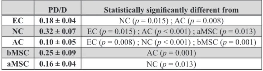
In order to determine the proliferation rate of cultured ECs, NCs, ACs, bMSCs and aMSCs, growth kinetics of three donors from each cell source were evaluated in monolayer expansion using the number of population doublings (PD) until subconluency and the time to reach passage four. Therefore, we have calculated the PD/D (Population Doublings per Day) by using the formula: PD/D = (ln(N2/N1)/ln (2))/D; where N1 was the number of cells at the beginning of each passage, N2 the number of cells at subconluency, and D the number of days to reach passage four.
Chondrogenic differentiation
each of the MSC-sources were suspended at a density of 4 x 10⁶ cells/mL in clinical grade 1.1 % low viscosity alginate solution dissolved in 0.9 % NaCl (Batch MG-004, CellMed, Alzenau, Germany). Afterwards, the cell-alginate mixture was transferred into a 10 mL sterile syringe from which the suspension was slowly passed through a 23-gauge needle to produce drops, which fell into a 102 mM CaCl2 solution. Following instantaneous gelation, the beads were allowed to further gelate for a period of 10 min in the CaCl2 solution. After being washed once with 0.9 % NaCl and HG-DMEM, the beads were transferred to 24-well plates. Controls were cultured in 150 μL/bead ‘control differentiation medium’ containing serum-free HG-DMEM supplemented with 50 µg/mL
gentamycin; 0.5 µg/mL Fungizone; 1 mM sodium pyruvate (Gibco); 40 μg/mL L-proline (Sigma-Aldrich); insulin transferrin selenium (ITS+; B&D Bioscience, Bedford, MA, USA); 10-7 M dexamethasone (Sigma-Aldrich); and
25 μg/mL L-ascorbic acid 2-phosphate. In the experimental condition (‘chondrogenic differentiation medium’), 10 ng/mL transforming growth factor β1 (TGFβ1; R&D Systems, Minneapolis, MN, USA) was added to induce chondrogenesis. Medium was changed twice a week. After two and ive weeks, alginate beads were processed for biochemical or gene-expression analysis as described below. For all in vitro experiments, four donors for the chondrocyte sources and six donors for the MSC sources were used, with at least duplicate samples per analyses for each individual donor.
To study in vivo functionality and stability of cartilage TE constructs after in vitro cell culture, larger lat constructs were created from cells of three donors of each cell source as previously described (Wong et al., 2001). In short, alginate suspensions containing 4 x 10⁶ cells/mL were injected into a custom-designed slab mould consisting of two calcium-permeable membranes (Durapore® 5.0 μm membrane ilters, Millipore, Amsterdam, the Netherlands) rigidly supported by stainless-steel meshes and separated
by a stainless-steel casting frame. Part of these constructs were harvested after ive weeks of cell culture for analysis and a part was implanted subcutaneously on the dorsal side of athymic mice. For the in vivo experiments, a total of six constructs per cell source were used, with duplicate samples for three different donors.
Subcutaneous implantation in vivo
In total, seventeen nine-week old, female NMRI nu/ nu mice (Charles River Laboratories, Wilmington,
Primers and probes
GAPDH Forward: ATGGGGAAGGTGAAGGTCG
Reverse: TAAAAGCAGCCCTGGTGACC Fam-CGCCCAATACGACCAAATCCGTTGAC
HPRT1 Forward: TATGGACAGGACTGAACGTCTTG
Reverse: CACACAGAGGGCTACAATGTG Fam-CGCCCAATACGACCAAATCCGTTGAC
ACAN Forward: TCGAGGACAGCGAGGCC
Reverse: TCGAGGGTGTAGCGTGTAGAGA Fam-ATGGAACACGATGCCTTTCACCACGA
COL2A1 Forward: GGCAATAGCAGGTTCACGTACA
Reverse: CGATAACAGTCTTGCCCCACTT Fam-CCGGTATGTTTCGTGCAGCCATCCT
COL10 Forward: CAAGGCACCATCTCCAGGAA
Reverse: AAAGGGTATTTGTGGCAGCATATT Fam-TCCAGCACGCAGAATCCATCTGA
ALP Forward: GACCCTTGACCCCCACAAT
Reverse: GCTCGACTGCATGTCCCCT
Fam-TGGACTACCTATTGGGTCTCTTCGAGCCA
MMP13 Forward: AAGGAGCATGGCGACTTCT
Reverse: TGGCCCAGGAGGAAAAGC Fam-CCCTCTGGCCTGCGGCTCA
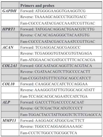
Table 1. Sequences of primers and probes for qRT-PCR.
Scoring categories Score
(1) Uniformity and darkness of the stain
No stain 0
Weak stain of poorly formed matrix 1
Moderately even stain 2
Even dark stain 3
(2) Distance between cells / amount of matrix accumulated
High cell densities with no matrix in between (no spacing between cells) 0
High cell densities with little matrix in between (cells <1 cell-size apart) 1
Moderate cell density with matrix (cells approximately 1 cell-size apart) 2
Low cell density with moderate distance between cells (>1 cell) and an extensive matrix 3
(3) Cell morphologies represented
Condensed/necrotic/pycnotic bodies 0
Spindle/ibrous 1
Mixed spindle/ibrous with rounded chondrogenic morphology 2
Majority rounded/chondrogenic 3
MAXIMUM SCORE 9
MA, USA) were used to evaluate the performance of constructs cultured with or without TGFβ1. Mice were placed under general anaesthesia using 2.5 % isolurane. Two separate subcutaneous incisions of approximately 10 mm were made along the central line of the spine (one at the shoulders and one at the hips), after which four separate subcutaneous pockets were prepared by blunt dissection of the subcutaneous tissue. For implantation, the alginate constructs were randomly assigned to these four pockets. Eight weeks after subcutaneous implantation, animals were sacriiced and samples were explanted for histological, biomechanical and biochemical analyses. Animal experiments were carried out with approval of the local Animal Experiments Committee of the Erasmus MC and were approved as outlined in the national Animals Act (EMC 2429).
Gene expression analyses
For total RNA isolation, alginate was dissolved in ice-cold 55 mM sodium citrate (150 μL/bead) and 20 mM ethylenediaminetetraacetate (EDTA) in 150 mM NaCl and centrifuged at 2.5 g for 8 min. Each pellet was subsequently suspended in 1 mL RNA-Bee™ (Tel-Test, Friendswood, TX, USA). RNA was extracted with chloroform and purified from the supernatant using the RNAeasy Micro Kit (Qiagen, Hilden, Germany) according to the manufacturer’s guidelines by on-column DNA-digestion. Extracted total RNA was quantiied using NanoDrop® ND-1000 Spectrophotometer (NanoDrop Technologies, Wilmington, DE, USA) at 260/280 nm. Total RNA of each sample was reverse transcribed into cDNA using RevertAid™ First Strand cDNA Synthesis Kit (MBI Fermentas, St. Leon-Rot, Germany).
For quantitative real-time polymerase chain reaction (qRT-PCR) analysis, forward and reverse primers were designed using PrimerExpress 2.0 software (Applied Biosystems, Foster City, CA, USA) to meet TaqMan or SYBR Green requirements. They were designed to bind separate exons to avoid co-ampliication of genomic DNA. Gene speciicity of all primers was guaranteed by basic local alignment search tool (BLASTN), as listed in Table 1. The following genes were analysed: aggrecan (ACAN), collagen type IIA1 (COL2A1), collagen type X (COL10), alkaline phosphatase (ALP), and matrix metalloproteinase-13 (MMP13). Glyceraldehyde 3-phosphate dehydrogenase (GAPDH), and hypoxanthine phosphoribosyltransferase 1 (HPRT1) were used as housekeeping genes. The expression of GAPDH and HPRT1 did not differ between cell sources and both were used to calculate the best housekeeper index (Pfafl et al., 2004). Using repeated pair-wise correlation analysis, data were normalised by calculating the ‘best housekeeper index’ (data not shown). Polymerase chain reactions were performed using TaqMan® Universal PCR Mastermix (Applied Biosystems) or qPCR Mastermix Plus for SYBR Green (Eurogentec, Nederland BV, Maastricht, the Netherlands) according to the manufacturers’ guidelines and using an ABIPRISM® 7000 with SDS software version 1.7 (Applied Biosystems, Nieuwerkerk a/d IJssel, the Netherlands). Ampliication eficiencies for all assays were between 90-110 %. Relative gene expressions of triplicate
samples of each donor were calculated by means of the 2-ΔCT formula.
Biochemical evaluation of the extracellular matrix Sample preparation
At room temperature, alginate beads and discs were dissolved in 55 mM sodium citrate and 20 mM EDTA in 150 mM NaCl. All samples were then digested overnight at 60 °C with papain buffer to a inal concentration of 250 μg/ mL papain (0.2 M NaH2PO4, 0.01 M EDTA, pH 6.0, and freshly added 250 μg/mL papain and 5 mM L-cysteine), and later subjected to biochemical analyses to determine the DNA, glycosaminoglycan, and hydroxyproline contents.
DNA content
The amount of DNA measured in each papain-digested sample was determined by ethidium bromide (Gibco), using calf thymus DNA (Sigma-Aldrich) as a standard. Samples were analysed with a spectroluorometer (Wallac 1420 Victor 2; Perkin-Elmer, Wellesley, MA, USA), using an extinction ilter (340 nm) and an emission ilter (590 nm).
Glycosaminoglycan content
Sulphated glycosaminoglycans (GAGs) were quantiied using the 1,9-dimethylmethylene blue (DMMB) dye-binding assay. To be suitable for cell cultures containing alginate, the DMMB-pH-level was decreased to pH 1.75, as described previously (Enobakhare et al., 1996). The metachromatic reaction of DMMB was monitored using a spectrophotometer. Absorption ratios of 540 and 595 nm were used to determine the GAG content with chondroitin sulphate C (shark; Sigma-Aldrich) as a standard. For each sample, the amount of GAG was corrected for the amount of DNA.
Hydroxyproline content
The hydroxyproline content was quantiied using a method described previously (Creemers et al., 1997). Briely, the
papain digests were hydrolysed with equal volumes of 12 M HCl at 108 °C for 18-20 h. Samples were then dried and re-dissolved in 150 µL water. Hydroxyproline contents were
measured using a colorimetric method (extinction, 570 nm),
with chloramine-T and dimethylaminobenzaldehyde as reagents and hydroxyproline (Merck, Darmstadt,
Germany) as a standard.
Histological evaluation of the extracellular matrix After eight weeks of subcutaneous implantation, alginate discs were harvested, set in 2 % agarose, ixed in 4 % formalin in PBS and embedded in paraffin. Paraffin-embedded sections (6 μm) were deparaffinised and rehydrated.
Immunohistochemistry for collagen type II, elastin and human vimentin
unwanted binding of the anti-mouse antibodies to mouse-immunoglobulins (Hierck et al., 1994). In short, primary antibodies were pre-coupled overnight with goat anti-mouse biotin at 4 °C (Jackson Laboratories, Bar Harbor, ME, USA), followed by a 2 h incubation in 0.1 % normal mouse serum (CLB, Amsterdam, the Netherlands) in order to capture the unbound second antibody.
Antigen retrieval for the collagen type II antibody (Developmental Studies Hybridoma Bank, Iowa City, IA, USA) was performed through incubation with 0.1 % pronase (Sigma-Aldrich) in PBS for 30 min at 37 °C, continued with a 30 min incubation with 1 % hyaluronidase (Sigma-Aldrich) in PBS at 37 °C. Antigen retrieval for elastin (BA4, Sigma-Aldrich) required incubation with 0.25 % trypsin (Sigma-Aldrich) in PBS for 20 min at 37 °C. Non-speciic binding sites were blocked with 10 % goat serum (Sigma-Aldrich) in PBS and sections were stained with the pre-treated primary antibodies against collagen type II (1:100) or elastin (1:1000) for 60 min. Sections were than incubated with enzyme-streptavidin conjugate (Label, 1:100, Biogenex, HK-321-UK, San Ramon, CA, USA) in PBS, 1 % BSA, followed by incubation with Neu Fuchsin substrate (Chroma, Köngen, Germany). Positive staining for collagen II and elastin was conirmed with the use of native ear cartilage. A mouse monoclonal negative control antibody (mIgG1: X0931, Dako, Eindhoven, the Netherlands) was used as an isotype control.
To study whether cells in the alginate constructs harvested after in vivo implantation were of human origin, a monoclonal mouse anti-human vimentin antibody was used (AMF-17b, Developmental Studies Hybridoma Bank), as described previously (Hellingman et al., 2011). In short, slides were incubated in 3 % aqueous hydrogen peroxidase solution, in order to inhibit endogenous peroxidase and allow for peroxidase-antiperoxidase staining. Antigen retrieval required incubation in Rodent Decloaker® for 60 min at 95 °C. Non-speciic binding sites were blocked with Rodent Block M® followed by a 30 min staining with vimentin (1:40, V6630, Sigma-Aldrich). Thereafter, the MM-polymer-HRP® secondary antibody was used, followed by incubation with 3’diaminobenzidine chromogen solution (Sigma-Aldrich). Tissue speciicity was conirmed by the absence of staining on sections of mouse liver tissue. A mouse monoclonal negative control antibody was used as an isotype control.
Von Kossa/Thionin/Resorcin-Fuchsin staining
To evaluate tissue calciication, a Von Kossa staining was performed. Slides were immersed in 5 % silver nitrate solution (Sigma-Aldrich) for 10 min, rinsed in MiliQ water and exposed to light for another 10 min. Sections were counterstained with Nuclear fast red (Merck, Rayway, NJ, USA).
GAGs were visualised using 0.4 % Thionin (Sigma-Aldrich) in 0.01 M aqueous sodium acetate (pH 4.5) for 5 min at room temperature. To check whether we stained GAGs rather than the remaining alginate, sections were pre-treated with 20 mM EDTA (Sigma-Aldrich). As EDTA
treatment did not change the intensity and/or localisation of thionin on our slides, we conirmed that alginate did not interfere with our GAG-staining protocol. The presence, as well as the arrangement of the elastic ibres, was visualised using Weigert’s Resorcin-Fuchsin staining (Klinipath, Duiven, the Netherlands).
We used a semi-quantitative scoring system – The Bern Score (Grogan et al., 2006) – to evaluate the chondrogenic capacity of alginate-encapsulated cells after subcutaneous implantation (Table 2). In short, the scoring system evaluates cartilage formation based on three elements: (1) the uniformity and/or intensity of the thionin and collagen type II staining; (2) the distance between cells and the extent of matrix produced; and (3) the cellular morphology. Each category has scores ranging from 0 to 3, resulting in a possible minimum collective score of 0 and a maximum of 9. Samples that were either not visible anymore after eight weeks of subcutaneous implantation or were dissolved during formalin ixation were scored 0.
Biomechanical analysis
For mechanical characterisation of engineered cartilage constructs after in vitro and in vivo cell culture, we used 2.5 mm thick and 5 mm diameter constructs. The samples were placed in a close-itting Ø5 mm stainless steel cylindrical wells. Mechanical testing was performed with a materials testing machine (Zwick Z005, Ulm, Germany) equipped with a 10 N load cell, a built-in displacement control, and a cylindrical, plane-ended, stainless steel indenter (Ø1.2 mm). During mechanical testing the samples were immersed in PBS. Stress-strain testing was performed: the samples were compressed to a inal height of 0.5 mm at a loading rate of 5 mm per min. An in-house Matlab® script was used to locate the sample surface and measure the sample thickness. The sample surface was identiied by detecting the corresponding slope discontinuity of the force-displacement curve using its second derivative. Force-displacement curves were then converted to stress-strain curves. Compressive modulus at 40 % strain (E40 %), deined as the derivative of the stress-strain curve at 40 % strain, was determined for every sample (n = 98).
Statistical analysis
All data were analysed with PASW Statistics 20.0 (SPSS
inc. Chicago, IL, USA). The mean and standard deviations
are presented. For statistical evaluation, a mixed linear
model was used. Cell source, time point and treatment
(TGFβ1) were deined as ixed factors in the model. Donor and sample number were treated as random factors. Values
of p < 0.05 were considered statistically signiicant. For histological scoring we used the Kruskal-Wallis followed
by the Mann-Whitney U tests for their statistical analysis
(p < 0.05). In order to determine whether mechanical
properties were enhanced by the deposition of matrix
Results
Cell expansion
The cell sources showed clear differences in growth rate. NCs proliferated signiicantly faster than ECs, ACs and aMSCs (p < 0.05). NCs had gone through 8.9 ± 1.7 population doublings (PD) in four passages taking 28 ± 5 d, ECs had gone through 6.8 ± 1.3 PD in 38 ± 6 d and ACs through 3.9 ± 1.1 PD in 44 ± 13 d. It took 39 ± 8 d for bMSCs and 48 ± 8 d for aMSCs to complete 4 passages (Table 3).
Chondrogenic differentiation in vitro
After cell-expansion, cells were encapsulated in clinical-grade alginate to promote chondrogenesis. Alginate beads cultured without TGFβ1 had maintained their DNA content after 5 weeks of culture. Addition of TGFβ1 signiicantly increased the total amount of DNA in alginate beads seeded with ECs and NCs (p < 0.001), which was also signiicantly higher compared to the other cell sources (p < 0.05), indicating that those cells were able to proliferate after encapsulation in alginate. The other cell conditions remained at a stable cell content (Fig. 1A).
Chondrocytes did express low levels of COL2A1 and ACAN without TGFβ1. After chondrogenic induction (with TGFβ1), the COL2A1 and ACAN gene expression levels increased in all cell sources used. Both genes were most highly expressed by ACs (p < 0.001), followed by bMSCs (Fig. 1B). This was already seen after 2 weeks of culture (data not shown), suggesting that chondrogenesis was not only enhanced but also accelerated.
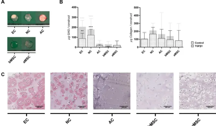
Matrix production was quantiied by GAG and collagen content of alginate beads during in vitro culture. Without TGFβ1 very little GAG was formed in vitro. Addition of TGFβ1 enhanced GAG production and after 5 weeks of culture ACs deposited signiicantly more GAGs (p < 0.01). When GAG content was adjusted to the amount of DNA, similar but more pronounced differences were observed (ACs produced most GAGs: 60.89 ± 53.04 μg GAG / μg DNA; p < 0.001). GAG content per alginate bead in constructs containing bMSCs, ECs, NCs and aMSCs was not signiicantly different, although a large donor variation was observed. Also with large variation between
donors, aMSCs performed worse. The amount of collagen deposited was just above background for all cell sources after 5 weeks of chondrogenic differentiation, being on average 1.53 ± 1.84 μg collagen per alginate bead (Fig. 1C).
Chondrogenic differentiation in vivo
To study the stability and quality of TE cartilage in vivo, alginate constructs were irst differentiated in vitro for 5 weeks and subsequently implanted subcutaneously on the dorsal side of athymic mice for an additional 8 weeks of culture. Constructs seeded with ECs or NCs, pre-cultured with TGFβ1, had a macroscopically white opaque appearance and were relatively strong on handling. Conversely, constructs pre-cultured without TGFβ1 or constructs encapsulating ACs, bMSCs or aMSCs, were fragile and also did not resemble cartilaginous tissue macroscopically (Figs. 2A and 3A).
Prior to implantation, constructs pre-cultured without TGFβ1 produced very little GAG in vitro, being on average 1.10 ± 1.20 µg GAG per construct. After in vivo implantation, these constructs greatly increased their production of matrix components, although they did not reach levels which equalled the matrix content found in constructs cultured with TGFβ1(Fig. 2B). After subcutaneous implantation preceded by chondrogenic culture (with TGFβ1), ACs, bMSCs and aMSCs retained their GAG content but did not further increase it. In contrast, ECs and NCs signiicantly enhanced matrix formation in vivo (EC 7.26-fold and NC 2.86-fold ; both p < 0.001) leading to a superior GAG deposition after implantation compared to the other cell sources (both p < 0.001). These results were further conirmed by Thionin-staining (data not shown). Total collagen deposition was hugely increased after implantation and no signiicant differences could be detected between the different cell sources (Fig. 3B). Constructs containing ACs, bMSCs or aMSCs exhibited a very weak staining for cartilage-speciic collagen type II (Fig. 3C), which was in contrast to the overall production of collagens (Fig. 3B), thus indicating that other collagens were also produced (e.g. collagen type I or type III). The cartilage matrix of constructs containing ECs and NCs showed a strong staining for collagen type II, although
PD/D Statistically signiicantly different from
EC 0.18 ± 0.04 NC (p = 0.015) ; AC (p = 0.008)
NC 0.32 ± 0.07 EC (p = 0.015) ; AC (p < 0.001) ; aMSC (p = 0.013)
AC 0.10 ± 0.05 EC (p = 0.008) ; NC (p < 0.001) ; bMSC (p = 0.001)
bMSC 0.25 ± 0.09 AC (p = 0.001)
aMSC 0.16 ± 0.04 NC (p = 0.013)
Table 3. Population doubling time of different cell types over four passages.
dissimilar distributions of collagen type-II ibres within the cartilage matrices were apparent. The semi-quantitative histological scores of constructs containing ECs or NCs were signiicantly better than the scores of the other cell sources (Fig. 3C).
The presence of elastin was determined to evaluate differentiation into elastic cartilage. There was no elastin detectable in any of the constructs with an elastin immunostaining after 5 weeks of in vitro cell culture (data not shown). After subcutaneous implantation, elastin was only present in alginate constructs containing ECs, and predominantly found in constructs which were pre-cultured with TGFβ1. Most elastin was located around the cell (Fig. 3D).
To ensure that these cartilage constructs were of human origin, a human-speciic vimentin stain was used on histological sections. It conirmed that the cartilage constructs were indeed of human origin (Fig. 3E), while the surrounding ibrous tissue was not (data not shown).
Cartilage stability
Hypertrophic differentiation is an unwanted phenomenon in cartilage regeneration, resulting in cartilage that can
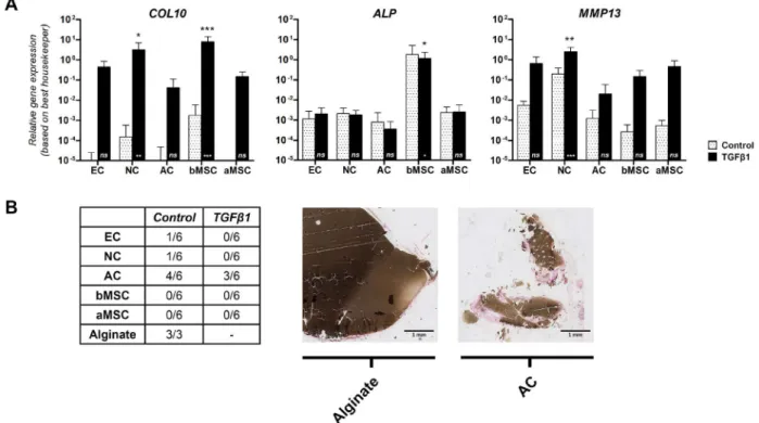
remodel into bone when implanted in vivo. To evaluate hypertrophy in vitro, we have studied gene expression of a panel of three hypertrophic markers during 5 weeks of cell culture (i.e. COL10, ALP and MMP13; Fig. 4A). Cultured with TGFβ1, COL10 expression was highest in NCs (p < 0.05) and bMSCs (p < 0.001), and was minimally expressed by ACs. MMP13 was expressed by all cells and signiicantly highest in NCs. ALP was signiicantly higher in constructs with bMSCs compared to the other cell sources (p < 0.05). In addition, constructs with bMSCs already expressed high COL10 and ALP after 2 weeks of culture, indicating early hypertrophic differentiation (data not shown).
Fig. 4. Stability of cartilage constructs in vitro and in vivo. (A) Hypertrophy in vitro. Relative gene expression levels of COL10, ALP and MMP13 were examined after 5 weeks of culture and corrected for the best housekeeper index. Hypertrophic genes were most highly expressed by bMSCs and NCs. Data are shown as mean ± SD. For statistical evaluation, a mixed model was used. *, ** or *** indicates p-values less than 0.05, 0.01 or 0.001, respectively, compared to the control condition (asterisk is shown in the bar) or compared to the other cell sources (asterisk is shown above the bar). EC, NC and AC: n = 4 donors each; bMSC, aMSC: n = 6 donors each. (B) Mineralisation in vivo. Von Kossa staining was used to evaluate construct calciication in vivo. MSCs did not calcify the construct in vivo. Non-seeded alginate andconstructs encapsulating ACs did calcify. For each cell source we had a total of 6 alginate constructs: duplicate samples of 3 different donors. EC = ear chondrocytes; NC = nasal chondrocytes; AC = articular chondrocytes; bMSC= bone marrow-derived mesenchymal stem cells; aMSC = adipose tissue-derived mesenchymal stem cells.
Cartilage structure and functionality
The elastic modulus of constructs was low in vitro, irrespective of the cell source used, being on average 7.42 ± 2.10 kPa. However, after subcutaneous implantation, mechanical properties improved in constructs containing either ECs (23.68 ± 10.20) or NCs (55.12 ± 59.25), but was not perceived in constructs containing ACs, bMSCs or aMSCs (Fig. 5). Since tissue calciication misrepresents the biomechanical properties of the cartilage matrix; we excluded calciied cartilage constructs from further analyses.
To determine whether the mechanical properties were enhanced by the deposition of matrix components, a multiple regression analyses was performed for all cell sources separately using GAG and collagen deposition as independent variables. Only for constructs containing NCs, matrix components signiicantly associated with the biomechanical functionality of the constructs (R2 = 0.477, F = 4.558, p = 0.039). For these constructs, only GAG deposition associated signiicantly with the biomechanical properties of the cartilage constructs independently (GAG: β = 0.689, p = 0.013; collagen: β = 0.044, p = 0.851).
Discussion
For successful regeneration of cartilage tissue, selection of the most appropriate cell source is crucial. This study demonstrates that cartilage matrix formation and functionality is cell source dependent; articular chondrocytes (ACs) possess the highest chondrogenic capacity in vitro, while ear chondrocytes (ECs) and nasal chondrocytes (NCs) are most potent for cartilage regeneration after subcutaneous implantation in vivo.
To date, we and others have evaluated the use of chondrocytes and mesenchymal stem cells (MSCs) from several anatomical locations for their applicability in cartilage regenerative medicine (Aizah et al., 2007; Asawa et al., 2009; Chung et al., 2008; Hellingman et al., 2011; Henderson et al., 2007; Isogai et al., 2006; Johnson et al., 2004; Kaienah et al., 2002; Karlsson et al., 2007; Kusuhara et al., 2009; Lohan et al., 2011; Malicev et al., 2009; Naumann et al., 2004; Panossian et al., 2001; Sakaguchi et al., 2005; Seda Tigli et al., 2009; Tay et al., 2004; van Osch et al., 2004; Vinardell et al., 2012; Xu et al., 2004; Yoshimura et al., 2007; Zhang and Spector, 2009). However, these studies often used non-expanded cells isolated from animals. Moreover, a detailed direct comparison between various chondrocyte and MSC- sources was lacking. Therefore, this study is the irst to systematically compare the quality and tissue stability of engineered cartilage constructs produced by culture-expanded ACs, NCs, ECs, bMSCs and aMSCs of human origin.
We have used culture-expanded human cells, to closely simulate the clinical situation. For clinical application, the use of autogeneic cells is favourable, since these cells do not elicit a tissue rejection response. However, it has been dificult to obtain appropriate numbers of cells, as donor
gel for cartilage regeneration, although future work needs to clarify which approach is required to exclude alginate calciication after in vivo implantation.
In addition to a 3D-culture environment, specific growth and differentiation factors will help to regain and induce a chondrocyte-like phenotype. In vitro, culture-expanded cells of all sources studied failed to differentiate towards the chondrogenic lineage in the absence of TGFβ1, as assessed by an almost negligible deposition of GAGs and the inferior expression of both ACAN and COL2A1 in alginate constructs. The presence of TGFβ1 induced chondrogenic differentiation in vitro, where ACs exhibit a superior chondrogenic capacity in vitro, compared to the other cell sources. The beneicial effect of culturing with TGFβ1 during in vivo chondrogenesis was present, although less obvious. Constructs cultured without TGFβ1 increased their production of matrix components after in vivo implantation, but were not able to reach levels found in constructs cultured with TGFβ1.
Even after four passages of culture expansion, chondrocytes demonstrated some clear subtype speciic differences. Firstly, ACs possessed the highest chondrogenic capacity in vitro, but were not able to further increase their cartilage matrix in vivo. The inability of ACs to promote cartilage formation in vivo may be due to the lack of mechanical loading or growth factor stimulation after subcutaneous implantation, which may have led to a loss of chondrogenic capacity. ACs, different from the other cell sources, are exposed to mechanical loading within native articular cartilage and unloading is known to induce GAG-release from the cartilage matrix and to reduce cell proliferation and GAG synthesis within the matrix (Tomiya et al., 2009). Secondly, chondrocytes from ear cartilage were able to form an elastin network after subcutaneous implantation in vivo. Elastin was predominantly found in constructs which were cultured with TGFβ1. In vitro culture did not demonstrate elastin deposition at all, which was in accordance with our previous work (Hellingman et al., 2011). The capability of culture-expanded ECs to produce elastin in vivo suggests that these cells retain their capability to form an elastic cartilage matrix. Both indings – the inability of ACs to promote cartilage formation in vivo without mechanical loading and the ability of cultured expanded ECs to produce elastin – indicate that both cell types preserved their subtype speciic phenotype after culture expansion, conirming our previous study where gene expression proiles of culture-expanded NCs and ECs displayed clear differences that were related to their developmental origin (Hellingman et al., 2011).
Besides chondrocytes, MSCs have been demonstrated to be an attractive cell source for cartilage TE (Barry et al., 2001; Caplan, 1991; Johnstone et al., 1998; Pittenger et al., 1999). Although bone marrow offers the most common source of MSCs, adipose tissue has been proven to be an attractive alternative in respect to the abundant and easily accessible pool of MSCs (Zuk et al., 2002; Zuk et al., 2001). We have demonstrated that both bMSCs and aMSCs underwent chondrogenic differentiation in vitro and in vivo, although matrix production was less than in constructs containing chondrocytes (ACs, NCs or ECs).
Constructs containing bMSCs had a higher chondrogenic potential than aMSCs, demonstrated by an increased ACAN and COL2A1 gene-expression and an improved GAG deposition. With the exception of a few studies (De Ugarte et al., 2003; Izadpanah et al., 2006; Kern et al., 2006; Lee et al., 2004), this conirms other studies (Aizah et al., 2007; Huang et al., 2005; Im et al., 2005; Liu et al., 2007; Mehlhorn et al., 2006; Noel et al., 2008; Rebelatto et al., 2008; Rider et al., 2008; Sakaguchi et al., 2005; Segawa et al., 2009; Winter et al., 2003). Nevertheless, the assumptions that MSCs are fundamentally less chondrogenic than chondrocytes, and that bMSCs are more in favour for cartilage regeneration than aMSCs, seems unjustiied. It appears that cell culture conditions for both bMSCs and aMSCs remain to be improved. For instance, it was found that another member of the TGFβ-superfamily, bone morphogenetic protein 6, is obligatory to improve chondrogenic differentiation in aMSCs (Estes et al., 2010).
Finally, in order to understand how the distribution and composition of matrix components resulted in a mechanically functional cartilage matrix, the compositional-biomechanical relationship of the cartilage constructs was evaluated. After in vivo implantation, mechanical properties increased in constructs containing ECs and NCs. Only for constructs containing NCs, matrix components were signiicantly correlated to their biomechanical functionality. It is already known that GAGs and collagens, the main components of the ECM, are both associated with the biomechanical properties of native cartilage: (1) the negatively charged GAGs provide an osmotic pressure within the tissue; and (2) the architecture of the collagen network captures the GAGs and prevents them from leaking out of the tissue (Han et al., 2011). In contrast, the elastic ibre network in constructs containing ECs might have inluenced the biomechanical properties in vivo as well, although the exact contribution of elastin to mechanical functionality is not yet fully understood. Besides the existence of matrix components, the quality of the matrix is not only determined by the amount of matrix components deposited, but also inluenced by the number of cross-links between matrix molecules (i.e. collagen cross-links) (Bastiaansen-Jenniskens et al., 2008a). The distribution of matrix components in the ECM was clearly different between cell sources: ECs deposited most matrix components pericellularly, whereas NCs deposited these matrix components homogenously throughout their matrix, which was clearly visible on the immunohistochemical collagen type II staining. It is well known that a heterogeneously distributed matrix alters the biomechanical properties of the matrix, as the physical properties are determined by the weakest point in the matrix (Mow and Guo, 2002).
density of 4 x 10⁶ cells per mL of alginate, since the size of our experimental set-up did not enable higher densities. Secondly, as mentioned before, we have culture-expanded all cells until passage four to obtain a suficient number of cells. In order to be able to compare all different cell sources, we have used standardised protocols for the culture expansion of both chondrocytes and MSCs. Differences in expansion rates between the different cells were obvious. While culture expansion is associated with chondrocyte dedifferentiation and replicative cell senescence, the enforcement of population doublings instead of culture passages might have been more appropriate, since population doublings more accurately relect cell growth and thus cell aging. However, since nasal chondrocytes had most doublings in four passages but still produced most cartilage, a direct link between doublings and cartilage formation seems unlikely. How all the parameters such as number of doublings, expansion speed, initial seeding density, growth factors present in the medium or produced by the cells themselves exactly determine dedifferentiation and possible loss of chondrogenic capacity during monolayer expansion remains to be elucidated. Finally, we have demonstrated large donor variation in constructs containing ACs or bMSCs. Nevertheless these differences were not based on a donor-age effect nor explained by the use of healthy versus diseased ACs. Moreover, donors for nasal cartilage appeared younger than other sources. Although there is a possibility that the donor age has inluenced the general outcome of our study, improved chondrogenic and proliferative capacity of nasal chondrocytes was also stated by others in the literature (Kaienah et al., 2002; Naumann et al., 2004; Tay et al., 2004).
In summary, we have demonstrated that cartilage matrix formation and functionality are cell source dependent. Articular chondrocytes possess the highest chondrogenic capacity in vitro, while ear and nasal chondrocytes are most potent for cartilage regeneration after subcutaneous implantation, making ear and nasal chondrocytes attractive cell sources for future cell-based cartilage repair. Only for constructs containing nasal chondrocytes, GAG and collagen content were associated with biomechanical functionality of the constructs, indicating the differences in matrix component assembly by different cell sources. The inability of articular chondrocytes to increase cartilage matrix in vivo may be due to a loss of chondrogenic capacity in the absence of mechanical loading or growth factor stimulation. Although mesenchymal stem cells are considered as a promising cell source for the reconstruction of cartilage defects, it appears that improvements in cell culture conditions for both bone-marrow-derived and adipose tissue-derived mesenchymal stem cells are needed.
Acknowledgments
The authors thank the Departments of Orthopaedic Surgery, Plastic and Reconstructive Surgery, and Otorhinolaryngology at Erasmus MC for their assistance in obtaining ear, nasal and articular cartilage as well as bone marrow and adipose tissue, and CellMed (Alzenau,
Germany) for providing the clinical grade alginate. The study was performed within the framework of EuroNanoMed (EAREG-406340-131009/1) and funded by SenterNovem (ENM09001). We wish to confirm that there are no known conlicts of interest associated with this publication and there has been no signiicant inancial support for this work that could have inluenced its outcome.
References
Aizah H, Yang Z, Hui JH, Ouyang HW, Lee EH (2007) A comparison between the chondrogenic potential of human bone marrow stem cells (BMSCs) and adipose-derived stem cells (ADSCs) taken from the same donors. Tissue Eng 13: 659-666.
Alsberg E, Anderson KW, Albeiruti A, Franceschi RT, Mooney DJ (2001) Cell-interactive alginate hydrogels for bone tissue engineering. J Dent Res 80: 2025-2029.
Asawa Y, Ogasawara T, Takahashi T, Yamaoka H, Nishizawa S, Matsudaira K, Mori Y, Takato T, Hoshi K (2009) Aptitude of auricular and nasoseptal chondrocytes cultured under a monolayer or three-dimensional condition for cartilage tissue engineering. Tissue Eng Part A 15: 1109-1118.
Barry F, Boynton RE, Liu B, Murphy JM (2001) Chondrogenic differentiation of mesenchymal stem cells from bone marrow: differentiation-dependent gene expression of matrix components. Exp Cell Res 268: 189-200.
Bastiaansen-Jenniskens YM, Koevoet W, De Bart AC, Zuurmond AM, Bank RA, Verhaar JA, DeGroot J, van Osch GJ (2008a) TGFbeta affects collagen cross-linking independent of chondrocyte phenotype but strongly depending on physical environment. Tissue Eng Part A 14: 1059-1066.
Caplan AI (1991) Mesenchymal stem cells. J Orthop Res 9: 641-650.
Chung C, Erickson IE, Mauck RL, Burdick JA (2008) Differential behavior of auricular and articular chondrocytes in hyaluronic acid hydrogels. Tissue Eng Part A 14: 1121-1131.
Creemers LB, Jansen DC, van Veen-Reurings A, van den Bos T, Everts V (1997) Microassay for the assessment of low levels of hydroxyproline. BioTechniques 22: 656-658.
De Ugarte DA, Morizono K, Elbarbary A, Alfonso Z, Zuk PA, Zhu M, Dragoo JL, Ashjian P, Thomas B, Benhaim P, Chen I, Fraser J, Hedrick MH (2003) Comparison of multi-lineage cells from human adipose tissue and bone marrow. Cells Tissues Organs 174: 101-109.
Enobakhare BO, Bader DL, Lee DA (1996) Quantiication of sulfated glycosaminoglycans in chondrocyte/alginate cultures, by use of 1,9-dimethylmethylene blue. Anal Biochem 243: 189-191.
JA, Weinans H, van Osch GJ (2009) Chondrogenic priming of human bone marrow stromal cells: a better route to bone repair? Tissue Eng Part C Methods 15: 285-295.
Grogan SP, Barbero A, Winkelmann V, Rieser F, Fitzsimmons JS, O’Driscoll S, Martin I, Mainil-Varlet P (2006) Visual histological grading system for the evaluation of in vitro-generated neocartilage. Tissue Eng 12: 2141-2149.
Han EH, Chen SS, Klisch SM, Sah RL (2011) Contribution of proteoglycan osmotic swelling pressure to the compressive properties of articular cartilage. Biophys J 101: 916-924.
Hauselmann HJ, Aydelotte MB, Schumacher BL, Kuettner KE, Gitelis SH, Thonar EJ (1992) Synthesis and turnover of proteoglycans by human and bovine adult articular chondrocytes cultured in alginate beads. Matrix 12: 116-129.
Hellingman CA, Verwiel ET, Slagt I, Koevoet W, Poublon RM, Nolst-Trenite GJ, Baatenburg de Jong RJ, Jahr H, van Osch GJ (2011) Differences in cartilage-forming capacity of expanded human chondrocytes from ear and nose and their gene expression proiles. Cell Transplant 20: 925-940.
Hellingman CA, Koevoet W, van Osch GJ (2012) Can one generate stable hyaline cartilage from adult mesenchymal stem cells? A developmental approach. J Tissue Eng Regen Med 6: e1-e11.
Henderson JH, Welter JF, Mansour JM, Niyibizi C, Caplan AI, Dennis JE (2007) Cartilage tissue engineering for laryngotracheal reconstruction: comparison of chondrocytes from three anatomic locations in the rabbit. Tissue Eng 13: 843-853.
Hierck BP, Iperen LV, Gittenberger-De Groot AC, Poelmann RE (1994) Modiied indirect immunodetection allows study of murine tissue with mouse monoclonal antibodies. J Histochem Cytochem 42: 1499-1502. Huang JI, Kazmi N, Durbhakula MM, Hering TM, Yoo JU, Johnstone B (2005) Chondrogenic potential of progenitor cells derived from human bone marrow and adipose tissue: a patient-matched comparison. J Orthop Res 23: 1383-1389.
Im GI, Shin YW, Lee KB (2005) Do adipose tissue-derived mesenchymal stem cells have the same osteogenic and chondrogenic potential as bone marrow-derived cells? Osteoarthritis Cartilage 13: 845-853.
Isogai N, Kusuhara H, Ikada Y, Ohtani H, Jacquet R, Hillyer J, Lowder E, Landis WJ (2006) Comparison of different chondrocytes for use in tissue engineering of cartilage model structures. Tissue Eng 12: 691-703. Izadpanah R, Trygg C, Patel B, Kriedt C, Dufour J, Gimble JM, Bunnell BA (2006) Biologic properties of mesenchymal stem cells derived from bone marrow and adipose tissue. J Cell Biochem 99: 1285-1297.
Johnson TS, Xu JW, Zaporojan VV, Mesa JM, Weinand C, Randolph MA, Bonassar LJ, Winograd JM, Yaremchuk MJ (2004) Integrative repair of cartilage with articular and nonarticular chondrocytes. Tissue Eng 10: 1308-1315. Johnstone B, Hering TM, Caplan AI, Goldberg VM, Yoo JU (1998) In vitro chondrogenesis of bone marrow-derived mesenchymal progenitor cells. Exp Cell Res 238: 265-272.
Kaienah W, Jakob M, Demarteau O, Frazer A, Barker MD, Martin I, Hollander AP (2002) Three-dimensional tissue engineering of hyaline cartilage: comparison of adult nasal and articular chondrocytes. Tissue Eng 8: 817-826. Kanczler JM, Oreffo RO (2008) Osteogenesis and angiogenesis: the potential for engineering bone. Eur Cell Mater 15: 100-114.
Karlsson C, Brantsing C, Svensson T, Brisby H, Asp J, Tallheden T, Lindahl A (2007) Differentiation of human mesenchymal stem cells and articular chondrocytes: analysis of chondrogenic potential and expression pattern of differentiation-related transcription factors. J Orthop Res 25: 152-163.
Kern S, Eichler H, Stoeve J, Kluter H, Bieback K (2006) Comparative analysis of mesenchymal stem cells from bone marrow, umbilical cord blood, or adipose tissue. Stem Cells 24: 1294-1301.
Kusuhara H, Isogai N, Enjo M, Otani H, Ikada Y, Jacquet R, Lowder E, Landis WJ (2009) Tissue engineering a model for the human ear: assessment of size, shape, morphology, and gene expression following seeding of different chondrocytes. Wound Repair Regen 17: 136-146. Lee RH, Kim B, Choi I, Kim H, Choi HS, Suh K, Bae YC, Jung JS (2004) Characterization and expression analysis of mesenchymal stem cells from human bone marrow and adipose tissue. Cell Physiol Biochem 14: 311-324.
Lee CS, Moyer HR, Gittens RA, Williams JK, Boskey AL, Boyan BD, Schwartz Z (2010) Regulating in vivo calciication of alginate microbeads. Biomaterials 31: 4926-4934.
Liu TM, Martina M, Hutmacher DW, Hui JH, Lee EH, Lim B (2007) Identiication of common pathways mediating differentiation of bone marrow- and adipose tissue-derived human mesenchymal stem cells into three mesenchymal lineages. Stem Cells 25: 750-760.
Lohan A, Marzahn U, El Sayed K, Haisch A, Kohl B, Muller RD, Ertel W, Schulze-Tanzil G, John T (2011) In vitro and in vivo neo-cartilage formation by heterotopic chondrocytes seeded on PGA scaffolds. Histochem Cell Biol 136: 57-69.
Malicev E, Kregar-Velikonja N, Barlic A, Alibegovic A, Drobnic M (2009) Comparison of articular and auricular cartilage as a cell source for the autologous chondrocyte implantation. J Orthop Res 27: 943-948.
Mehlhorn AT, Niemeyer P, Kaiser S, Finkenzeller G, Stark GB, Sudkamp NP, Schmal H (2006) Differential expression pattern of extracellular matrix molecules during chondrogenesis of mesenchymal stem cells from bone marrow and adipose tissue. Tissue Eng 12: 2853-2862.
Mow VC, Guo XE (2002) Mechano-electrochemical properties of articular cartilage: their inhomogeneities and anisotropies. Annu Rev Biomed Eng 4: 175-209.
Muir H (1995) The chondrocyte, architect of cartilage. Biomechanics, structure, function and molecular biology of cartilage matrix macromolecules. Bioessays 17: 1039-1048.
three-dimensional in vitro macroaggregate culture system. Tissue Eng 10: 1695-1706.
Noel D, Caton D, Roche S, Bony C, Lehmann S, Casteilla L, Jorgensen C, Cousin B (2008) Cell speciic differences between human adipose-derived and mesenchymal-stromal cells despite similar differentiation potentials. Exp Cell Res 314: 1575-1584.
Panossian A, Ashiku S, Kirchhoff CH, Randolph MA, Yaremchuk MJ (2001) Effects of cell concentration and growth period on articular and ear chondrocyte transplants for tissue engineering. Plast Reconstr Surg 108: 392-402. Pfafl MW, Tichopad A, Prgomet C, Neuvians TP (2004) Determination of stable housekeeping genes, differentially regulated target genes and sample integrity: BestKeeper – Excel-based tool using pair-wise correlations. Biotechnol Lett 26: 509-515.
Pittenger MF, Mackay AM, Beck SC, Jaiswal RK, Douglas R, Mosca JD, Moorman MA, Simonetti DW, Craig S, Marshak DR (1999) Multilineage potential of adult human mesenchymal stem cells. Science 284: 143-147. Puelacher WC, Kim SW, Vacanti JP, Schloo B, Mooney D, Vacanti CA (1994) Tissue-engineered growth of cartilage: the effect of varying the concentration of chondrocytes seeded onto synthetic polymer matrices. Int J Oral Maxillofac Surg 23: 49-53.
Rebelatto CK, Aguiar AM, Moretao MP, Senegaglia AC, Hansen P, Barchiki F, Oliveira J, Martins J, Kuligovski C, Mansur F, Christofis A, Amaral VF, Brofman PS, Goldenberg S, Nakao LS, Correa A (2008) Dissimilar differentiation of mesenchymal stem cells from bone marrow, umbilical cord blood, and adipose tissue. Exp Biol Med 233: 901-913.
Rider DA, Dombrowski C, Sawyer AA, Ng GH, Leong D, Hutmacher DW, Nurcombe V, Cool SM (2008) Autocrine ibroblast growth factor 2 increases the multipotentiality of human adipose-derived mesenchymal stem cells. Stem Cells 26: 1598-1608.
Sakaguchi Y, Sekiya I, Yagishita K, Muneta T (2005) Comparison of human stem cells derived from various mesenchymal tissues: superiority of synovium as a cell source. Arthritis Rheum 52: 2521-2529.
Seda Tigli R, Ghosh S, Laha MM, Shevde NK, Daheron L, Gimble J, Gumusderelioglu M, Kaplan DL (2009) Comparative chondrogenesis of human cell sources in 3D scaffolds. J Tissue Eng Regen Med 3: 348-360.
Segawa Y, Muneta T, Makino H, Nimura A, Mochizuki T, Ju YJ, Ezura Y, Umezawa A, Sekiya I (2009) Mesenchymal stem cells derived from synovium, meniscus, anterior cruciate ligament, and articular chondrocytes share similar gene expression proiles. J Orthop Res 27: 435-441. Stoddart MJ, Grad S, Eglin D, Alini M (2009) Cells and biomaterials in cartilage tissue engineering. Regen Med 4: 81-98.
Tay AG, Farhadi J, Suetterlin R, Pierer G, Heberer M, Martin I (2004) Cell yield, proliferation, and postexpansion differentiation capacity of human ear, nasal, and rib chondrocytes. Tissue Eng 10: 762-770.
Tomiya M, Fujikawa K, Ichimura S, Kikuchi T, Yoshihara Y, Nemoto K (2009) Skeletal unloading induces a full-thickness patellar cartilage defect with increase of
urinary collagen II CTx degradation marker in growing rats. Bone 44: 295-305.
van Osch GJVM, Mandl EW, Jahr H, Koevoet W, Nolst-Trenite G, Verhaar JA (2004) Considerations on the use of ear chondrocytes as donor chondrocytes for cartilage tissue engineering. Biorheology 41: 411-421.
Vinardell T, Sheehy EJ, Buckley CT, Kelly DJ (2012) A comparison of the functionality and in vivo phenotypic stability of cartilaginous tissues engineered from different stem cell sources. Tissue Eng Part A 18: 1161-1170. Winter A, Breit S, Parsch D, Benz K, Steck E, Hauner H, Weber RM, Ewerbeck V, Richter W (2003) Cartilage-like gene expression in differentiated human stem cell spheroids: a comparison of bone marrow-derived and adipose tissue-derived stromal cells. Arthritis Rheum 48: 418-429.
Wong M, Siegrist M, Wang X, Hunziker E (2001) Development of mechanically stable alginate/chondrocyte constructs: effects of guluronic acid content and matrix synthesis. J Orthop Res 19: 493-499.
Xu JW, Zaporojan V, Peretti GM, Roses RE, Morse KB, Roy AK, Mesa JM, Randolph MA, Bonassar LJ, Yaremchuk MJ (2004) Injectable tissue-engineered cartilage with different chondrocyte sources. Plast Reconstr Surg 113: 1361-1371.
Yaeger PC, Masi TL, de Ortiz JL, Binette F, Tubo R, McPherson JM (1997) Synergistic action of transforming growth factor-beta and insulin-like growth factor-I induces expression of type II collagen and aggrecan genes in adult human articular chondrocytes. Exp Cell Res 237: 318-325.
Yoshimura H, Muneta T, Nimura A, Yokoyama A, Koga H, Sekiya I (2007) Comparison of rat mesenchymal stem cells derived from bone marrow, synovium, periosteum, adipose tissue, and muscle. Cell Tissue Res 327: 449-462.
Zhang L, Spector M (2009) Comparison of three types of chondrocytes in collagen scaffolds for cartilage tissue engineering. Biomed Mater 4: 045012.
Zuk PA, Zhu M, Ashjian P, De Ugarte DA, Huang JI, Mizuno H, Alfonso ZC, Fraser JK, Benhaim P, Hedrick MH (2002) Human adipose tissue is a source of multipotent stem cells. Mol Biol Cell 13: 4279-4295.
Zuk PA, Zhu M, Mizuno H, Huang J, Futrell JW, Katz AJ, Benhaim P, Lorenz HP, Hedrick MH (2001) Multilineage cells from human adipose tissue: implications for cell-based therapies. Tissue Eng 7: 211-228.
Discussion with Reviewers
the absence of bone formation 8 weeks after subcutaneous implantation.
We agree with the reviewer that the biomechanical properties of the alginate used were low and inferior to that of native cartilages (e.g. ear cartilage: Eeq 3.3 ± 1.3 MPa (Nimeskern et al., 2013); articular cartilage: Eeq 10.9 ± 5.0 MPa (Richard et al., 2013)). However, alginate was chosen since it is being used as a cell carrier for clinical applications in cell-based cartilage repair (Almqvist et al., 2009; Selmi et al., 2008). Injected into a mechanically stable scaffold, alginate enables a homogeneous cell distribution into a mechanically stable environment. Moreover, it prevents cells from loating out while permitting nutrient diffusion and oxygen transfer to the encapsulated cells and thereby promotes the synthesis of cartilage-speciic matrix components, such as GAGs and collagen type II (Hauselmann et al., 1992). Therefore, in our opinion, alginate injected into a mechanically stable scaffold could be an excellent therapy for cell-based cartilage repair. By the time alginate is completely gone (which will depend on the type of alginate and can take months), we hope the cartilage formed is mature and stable and will not undergo endochondral ossiication anymore. However, this remains to be evaluated with long term in vivo experiments.
Reviewer I: What does it mean when large areas are negative for vimentin in Fig. 3E? Are some of the cells of host origin or is vimentin expression very variable? Authors: Vimentins are class-III intermediate ilaments and primarily expressed in cells of mesenchymal origin. Vimentins are attached to the nucleus, endoplasmic reticulum, and mitochondria. Strong positivity for the vimentin antibody is therefore demonstrated by a brown staining of the cell nucleus and cytoplasm. In our positive control of human cartilage, all cells were positive. The large area negative for vimentin is in our opinion the extracellular matrix, but we agree with the reviewer that some background stain was visible.
Reviewer I: The influence of alginate on construct ‘ossiication’ should be questioned.
Authors: We have demonstrated that cell-free alginate constructs did mineralise in vivo, which was an unexpected finding and obviously not related to hypertrophic differentiation. It seemed that less metabolically active (meaning the colour of the culture medium was neutral to alkaline) alginate constructs were likely to mineralise in vivo. This is obviously a typical characteristic of alginate itself and it very valuable to know since alginate is actually being used in (experimental) clinical applications. Furthermore, cell-alginate constructs expressing hypertrophic genes in vitro, never calciied nor formed bone in vivo. This is a very promising outcome and indicates a possible beneit for the use of alginate as a cell-carrier for cartilage reconstruction.
Reviewer II: Unlike previous studies from the authors using MSCs seeded onto collagen scaffolds or in pellets, MSC-alginate constructs did not undergo endochondral
ossiication in vivo. Can the authors speculate as to why they believe this to be the case?
Authors: In our opinion, endochondral ossification requires 1) proper chondrogenic differentiation, 2) hypertrophic differentiation, and 3) a matrix that allows vessel ingrowth. We demonstrated that after chondrogenic induction (with TGFβ1), alginate constructs containing bone marrow-derived MSCs (bMSCs) chondrogenically differentiated and expressed hypertrophic markers in vitro, but did not form bone after subcutaneous implantation. Therefore, we believe that alginate did not necessarily inhibit hypertrophic differentiation of bMSCs, but prevented vessel ingrowth and subsequent bone formation. Vessel ingrowth is extremely important for endochondral ossiication. Angiogenesis in alginate is unlikely since vessels are unable to invade it. Although, alginate and other hydrogels have been used for research on bone regeneration (Park, 2011), modiication of hydrogels with bioactive molecules (e.g. RGDs) seemed important allowing cell attachment and thereby vessel ingrowth.
To conclude, we believe that alginate differs from pellets or collagen scaffolds by preventing vascular ingrowth, thus resulting in the absence of bone formation 8 weeks after subcutaneous implantation.
Reviewer II: Why do the authors believe that they saw no difference in tissue mechanical properties in vitro despite differences in ECM accumulation between the different groups?
Authors: Extracellular matrix formation, and in particular collagen network formation, is a slow process in vitro. We have shown before that the mechanical properties of the matrix are not only determined by the amount of matrix components deposited, but also influenced by the cross-links between matrix molecules (i.e. collagen cross-links) (Bastiaansen-Jenniskens et al., 2008b). Moreover, the addition of TGFβ to chondrocytes has previously been demonstrated to result in less collagen cross-links in alginate, although it induced collagen cross-links in monolayer (Bastiaansen-Jenniskens et al., 2008a; Jenniskens et al., 2006). The optimal use of TGFβ, and maybe the removal of it for a certain period before implantation, will be an interesting topic to consider in future research.
Reviewer III: The major issue I have with this study is how meaningful it is to culture and ‘re-differentiate’ articular chondrocytes as if they were a chondro-progenitor population. They’ve already negotiated the chondrogenesis process.
in vitro. To be able to compare both chondrocytes and MSCs, we decided to expose all cell sources to an identical chondrogenic culture medium in vitro, that contained TGFβ1. Our experiments nicely conirmed that cartilage formation was hugely decreased for all cell sources in a subcutaneous environment when cultured without TGFβ1.
Reviewer III: The other interesting aspects of the study are the disparities between the matrix protein data and the modulus values, and between the hypertrophic marker expression and construct mineralisation. Unfortunately these disparities undermine the overall value of the manuscript.
Authors: There is a good possibility that the biochemical data are not directly correlated to the biomechanical data in these developing cartilage constructs, since the quality of the matrix is not only determined by the amount of matrix components deposited, but also influenced by the number of cross-links between matrix molecules (i.e. collagen cross-links) (Bastiaansen-Jenniskens et al., 2008b). Furthermore, we have performed a multiple regression analysis for all cell sources separately using only GAG and collagen deposition as independent variables. Presumably, other components such as the elastic ibre network in constructs containing ECs have inluence on the biomechanical properties in vivo as well, although the exact contribution of these components to mechanical functionality is not fully understood in the literature. We believe the lack of correlation between the hypertrophic differentiation and scaffold calciication is very interesting and important to realise. It teaches us some valuable things for future applications. Firstly, we have demonstrated that cell-free alginate constructs did mineralise in vivo, which was an unexpected inding and obviously not related to hypertrophic differentiation. It seemed that less metabolically active (meaning the colour of the culture medium was neutral to alkaline) alginate constructs were likely to mineralise in vivo. This is obviously a typical characteristic of alginate and it is very valuable to know because alginate is actually being using in (experimental) clinical applications. Furthermore, cell-alginate constructs expressing hypertrophic genes in vitro, never calciied nor formed bone in vivo. This is a very promising outcome and indicates a possible beneit for the use of alginate as a cell carrier for cartilage reconstruction.
Reviewer III: On a practical level, considering the doubling times, retention of chondrogenic potential, and the amount of donor tissue available, which source of
chondrogenic cells do you think is the superior choice for clinical applications?
Authors: At the end of our manuscript we conclude that articular chondrocytes possess the highest chondrogenic capacity in vitro, while ear and nasal chondrocytes are most potent for cartilage regeneration after subcutaneous implantation, making them attractive cell sources for future cell-based cartilage repair.
Additional References
Almqvist KF, Dhollander AAM, Verdonk PCM, Forsyth R, Verdonk R, Verbruggen G (2009) Treatment of cartilage defects in the knee using alginate beads containing human mature allogenic chondrocytes. Am J Sport Med 37: 1920-1929.
Bastiaansen-Jenniskens YM, Koevoet W, de Bart AC, van der Linden JC, Zuurmond AM, Weinans H, Verhaar JA, van Osch GJ, Degroot J (2008b) Contribution of collagen network features to functional properties of engineered cartilage. Osteoarthritis Cartilage 16: 359-366.
Jenniskens YM, Koevoet W, de Bart AC, Weinans H, Jahr H, Verhaar JA, DeGroot J, van Osch GJ (2006) Biochemical and functional modulation of the cartilage collagen network by IGF1, TGFbeta2 and FGF2. Osteoarthritis Cartilage 14: 1136-1146.
Marijnissen WJ, van Osch GJ, Aigner J, Verwoerd-Verhoef HL, Verhaar JA (2000) Tissue-engineered cartilage using serially passaged articular chondrocytes. Chondrocytes in alginate, combined in vivo with a synthetic (E210) or biologic biodegradable carrier (DBM). Biomaterials 21: 571-580.
Nimeskern L, Martinez Avila H, Sundberg J, Gatenholm P, Muller R, Stok KS (2013) Mechanical evaluation of bacterial nanocellulose as an implant material for ear cartilage replacement. J Mech Behav Biomed Mater 22: 12-21.