amongst Isolates of the
Cryptococcus neoformans
/
C.
gattii
Species Complex in Thailand
Sirada Kaocharoen1,2, Popchai Ngamskulrungroj2,3, Carolina Firacative2,4, Luciana Trilles2,5, Dumrongdej Piyabongkarn1, Wijit Banlunara6, Natteewan Poonwan7, Angkana Chaiprasert3, Wieland Meyer2, Ariya Chindamporn1*
1Mycology Laboratory, King Chulalongkorn Memorial Hospital, Faculty of Medicine, Chulalongkorn University, Bangkok, Thailand,2Molecular Mycology Research Laboratory, CIDM, Sydney Medical School - Westmead Hospital, The University of Sydney, Westmead Millennium Institute, Westmead, New South Wales, Australia, 3Department of Microbiology, Faculty of Medicine Siriraj Hospital, Mahidol University, Bangkok, Thailand,4Grupo de Microbiologı´a, Instituto Nacional de Salud, Bogota´, Colombia,5Laborato´rio de Micologia, Instituto de Pesquisa Clı´nica Evandro Chagas, Fundac¸a˜o Oswaldo Cruz, Rio de Janeiro, Brazil,6Department of Pathology, Faculty of Veterinary Sciences, Chulalongkorn University, Bangkok, Thailand,7Mycology Laboratory, National Institute of Health, Nonthaburi, Thailand
Abstract
To gain a more detailed picture of cryptococcosis in Thailand, a retrospective study of 498C. neoformans and C. gattii isolates has been conducted. Among these, 386, 83 and 29 strains were from clinical, environmental and veterinary sources, respectively. A total of 485C. neoformans and 13C. gattiistrains were studied. The majority of the strains (68.9%) were isolated from males (mean age of 37.97 years), 88.5% ofC. neoformansand only 37.5% ofC. gattiistrains were from HIV patients.URA5-RFLP and/or M13 PCR-fingerprinting analysis revealed that the majority of the isolates wereC. neoformans molecular type VNI regardless of their sources (94.8%; 94.6% of the clinical, 98.8% of the environmental and 86.2% of the veterinary isolates). In addition, the molecular types VNII (2.4%; 66.7% of the clinical and 33.3% of the veterinary isolates), VNIV (0.2%; 100% environmental isolate), VGI (0.2%; 100% clinical isolate) and VGII (2.4%; 100% clinical isolates) were found less frequently. Multilocus Sequence Type (MLST) analysis using the ISHAM consensus MLST scheme for theC. neoformans/ C. gattiispecies complex identified a total of 20 sequence types (ST) in Thailand combining current and previous data. The Thai isolates are an integrated part of the global cryptococcal population genetic structure, with ST30 forC. gattiiand ST82, ST83, ST137, ST141, ST172 and ST173 for C. neoformans being unique to Thailand. Most of the C. gattiiisolates were ST7 = VGIIb, which is identical to the less virulent minor Vancouver island outbreak genotype, indicating Thailand as a stepping stone in the global spread of this outbreak strain. The current study revealed a greater genetic diversity and a wider range of major molecular types being present amongst Thai cryptococcal isolates than previously reported.
Citation:Kaocharoen S, Ngamskulrungroj P, Firacative C, Trilles L, Piyabongkarn D, et al. (2013) Molecular Epidemiology Reveals Genetic Diversity amongst Isolates of theCryptococcus neoformans/C. gattiiSpecies Complex in Thailand. PLoS Negl Trop Dis 7(7): e2297. doi:10.1371/journal.pntd.0002297
Editor:Bodo Wanke, Fundac¸a˜o Oswaldo Cruz, Brazil, United States of America
ReceivedJanuary 15, 2013;AcceptedMay 23, 2013;PublishedJuly 4, 2013
Copyright:ß2013 Kaocharoen et al. This is an open-access article distributed under the terms of the Creative Commons Attribution License, which permits unrestricted use, distribution, and reproduction in any medium, provided the original author and source are credited.
Funding:S. Kaocharoen was supported by Chulalongkorn University Graduate Scholarship to commemorate the 72nd Anniversary of His Majesty King Bhumibol Adulyadej. This work was also supported by the 90th Anniversary of Chulalongkorn University Fund (Ratchadaphiseksomphot Endowment Fund)#2/2551 to A. Chindamporn and S. Kaocharoen, a New Researcher Scholarship of CSTS, NSTDA Ministry of Science and Technology, to P. Ngamskulrungroj and an NH&MRC project grants#352303 and APP1031943 to W. Meyer. The funders had no role in study design, data collection and analysis, decision to publish, or preparation of the manuscript.
Competing Interests:The authors have declared that no competing interests exist.
* E-mail: [email protected]
Introduction
The members of the Cryptococcus neoformans/C. gattii species complex are the causative agent of cryptococcosis, which is a systemic mycosis, in a wide range of animals and humans [1–4]. Inhalation of infectious propagules (basidiospores or blastoconidia) are proposed to be the source of the infection [1]. C. neoformans comprises two varieties and three serotypes:C. neoformansvar.grubii (serotype A),C. neoformansvar.neoformans(serotype D) and a hybrid (serotype AD), whereasC. gattiicomprises two serotypes, B and C [1,3].
Extensive surveys of the yeast have shown that the ecological niches of both species are different. C. neoformans has been associated worldwide with soil enriched with pigeon excreta and decaying wood [1,5,6]. On the contrary,C. gattiiwas until recently
thought to be geographically restricted to tropical and subtropical regions and thought to be related mainly to eucalyptus trees [7–9]. Further environmental studies in South America and Asia pointed out several species of tropical trees as the natural habitat ofC. gattii such as oiti (Licania tomentosa), almond trees (Terminalia cattapa), cassia (Cassia grandis), pottery tree (Ficus microcarpa) and Syzygium cumini [10–12]. A recent ongoing outbreak of C. gattii on Vancouver Island, Canada, a temperate area, indicated an environmental shift of this species [13]. Moreover, in contrast to previous known environmental sources of this species,C. gattiihas been found in association with a number of native tree species (Douglas fir, alder, maple and Garry oak) on Vancouver Island rather than with eucalypt trees [13].
species complex [14–17], providing more discriminatory power than conventional techniques [1]. Using M13-PCR fingerprinting, eight major molecular types have been established [18,19]. The major molecular types VNI/AFLP1 and VNII/AFLP1A corre-spond to C. neoformans var. grubii, serotype A; VNIII/AFLP2 corresponds to the AD hybrid; serotype AD, and VNIV/AFLP3 corresponds to C. neoformans var. neoformans, serotype D. The molecular types VGI/AFLP4, VGII/AFLP6, VGIII/AFLP5 and VGIV/AFLP7 correspond all toC. gattii, serotypes B and C. These eight major molecular types have been subsequently confirmed using restriction fragment length polymorphism (RFLP) analysis of the orotidine monophosphate pyrophosphorylase (URA5) or phospholipase B (PLB1) genes, Amplified Fragment Length Polymorphism (AFLP) and Multi-Locus Sequence Typing (MLST) analysis [16,19–23].
Cryptococcosis is among the most prevalent life-threatening mycoses, especially in hosts with an impaired immune system such as HIV positive patients.C. neoformanshas been known to be the major cause of the infection in immunocompromised hosts, while the immunocompetent hosts were virtually always affected byC. gattii[1]. Previous studies showed that VNI is the major cause of cryptococcosis in AIDS patients, whereas VGI has been the most prevalent genotype of infections due toC. gattii[19]. However, in temperate climates, especially in the American continent, VGII is found to be on the rise [13,24].
Before the AIDS era, cryptococcosis in Thailand was mainly caused by C. gattii, serotypes B and C [25]. At that time, the serotypes A and D were reported as opportunistic fungal pathogens, which were mainly related to pet bird excreta [26]. C. gattiihas never been reported from the environment in Thailand [25–27]. The prevalence of cryptococcosis has increased dramat-ically since 1992 due to a rising number of AIDS patients, withC. neoformansvar.grubiiserotype A being the major cause of infection [27–31]. Two typing methods, random amplification of polymor-phic DNA (RAPD) [27,30,32] and Pulsed Field Gel Electropho-resis (PFGE) [32], were previously applied for the genotyping of Thai isolates, which revealed the prevalence of serotype A strains. A recent molecular epidemiological study of 183 Thai isolates using MLST analysis showed that almost all of the isolates
belonged to the VNI molecular type, with only one isolate being found to be VNII [33]. However, the genetic diversity of Thai isolates is expected to be more diverse since C. neoformans var. neoformans (VNIV) and C. gattii (VG) strains had been isolated before in Thailand [26,30]. Thus, the current study aimed to expend the current epidemiological knowledge by including a further 498 clinical, environmental and veterinary isolates of theC. neoformans/C. gattiispecies complex.
Methods
Strains and media
The 498 cryptococcal isolates were recovered from the culture collections of the Molecular Mycology and Mycobacteriology Laboratory, Faculty of Medicine, Siriraj Hospital, Mahidol University, the Mycology Unit Laboratory, King Chulalongkorn Memorial Hospital, Faculty of Medicine, Chulalongkorn Univer-sity, the Mycology Laboratory of the National Institute of Health, Nonthaburi, Thailand [30] and the Molecular Mycology Research Laboratory, Westmead Hospital, Westmead, NSW, Australia (see Table S1). All strains were maintained in glycerol at280uC.
Environmental sampling
The environmental strains were collected from pigeon drop-pings in Bangkok during the years 2002–2005 using a method described previously [26]. Briefly, the pigeon droppings were collected into sterile zip-lock bags from 21 districts in Bangkok and transferred to the Mycology Unit Laboratory, King Chulalong-korn Memorial Hospital for isolation and identification. The pigeon droppings were dissolved in 0.85% normal saline, vigorously vortex and centrifuged. The supernatant was diluted, then spread onto Sabouraud Dextrose Agar plates supplemented with 40mg/mL chloramphenicol and incubated at 37uC and observed for growth of yeast colonies every day. The grown yeast colonies were transferred to new media and identified as C. neoformansvia india-ink preparation, urease test and phenoloxidase production test [34]. If the isolates were positive for all three tests they were preliminarily identified as belonging to theC. neoformans/ C. gattii species complex and included into the study. Two additional environmental strains isolated from pigeon droppings were kindly provided from the mycology laboratory, Chiang Mai University. The strain information is listed in Table S1.
URA5-RFLP
Genomic DNA was isolated as described previously [19]. The major molecular types were determined byURA5-RFLP analysis as previously described [19]. Briefly, theURA5gene was amplified with the following primers URA5
(59ATGTCCTCC-CAAGCCCTCGACTCCG39) and SJ01 (59TTAAGACC
TCTGAACACCGTACTC39). The obtained amplification prod-ucts were digested with the restriction enzymesHhaI andSau96I. The digested PCR products were visualized and compared to the reference strains on a 3% agarose gel after staining with ethidium bromide.
PCR-fingerprinting
PCR-fingerprinting was carried out as described previously [18] using the microsatellite specific primer M13 (59 GAGGGTGGCGGTTCT 39). The PCR-fingerprinting profiles were visualized and compared on 1.4% agarose gels containing ethidium bromide using the 1D gel analysis module BioGalaxy in the software package BioloMICS ver. 7.5.30 (BioAware, Hannut, Belgium). Strains with identical M13 PCR-fingerprints were grouped in the same M13 type (see Table S1).
Author Summary
MLST
VNI strains representative of the different M13 types identified by PCR-fingerprinting and all VNII, VNIV and VGII strains were chosen for MLST analysis. Using the ISHAM consensus MLST typing scheme, seven unlinked genetic loci, including conserved and variable regions ofCAP59,GPD1,LAC1,PLB1,SOD1,URA5 and the IGS1 region, were amplified using the primers and amplification parameters described by the ISHAM Cryptococcal Working Group [21], sequenced and analyzed as reported previously [35,36]. To put the newly identified molecular patterns into context with previous Thai studies, sequences of additional strains of theC. neoformans/C. gattiispecies complex were retrieved from those studies [33,37] (see Tables S2 and S3). The previously published Thai sequence types [33] were downloaded from the mlst.mycologylab.org webpage. The generated sequences were manually edited using the software Sequencher 4.9 (Gene Codes Corporation, MI, USA) and aligned using Clustal W [38], part of the program Bioedit 7.0.9.0 [39]. The concatenated alignments were then imported to the program MEGA 5.03 [40] and analyzed using the neighbor-joining method with p-distance [41]. Bootstrap analysis [42] using 1000 replicates was used to estimate support for clades of the concatenate dataset. The genetic network analysis was performed using the software Network 4.5.1.6 (Fluxus Technologies Ltd., Suffolk, UK). All allele types and subsequently the combined sequence types were assigned using the ISHAM consensus database at mlst.mycologylab.org, as described previ-ously [35]. All MLST sequences are deposited at mlst.mycology-lab.org. The sequences of the herein determined alleles of the seven MLST loci are deposited in GenBank (Table S4).
Reference strains
The following set of laboratory standard reference strains representing each of the eight major molecular type of the Cryptococcus neoformans/C. gattiispecies complex were used: WM 148 (serotype A, VNI/AFLP1), WM 626 (serotype A, VNII/AFLP1A), WM 628 (serotype AD, VNIII/AFLP2), WM629 (serotype D, VNIV/AFLP3), WM 179 (serotype B, VGI/AFLP4), WM 178 (serotype B, VGII/AFLP6), WM 175 (serotype B, VGIII/AFLP5) WM 779 (serotype C, VGIV/AFLP7), R265 (VGIIa) and R272 (VGIIb) [19,35].
Patient data
Demographic and clinical data for each isolate, including isolation site and date, patient’s residence, age, gender and HIV status, were retrieved from the clinical records if they were available and are listed in Table S1. These isolates are anonymous and the data cannot be used to trace back to individuals
Data analysis
Statistical analysis was performed using the SPSS software package ver. 18.0.0 (IBM, Armonk, New York). Unknown data were regarded as missing data and excluded from the calculations.
Results
Demographic data
Among the 498 strains collected, 386, 83 and 29 strains were from clinical, environmental and veterinary sources, respectively. Of the clinical strains, 68.9% were from male and 31.1% from female patients, with an average age of 37.97 years. A total of 485 C. neoformansand 13C. gattiistrains were studied. Most of theC. neoformansclinical strains were from HIV positive patients (88.5%). In comparison, only 37.5% of theC. gattiistrains were from HIV positive patients. The clinical strains were collected from all areas of Thailand with most strains originating from Bangkok (47.3%) and the central part of Thailand (27.0%). From the clinical strains, 80% were recovered from CSF, 17.5% from blood and 2.5% from other sites. All environmental isolates were obtained from pigeon droppings. The most common site of cryptococcal isolation in veterinary cases was the nasal cavity of cats (72.4%). For further information, see Table S1.
Major molecular types
To examine the genetic diversity of Thai cryptococcal strains, the major molecular types were determined by M13 PCR-fingerprinting and/orURA5-RFLP analysis [19]. As seen globally [43], VNI was the most common molecular type among Thai human (94.6%), environmental (98.8%) and animal (86.2%) isolates, though less frequent, VNII, VNIV, VGI and VGII were also found (Table 1).
M13 PCR-fingerprinting of VNI Thai isolates showed six different subtypes
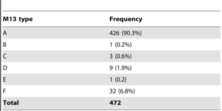
As the majority of VNI Thai isolates were reported previously to be clonal [33], M13 PCR fingerprinting analysis, which has shown to differentiate cryptococcal molecular subtypes in several previous studies [13,18,37], was performed to check for clonality amongst the collected Thai VNI isolates. The obtained PCR fingerprinting patterns were assigned with a M13 type. Six M13 types (A, B, C, D, E, and F) were identified amongst all studied isolates, with type A being the most common type identified (90.3%) (Table 2). The genetic diversity identified by M13 PCR-fingerprinting analysis suggested that the Thai isolates are more genetically diversified than previously reported [33].
Table 1.Distribution of the major molecular types among strains ofCryptococcus neoformansandC. gattiifrom different sources in Thailand.
Source Molecular type Total
VNI VNII VNIV VGI VGII
Human 365 (94.6%) 8 (2.1%) 0 1 (0.3%) 12 (3.1%) 386
Environ-ment
82 (98.8%) 0 1 (1.2%) 0 0 83
Animal 25 (86.2%) 4 (13.8%) 0 0 0 29
Total 472 (94.8%) 12 (2.4%) 1 (0.2%) 1 (0.2%) 12 (2.4%) 498
doi:10.1371/journal.pntd.0002297.t001
Table 2.Distribution of the M13 types among Thai VNI isolates.
M13 type Frequency
A 426 (90.3%)
B 1 (0.2%)
C 3 (0.6%)
D 9 (1.9%)
E 1 (0.2)
F 32 (6.8%)
Total 472
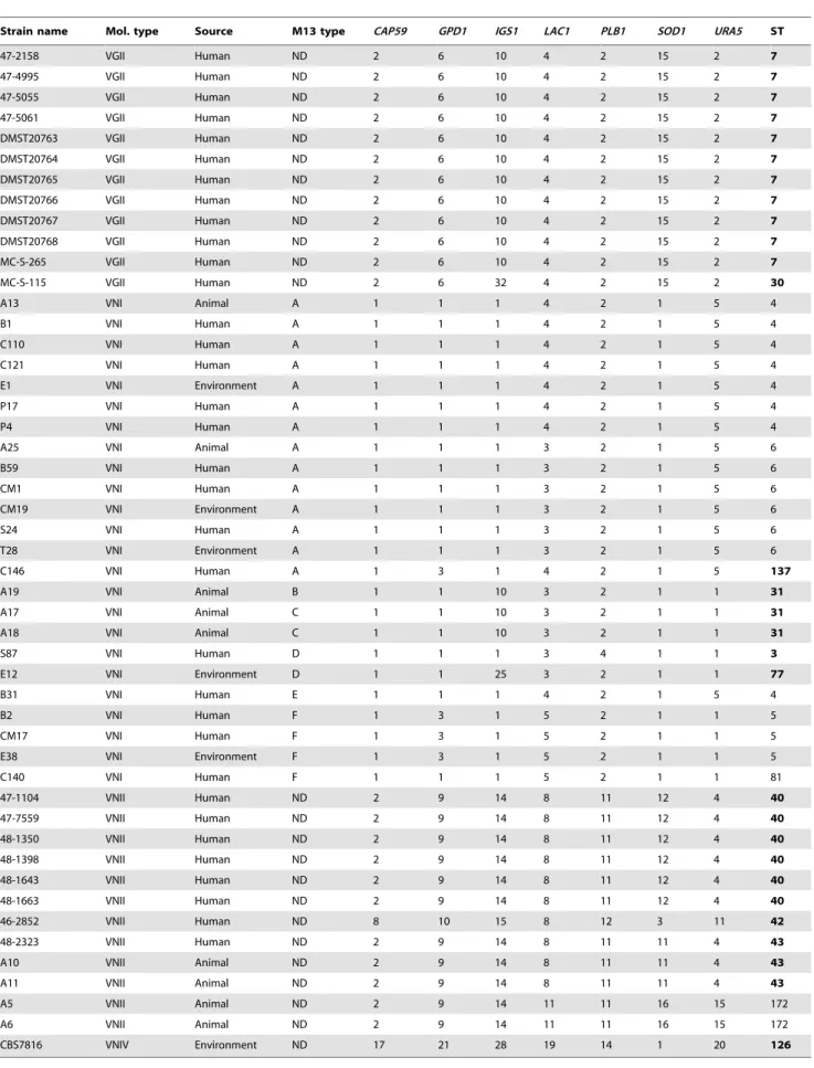
Table 3.Allele types and sequence types of selected Thai cryptococcal isolates.
Strain name Mol. type Source M13 type CAP59 GPD1 IGS1 LAC1 PLB1 SOD1 URA5 ST
47-2158 VGII Human ND 2 6 10 4 2 15 2 7
47-4995 VGII Human ND 2 6 10 4 2 15 2 7
47-5055 VGII Human ND 2 6 10 4 2 15 2 7
47-5061 VGII Human ND 2 6 10 4 2 15 2 7
DMST20763 VGII Human ND 2 6 10 4 2 15 2 7
DMST20764 VGII Human ND 2 6 10 4 2 15 2 7
DMST20765 VGII Human ND 2 6 10 4 2 15 2 7
DMST20766 VGII Human ND 2 6 10 4 2 15 2 7
DMST20767 VGII Human ND 2 6 10 4 2 15 2 7
DMST20768 VGII Human ND 2 6 10 4 2 15 2 7
MC-S-265 VGII Human ND 2 6 10 4 2 15 2 7
MC-S-115 VGII Human ND 2 6 32 4 2 15 2 30
A13 VNI Animal A 1 1 1 4 2 1 5 4
B1 VNI Human A 1 1 1 4 2 1 5 4
C110 VNI Human A 1 1 1 4 2 1 5 4
C121 VNI Human A 1 1 1 4 2 1 5 4
E1 VNI Environment A 1 1 1 4 2 1 5 4
P17 VNI Human A 1 1 1 4 2 1 5 4
P4 VNI Human A 1 1 1 4 2 1 5 4
A25 VNI Animal A 1 1 1 3 2 1 5 6
B59 VNI Human A 1 1 1 3 2 1 5 6
CM1 VNI Human A 1 1 1 3 2 1 5 6
CM19 VNI Environment A 1 1 1 3 2 1 5 6
S24 VNI Human A 1 1 1 3 2 1 5 6
T28 VNI Environment A 1 1 1 3 2 1 5 6
C146 VNI Human A 1 3 1 4 2 1 5 137
A19 VNI Animal B 1 1 10 3 2 1 1 31
A17 VNI Animal C 1 1 10 3 2 1 1 31
A18 VNI Animal C 1 1 10 3 2 1 1 31
S87 VNI Human D 1 1 1 3 4 1 1 3
E12 VNI Environment D 1 1 25 3 2 1 1 77
B31 VNI Human E 1 1 1 4 2 1 5 4
B2 VNI Human F 1 3 1 5 2 1 1 5
CM17 VNI Human F 1 3 1 5 2 1 1 5
E38 VNI Environment F 1 3 1 5 2 1 1 5
C140 VNI Human F 1 1 1 5 2 1 1 81
47-1104 VNII Human ND 2 9 14 8 11 12 4 40
47-7559 VNII Human ND 2 9 14 8 11 12 4 40
48-1350 VNII Human ND 2 9 14 8 11 12 4 40
48-1398 VNII Human ND 2 9 14 8 11 12 4 40
48-1643 VNII Human ND 2 9 14 8 11 12 4 40
48-1663 VNII Human ND 2 9 14 8 11 12 4 40
46-2852 VNII Human ND 8 10 15 8 12 3 11 42
48-2323 VNII Human ND 2 9 14 8 11 11 4 43
A10 VNII Animal ND 2 9 14 8 11 11 4 43
A11 VNII Animal ND 2 9 14 8 11 11 4 43
A5 VNII Animal ND 2 9 14 11 11 16 15 172
A6 VNII Animal ND 2 9 14 11 11 16 15 172
CBS7816 VNIV Environment ND 17 21 28 19 14 1 20 126
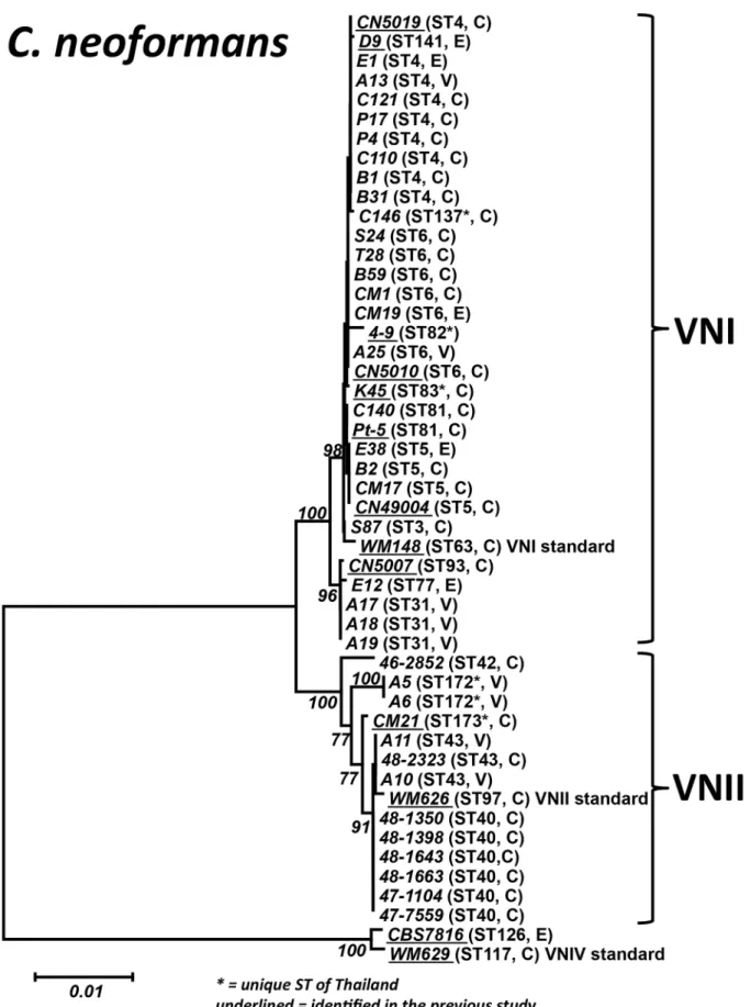
Figure 1. Phylogram of the ThaiC. neoformansisolates.Phylogram depicting the genetic relationships between the ThaiC. neoformansisolates based on neighbor joining analysis of the concatenated seven ISHAM consensus MLST loci using the program MEGA 5.03. Bold numbers on the branches indicate bootstrap support above 75%. Underlined strain numbers indicate STs identified in a previous study [33]. C = clinical, E = environmental, V = veterinary.
ThaiC. neoformans isolates are genetically diversified To verify the obtained diversity and to enable comparison with previous studies, MLST analysis, which has a superior discrimi-natory power and reproducibility over M13 PCR-fingerprinting [37], was performed. MLST analysis was performed on represen-tative strains of each M13 type from the VNI isolates (14 strains of M13 type A, 1 of M13 type B, 2 of M13 type C, 2 of M13 type D, 1 of M13 type E, and 4 of M13 F) and for all VNII and VNIV isolates. Eight additional sequence types (STs), with ST3, ST31, ST77, and ST137 for VNI and ST40, ST42, ST43 and ST172 for VNII (Table 3, Table S1 and Figure 1), were identified amongst the studiedC. neoformansisolates when compared with a previous report, which had identified the following STs: ST4, ST5, ST6, ST81, ST82, ST83, ST85, ST93 and ST141 for VNI; and ST173 for VNII [33]. Network analysis showed that the ThaiC. neoformans isolates are an integral part of the global population structure of this species, with nine ST’s being unique to Thailand, but closely related to other global isolates (Figure 2).
Almost all clinical VGII isolates belong to the same ST as the low virulent Vancouver Island outbreak strain
As VGII isolates are the causative agent of the ongoing outbreaks on Vancouver Island, Canada and Pacific Northwest region of USA
[35,44], MLST analysis was performed to determine the relation-ship of the Thai VGII isolates to the outbreak strains. Surprisingly, 11 out of the 12 VGII isolates were identical to the genotype of the low virulent Vancouver Island outbreak strains, VGIIb/ST7 (Figure 3). One isolate had a ST, which was unique to Thailand (ST30) (see Tables 3 and S1 and Figure 3).
Discussion
The obtained data concerning the demographics and the HIV status of the patients were in line with previous reports of cryptococcosis from Thailand [25,31]. Besides some missing demographic data it is clear from the available data that C. neoformans was the most common species identified among HIV positive patients, whileC. gattiiwas mainly a primary pathogen in immunocompetent patients, which is in accordance with a previous global study [18]. The fact that most isolates were recovered from male HIV positive patients with an average age of infection of 37.97 years represents the HIV demography in Thailand, with 60% of the HIV infected patients being male with an average age of 30–34 years [45].
C. neoformans has been found worldwide, with VNI being the most common molecular type, including recent reports from
Figure 2. Gene network placing the ThaiC. neoformansisolates in global context.Gene network constructed from allC. neoformansST types identified by MLST analysis in the current study in-cooperating the STs previously obtained from Thai cryptococcal isolates [33] and standard strains based on the combined seven ISHAM consensus MLST loci using the program Network 4.5.1.6, showing the close relationships between STs present in Thailand and globally.
Thailand [33]. The molecular typing in the current study confirmed this paradigm, where VNI is predominant regardless of the isolate source. Moreover, the rare molecular type VNII, for which only one isolate had been reported previously from Thailand [33], has now been identified from several strains from both humans and animals. As the natural reservoir of VNII has never been reported, the herein presented data allow to suggest: that a close relationship between animal and human VNII isolates may exist, as strains from humans and animals share the same genotype, ST43 (Figure 1 and Table 3). However, further studies are needed to draw a definite conclusion as the numbers of the studied VNII strains are very small and other human VNII strains showed no relationships with animal strains. On the other hand, a strong relationship between VNI clinical and environmental strains is evident, as they share the same STs (Figure 1 and Table 3).
The correlation betweenC. neoformansand HIV in Thailand is supported by the low prevalence of the genotype VNIc/M5, corresponding to M13 type F in the current study, which is known to be associated with non-HIV patients in China [46], Korea [37] and Japan [36]. In fact, only 6% (23 out of 386 isolates) of the clinical cases were of M13 type F (VNIc/M5) (Figure S1) and only one of them (P21) had been isolated from a HIV positive patient. All other cryptococcal isolates form HIV positive patients had either the M13 type A or D (Table S1).
The herein obtained MLST data when combined with data previously reported [33] showed clearly, that the STs present in Thailand are an integral part of the global population genetic
structure, and are not as unique as previously reported [33]. For theC. neoformansmolecular type VNI, seven of the STs are shared with global strains and six are unique for Thailand (Figure 2). All of the isolates form a close network with a number of Thai specific STs and are directly linked to other globally present STs (Figure 2). The current study describes for the first time molecular typing of C. gattiiisolates from Thailand, taken into account the literature since the 1990’s [30,31]. The high percentage of the VGII molecular type (92.3%) amongst the studiedC. gattiiisolates is in contrast to a report from a neighboring country, Malaysia, where 76.5% of theC. gattiiisolates belonged to the molecular type VGI [47]. No VGIII or VGIV isolates have been found in the current study and the fact that they have never been reported from this region may suggest that those molecular types are not endemic in this area. The geographically closest related place from which VGIII and VGIV isolates have been reported is India [43,48].
Before the AIDS epidemic, a predominance ofC. gattiias the causative agent of cryptococcosis was found in Thailand, which was possibly related to non-HIV immunocompromised conditions [25,26]. A recent study on non-HIV cryptococcosis cases suggested that the disease was not such a rare event in HIV negative patients and is also associated with high mortality rates [49,50], a fact also seen with the cases of C. gattii infection investigated in the current study.
Moreover, the predominance of the VGII molecular type in this tropical region revealed in the current study is of special interest, as a similar situation was only described form the northern part of Brazil [51], which is in contrast to most of the described isolations
Figure 3. Phylogram of the ThaiC. gattiiisolates.Phylogram depicting the genetic relationships between the ThaiC. gattiiisolates based on neighbor joining analysis of the concatenated seven ISHAM consensus MLST loci using the program MEGA 5.03. Bold numbers on the branches indicate bootstrap support above 75%. C = clinical, E = environmental.
which are associated mainly with areas of temperate climate or in the high mountain regions of Colombia [24,52,53]. In addition, the fact that several strains (the DMST strains) were isolated more than 10 years ago (Table S1), [30,54] suggests that this molecular type is prevalent in Thailand, as it is in South America [19], but unlike Australia [55] or Europe [56] where the molecular type VGI is predominant.
The fact that 11 out of the 12C. gattiistrains studied showed an identical ST type to the one of the Vancouver Island outbreak strain, VGIIb = ST7 is remarkable. It reveals the high clonality that this VGIIb C. gattii population has in Thailand, which is similar to the situation described in Australia [35]. It confirms the point previously made, that this low virulent outbreak strain is globally present, with Australia and Thailand being important stepping-stones in the global spread of this outbreak strain, linking South America, via Australia with North America and Europe.
In summary, as in other worldwide studies, the same distribution of cryptococcal genotypes has been found in Thailand, with a predominance of C. neoformansvar. grubii, molecular type VNI, isolated from HIV positive patients. Our study suggests a greater genetic diversity among Thai cryptococcal isolates especially amongst VNI strains with 13 different STs than reported previously [33]. The majority of Thai C. gattiiisolates are clonal and identical to the Vancouver Island outbreak strain with VGIIb = ST7, identifying Thailand as a stepping-stone in its global spread. In addition, a strong linkage between environmental and clinical strains was found for the VNI isolates. A connection between other rare molecular types, such as VNII forC. neoformans or VGI and VGII forC. gattiiand the environment in Thailand could still not be found and needs further investigation. Extensive environmental and veterinary sampling would be of great help to fill this gap. Moreover, despite an advanced development of HIV treatment, cryptococcosis is still a major problem as an opportu-nistic infection in Thailand, making further studies, concerning the epidemiology and virulence of the Cryptococcus neoformans/C. gattii species complex mandatory for a proper management of the disease in the future.
Supporting Information
Figure S1 Phylogram correlating the newly identified Thai C. neoformans sequence types with previously reported types.Phylogram depicting the genetic relationships between the Thai VNI isolates studied herein in combination with previously published data representing the following M13
PCR-fingerprinting patterns VNIa, VNIb, VNIc/M5 and VNId (32) based on neighbor joining analysis of the concatenated seven ISHAM consensus MLST loci. Bold numbers on the branches indicate bootstrap support above 75%. Letters in brackets indicate the M13 type. WM148 = VNI standard, WM626 = VNII stan-dard.
(TIF)
Table S1 Strains used in this study and associated demographic and molecular data.
(DOC)
Table S2 Correlation between old and new allele and sequence type numbering from the Simwamiet al.2011 (28) publication and the newC. gattiiMLST database at mlst.mycologylab.com for the MLST data used in the current study.
(DOC)
Table S3 MLST data for the additional published C. neoformansstrains used in this study.
(DOC)
Table S4 GenBank accession numbers for all sequences of the MLST alleles obtained from ThaiC. neoformans andC. gattiiisolates used in this study.
(DOC)
Acknowledgments
The authors would like to thank the staff and students of the Molecular Mycology and Mycobacteriology Laboratory, Faculty of Medicine Siriraj Hospital, Mahidol University and the Mycology Unit Laboratory, Faculty of Medicine, King Chulalongkorn Memorial Hospital for the preparation of the strains and providing the patient demographic data for this study and the Mycology Laboratory, Faculty of Medicine, Chiang Mai University, Chiang Mai, Thailand for kindly providing the two environmental strains.
Author Contributions
Conceived and designed the experiments: S. Kaocharoen, P. Ngamskul-rungroj, A. Chindamporn, W. Meyer. Performed the experiments: S. Kaocharoen, P. Ngamskulrungroj, D. Piyabongkarn. Analyzed the data: S. Kaocharoen, P. Ngamskulrungroj, C. Firacative, L. Trilles, W. Meyer. Contributed reagents/materials/analysis tools: A. Chindamporn, A. Chaiprasert, W. Meyer, P. Ngamskulrungroj, N. Poonwan. Wrote the paper: S. Kaocharoen, P. Ngamskulrungroj, C. Firacative, A. Chindam-porn, W. Meyer. Supplied strains: P. Ngamskulrungroj, D. Piyabongkarn, W. Banlunara, N. Poonwan, A. Chindamporn, A. Chaiprasert, W. Meyer.
References
1. Casadevall A, Perfect JR (1998)Cryptococcus neoformans. Washington, DC: ASM press.
2. Krockenberger MB, Canfield PJ, Malik R (2003)Cryptococcus neoformansvar.gattii in the koala (Phascolarctos cinereus): a review of 43 cases of cryptococcosis. Med Mycol 41: 225–234.
3. Kwon-Chung KJ, Boekhout T, Fell JW, Diaz M (2002) (1557) Proposal to conserve the name Cryptococcus gattii against C.hondurianusand C.basillisporus (Basidiomycota, Hymenomycetes, Tremellomycetidae). Taxon 51: 804–806. 4. Stephen C, Lester S, Black W, Fyfe M, Raverty S (2002) Multispecies outbreak
of cryptococcosis on southern Vancouver Island, British Columbia. Can Vet J 43: 792–794.
5. Lazera MS, Pires FD, Camillo-Coura L, Nishikawa MM, Bezerra CC, et al. (1996) Natural habitat ofCryptococcus neoformansvar.neoformansin decaying wood forming hollows in living trees. J Med Vet Mycol 34: 127–131.
6. Emmons CW (1951) Isolation ofCryptococcus neoformansfrom soil. J Bacteriol 62: 685–690.
7. Ellis D, Pfeiffer T (1992) The ecology ofCryptococcus neoformans. Eur J Epidemiol 8: 321–325.
8. Kwon-Chung KJ, Bennett JE (1984) High prevalence ofCryptococcus neoformans var.gattiiin tropical and subtropical regions. Zentralbl Bakteriol Mikrobiol Hyg [A] 257: 213–218.
9. Sorrell TC (2001) Cryptococcus neoformans variety gattii. Med Mycol 39: 155–168. 10. Granados DP, Castaneda E (2006) Influence of climatic conditions on the isolation of members of theCryptococcus neoformansspecies complex from trees in Colombia from 1992–2004. FEMS Yeast Res 6: 636–644.
11. Lazera MS, Cavalcanti MA, Trilles L, Nishikawa MM, Wanke B (1998) Cryptococcus neoformansvar.gattii–evidence for a natural habitat related to decaying wood in a pottery tree hollow. Med Mycol 36: 119–122.
12. Randhawa HS, Kowshik T, Preeti Sinha K, Chowdhary A, Khan ZU, et al. (2006) Distribution ofCryptococcus gattiiandCryptococcus neoformans in decayed trunk wood of Syzygium cumini trees in north-western India. Med Mycol 44: 623–630.
14. Perfect JR, Ketabchi N, Cox GM, Ingram CW, Beiser CL (1993) Karyotyping ofCryptococcus neoformansas an epidemiological tool. J Clin Microbiol 31: 3305– 3309.
15. Sorrell TC, Chen SC, Ruma P, Meyer W, Pfeiffer TJ, et al. (1996) Concordance of clinical and environmental isolates ofCryptococcus neoformansvar. gattiiby random amplification of polymorphic DNA analysis and PCR fingerprinting. J Clin Microbiol 34: 1253–1260.
16. Boekhout T, Theelen B, Diaz M, Fell JW, Hop WC, et al. (2001) Hybrid genotypes in the pathogenic yeastCryptococcus neoformans. Microbiology 147: 891– 907.
17. Hanafy A, Kaocharoen S, Jover-Botella A, Katsu M, Iida S, et al. (2008) Multilocus microsatellite typing forCryptococcus neoformansvar.grubii. Med Mycol 46: 685–696.
18. Meyer W, Marszewska K, Amirmostofian M, Igreja RP, Hardtke C, et al. (1999) Molecular typing of global isolates ofCryptococcus neoformansvar.neoformansby polymerase chain reaction fingerprinting and randomly amplified polymorphic DNA-a pilot study to standardize techniques on which to base a detailed epidemiological survey. Electrophoresis 20: 1790–1799.
19. Meyer W, Castaneda A, Jackson S, Huynh M, Castaneda E (2003) Molecular typing of IberoAmericanCryptococcus neoformansisolates. Emerg Infect Dis 9: 189– 195.
20. Latouche GN, Huynh M, Sorrell TC, Meyer W (2003) PCR-restriction fragment length polymorphism analysis of the phospholipase B (PLB1) gene for subtyping ofCryptococcus neoformansisolates. Appl Environ Microbiol 69: 2080– 2086.
21. Meyer W, Aanensen DM, Boekhout T, Cogliati M, Diaz MR, et al. (2009) Consensus multi-locus sequence typing scheme forCryptococcus neoformansand Cryptococcus gattii. Med Mycol 47: 561–570.
22. Litvintseva AP, Thakur R, Vilgalys R, Mitchell TG (2006) Multilocus sequence typing reveals three genetic subpopulations ofCryptococcus neoformansvar.grubii (serotype A), including a unique population in Botswana. Genetics 172: 2223– 2238.
23. Litvintseva AP, Carbone I, Rossouw J, Thakur R, Govender NP, et al. (2011) Evidence that the human pathogenic fungusCryptococcus neoformansvar.grubiimay have evolved in Africa. PLoS One 6: e19688.
24. Escandon P, Sanchez A, Martinez M, Meyer W, Castaneda E (2006) Molecular epidemiology of clinical and environmental isolates of theCryptococcus neoformans species complex reveals a high genetic diversity and the presence of the molecular type VGII mating type a in Colombia. FEMS Yeast Res 6: 625–635. 25. Sukroongreung S, Nilakul C, Ruangsomboon O, Chuakul W, Eampokalap B (1996) Serotypes ofCryptococcus neoformansisolated from patients prior to and during the AIDS era in Thailand. Mycopathologia 135: 75–78.
26. Imwidthaya P, Dithaprasop P, Egtasaeng C (1989) Clinical and environmental isolates ofCryptococcus neoformansin Bangkok (Thailand). Mycopathologia 108: 65–67.
27. Sriburee P, Khayhan S, Khamwan C, Panjaisee S, Tharavichitkul P (2004) Serotype and PCR-fingerprints of clinical and environmental isolates of Cryptococcus neoformansin Chiang Mai, Thailand. Mycopathologia 158: 25–31. 28. Imwidthaya P (1994) Systemic fungal infections in Thailand. J Med Vet Mycol
32: 395–399.
29. Vithayasai P, Vithayasai V (1993) Clinical manifestations of 174 AIDS cases in Maharaj Nakorn Chiang Mai Hospital. J Dermatol 20: 389–393.
30. Poonwan N, Mikami Y, Poosuwan S, Boon-Long J, Mekha N, et al. (1997) Serotyping ofCryptococcus neoformansstrains isolated from clinical specimens in Thailand and their susceptibility to various antifungal agents. Eur J Epidemiol 13: 335–340.
31. Imwidthaya P, Poungvarin N (2000) Cryptococcosis in AIDS. Postgrad Med J 76: 85–88.
32. Ngamwongsatit P, Sukroongreung S, Nilakul C, Prachayasittikul V, Tantima-vanich S (2005) Electrophoretic karyotypes ofC. neoformansserotype A recovered from Thai patients with AIDS. Mycopathologia 159: 189–197.
33. Simwami SP, Khayhan K, Henk DA, Aanensen DM, Boekhout T, et al. (2011) Low diversityCryptococcus neoformansvarietygrubiimultilocus sequence types from Thailand are consistent with an ancestral African origin. PLoS Pathog 7: e1001343.
34. Kwon-Chung JK, Bennett JE (1992) Cryptococcosis. In: Kwon-Chung JK, Bennett JE, editors. Medical Mycology. Pennsylvania: Lea & Febiger. pp. 397– 446.
35. Carriconde F, Gilgado F, Arthur I, Ellis D, Malik R, et al. (2011) Clonality and alpha-a recombination in the AustralianCryptococcus gattiiVGII population–an emerging outbreak in Australia. PLoS One 6: e16936.
36. Mihara T, Izumikawa K, Kakeya H, Ngamskulrungroj P, Umeyama T, et al. (2012) Multilocus sequence typing of Cryptococcus neoformans in non-HIV associated cryptococcosis in Nagasaki, Japan. Med Mycol 51: 252–60. 37. Choi YH, Ngamskulrungroj P, Varma A, Sionov E, Hwang SM, et al. (2010)
Prevalence of the VNIc genotype ofCryptococcus neoformansin non-HIV-associated cryptococcosis in the Republic of Korea. FEMS Yeast Res 10: 769–778. 38. Thompson JD, Higgins DG, Gibson TJ (1994) CLUSTAL W: improving the
sensitivity of progressive multiple sequence alignments through sequence weighting, position specific gap penalties and weight matrix choice. Nucl Acids Res 22: 4673–4680.
39. Hall T (1999) BioEdit: a user-friendly biological sequence alignment editor and analysis program for Windows 95/98/NT. Nucl Acids Symp Ser 41: 95–98. 40. Tamura K, Dudley J, Nei M, Kumar S (2007) MEGA4: Molecular Evolutionary
Genetics Analysis (MEGA) software version 4.0. Mol Biol Evol 24: 1596–1599. 41. Saitou N, Nei M (1987) The neighbor-joining method: a new method for
reconstructing phylogenetic trees. Mol Biol Evol 4: 406–425.
42. Felsenstein J (1985) Confidence-Limits on Phylogenies - an Approach Using the Bootstrap. Evolution 39: 783–791.
43. Meyer W, Gilgado F, Ngamskulrungroj P, Trilles L, Castan˜eda E, et al. (2011) Molecular typing of the Cryptococcus neoformans/C. gattiispecies complex. In: Heitman J, Kozel TR, Kwon-Chung J, Perfect JR, Casadevall A, editors. Cryptococcus: From Human Pathogen to Model Yeast. Washington DC: ASM press. pp. 327–358.
44. Fraser JA, Giles SS, Wenink EC, Geunes-Boyer SG, Wright JR, et al. (2005) Same-sex mating and the origin of the Vancouver Island Cryptococcus gattii outbreak. Nature 437: 1360–1364.
45. Khongphatthanayothin M, Tantipaibulvut S, Nookai S, Chumchee P, Kaldor J, et al. (2006) Demographic predictors of a positive HIV test result among clients attending a large metropolitan voluntary counselling and testing centre in Thailand. HIV Med 7: 281–284.
46. Chen J, Varma A, Diaz MR, Litvintseva AP, Wollenberg KK, et al. (2008) Cryptococcus neoformans strains and infection in apparently immunocompetent patients, China. Emerg Infect Dis 14: 755–762.
47. Tay ST, Lim HC, Tajuddin TH, Rohani MY, Hamimah H, et al. (2006) Determination of molecular types and genetic heterogeneity of Cryptococcus neoformansandC. gattiiin Malaysia. Med Mycol 44: 617–622.
48. Cogliati M, Chandrashekar N, Esposto MC, Chandramuki A, Petrini B, et al. (2012) Cryptococcus gattiiserotype-C strains isolated in Bangalore, Karnataka, India. Mycoses 55: 262–268.
49. Kiertiburanakul S, Wirojtananugoon S, Pracharktam R, Sungkanuparph S (2006) Cryptococcosis in human immunodeficiency virus-negative patients. Int J Infect Dis 10: 72–78.
50. Jongwutiwes U, Sungkanuparph S, Kiertiburanakul S (2008) Comparison of clinical features and survival between cryptococcosis in human immunodefi-ciency virus (HIV)-positive and HIV-negative patients. Jpn J Infect Dis 61: 111– 115.
51. Trilles L, Lazera Mdos S, Wanke B, Oliveira RV, Barbosa GG, et al. (2008) Regional pattern of the molecular types ofCryptococcus neoformansandCryptococcus gattiiin Brazil. Mem Inst Oswaldo Cruz 103: 455–462.
52. Kidd SE, Chow Y, Mak S, Bach PJ, Chen H, et al. (2007) Characterization of environmental sources of the human and animal pathogenCryptococcus gattiiin British Columbia, Canada, and the Pacific Northwest of the United States. Appl Environ Microbiol 73: 1433–1443.
53. Granados DP, Castaneda E (2005) Isolation and characterization ofCryptococcus neoformansvarieties recovered from natural sources in Bogota, Colombia, and study of ecological conditions in the area. Microb Ecol 49: 282–290. 54. Ngamskulrungroj P, Sorrell TC, Chindamporn A, Chaiprasert A, Poonwan N,
et al. (2008) Association between fertility and molecular sub-type of global isolates ofCryptococcus gattiimolecular type VGII. Med Mycol 46: 665–673. 55. Chen S, Sorrell T, Nimmo G, Speed B, Currie B, et al. (2000) Epidemiology and
host- and variety-dependent characteristics of infection due to Cryptococcus neoformans in Australia and New Zealand. Australasian Cryptococcal Study Group. Clin Infect Dis 31: 499–508.