Human N-Glycome Identifies HNF1
a
as a Master
Regulator of Plasma Protein Fucosylation
Gordan Lauc1,2., Abdelkader Essafi3., Jennifer E. Huffman3., Caroline Hayward3., Ana Knezˇevic´2 , Jayesh J. Kattla4,5, Ozren Polasˇek6,7, Olga Gornik2, Veronique Vitart3, Jodie L. Abrahams4,5, Maja Pucˇic´1, Mislav Novokmet1, Irma Redzˇic´2, Susan Campbell3, Sarah H. Wild8, Fran Borovecˇki7, Wei Wang9,10,11, Ivana Kolcˇic´7, Lina Zgaga7, Ulf Gyllensten12, James F. Wilson8", Alan F. Wright3", Nicholas D. Hastie3", Harry Campbell8", Pauline M. Rudd4,5", Igor Rudan8,11"*
1Glycobiology Laboratory, Genos Ltd., Zagreb, Croatia,2Department of Biochemistry and Molecular Biology, University of Zagreb, Faculty of Pharmacy and Biochemistry, Zagreb, Croatia,3Medical Research Council Human Genetics Unit, Institute of Genetics and Molecular Medicine, Western General Hospital, Edinburgh, United Kingdom, 4National Institute for Bioprocessing Research and Training, Dublin-Oxford Glycobiology Lab, Conway Institute, University College Dublin, Dublin, Ireland,5Conway Institute, University College Dublin, Dublin, Ireland,6Gen Info Ltd., Zagreb, Croatia,7Medical School, University of Zagreb, Zagreb, Croatia,8Centre for Population Health Sciences, The University of Edinburgh Medical School, Edinburgh, United Kingdom,9School of Public Health and Family Medicine, Capital Medical University, Beijing, China,10Graduate School of the Chinese Academy of Sciences, Beijing, China,11Croatian Centre for Global Health, University of Split Medical School, Split, Croatia, 12Department of Genetics and Pathology, Rudbeck Laboratory, Uppsala University, Uppsala, Sweden
Abstract
Over half of all proteins are glycosylated, and alterations in glycosylation have been observed in numerous physiological and pathological processes. Attached glycans significantly affect protein function; but, contrary to polypeptides, they are not directly encoded by genes, and the complex processes that regulate their assembly are poorly understood. A novel approach combining genome-wide association and high-throughput glycomics analysis of 2,705 individuals in three population cohorts showed that common variants in the Hepatocyte Nuclear Factor 1a (HNF1a) and fucosyltransferase genesFUT6andFUT8influence N-glycan levels in human plasma. We show that HNF1aand its downstream target HNF4a
regulate the expression of key fucosyltransferase and fucose biosynthesis genes. Moreover, we show that HNF1a is both necessary and sufficient to drive the expression of these genes in hepatic cells. These results reveal a new role for HNF1aas a master transcriptional regulator of multiple stages in the fucosylation process. This mechanism has implications for the regulation of immunity, embryonic development, and protein folding, as well as for our understanding of the molecular mechanisms underlying cancer, coronary heart disease, and metabolic and inflammatory disorders.
Citation:Lauc G, Essafi A, Huffman JE, Hayward C, Knezˇevic´ A, et al. (2010) Genomics Meets Glycomics—The First GWAS Study of Human N-Glycome Identifies HNF1aas a Master Regulator of Plasma Protein Fucosylation. PLoS Genet 6(12): e1001256. doi:10.1371/journal.pgen.1001256
Editor:Greg Gibson, Georgia Institute of Technology, United States of America ReceivedJuly 7, 2010;AcceptedNovember 19, 2010;PublishedDecember 23, 2010
Copyright:ß2010 Lauc et al. This is an open-access article distributed under the terms of the Creative Commons Attribution License, which permits unrestricted use, distribution, and reproduction in any medium, provided the original author and source are credited.
Funding:The work is supported by grants#309-0061194-2023 (to GL) and#216-1080315-0302 (to I Rudan) from the Croatian Ministry of Science, Education, and Sport; by European Commission EUROPHARM, EUROSPAN, and EUROGLYCOARRAYS grants; by the National Institute for Bioprocessing Research and Training, Ireland; and by Eurocarb DB (Contract: RIDS Contract number 011952). Studies carried out in the Croatian islands of Vis and Korcula were supported by Medical Research Council UK (to H Campbell, AF Wright, ND Hastie, and I Rudan). ORCADES was supported by the Scottish Executive Health Department and the Royal Society and the European Union framework program 6 EUROSPAN project (contract no. LSHG-CT-2006-018947). The funders had no role in study design, data collection and analysis, decision to publish, or preparation of the manuscript.
Competing Interests:The authors have declared that no competing interests exist. * E-mail: irudan@hotmail.com
.These authors contributed equally to this work. "These authors also contributed equally to this work.
Introduction
Glycosylation is a post-translational modification that enriches protein complexity and function. Over half of all known proteins are modified by covalently bound glycans, which are important for normal physiological processes, including protein folding, degra-dation and secretion, cell signalling, immune function and transcription [1–4]. Configuration and composition of attached glycans significantly change the structure and activity of polypeptide portions of glycoproteins [5] and since this process is not template driven, complexity of the glycoproteome is
Due to experimental limitations in quantifying glycans in complex biological samples, our understanding of the genetic regulation of glycosylation is currently very limited [12]. However, recent technological advances have allowed reliable, high-throughput quantification of N-glycans [13], which now permits investigation of the genetic regulation and biological roles of glycan structures and brings glycomics into line with genomics, proteomics and metabolomics [14]. Recently we completed the first comprehensive population study of human plasma N-glycome which revealed variability that by far exceeds the variability of proteins and DNA [15]. However, within a single individual composition of plasma glycome is rather stable [16] and environmental factors have limited impact on the majority of glycans [17]. Specific altered glyco-phenotypes that can be associated with specific pathologies were also identified to exist in a population [18].
Variations in glycosylation are of great physiological signifi-cance as alterations in glycans significantly change the structure and function of polypeptide parts of glycoproteins [5]. A particularly interesting element of protein glycosylation is the addition of fucose to non-reducing ends of N-glycans. Fucose is a relatively novel sugar in evolutionary terms with two important structural features that distinguishes it from all other mammalian six-carbon monosaccharides; it lacks a hydroxyl group on the carbon at the 6-position and is the only monosaccharide that is in the L-configuration. The conversion of mannose to GDP-fucose is catalyzed by two enzymes (GMD and FX) that display remarkable evolutionary conservation [19,20]. On the other hand, the large family of genes that add fucose to proteins and lipids (fucosyltranferases, FUTs) has a very complex evolutionary history, including several more recent events specific to primates [21]. In mammals, fucose-containing glycans have important roles in blood transfusion reactions, in the selectin-mediated leukocyte-endothelial adhesion that initiates an inflammatory response, in host-microbe interactions, and numerous ontogenic events [10,19]. Acute phase proteins have altered fucosylation in many diseases [22] and changes in the levels of fucosylated glycans have been shown to be associated with several important pathological processes, including cancer [23].
Hepatocyte nuclear factor 1a (HNF1a) and its downstream target HNF4a are transcription factors that regulate gene expression in both the liver and pancreas in a tissue-specific manner and are key regulators of metabolic genes [24]. Mutations in the encoding genesHNF1aandHNF4acause Maturity Onset
Diabetes of the Young (MODY) types 3 and 1 respectively [25,26]. Recently, HNF1a single nucleotide polymorphisms (SNPs) have
been associated with plasma C-reactive protein (CRP) [27], LDL cholesterol and gamma glutamyltransferase (GGT) [28], and coronary heart disease [29].HNF4avariants have been associated
with ulcerative colitis [30] and with the plasma concentrations of CRP and apolipoprotein A1 (APOA1) [31]. Currently there is little evidence to link these transcription factors with fucose metabolism and the upstream mechanisms regulating fucosylation pathways are unknown.
Results
Common variants in fucosyltransferase genes affect the relative proportions of plasma N-glycans
We performed the first systematic analysis of the genetic regulation of individual N-glycans in plasma from 2,705 individuals in three population cohorts, from Croatia and Scotland, which have previously been characterized in great detail [32]. Desialylated 2AB-labelled human plasma N-glycans were separated into 13 structurally related groups of glycans, referred to as DG1–DG13 (see Table S1 for a list of specific glycans found within each DG group) [13]. The concentration of plasma N-glycans measured in each of these groups was then expressed as a proportion of the total plasma N-glycome to obtain 13 quantitative variables in each examinee. All glycans contain two core N-acetylglucosamine (GlcNAc) residues, to which a ‘‘core’’ fucose can bea1,6-linked to the inner GlcNAc, which is directly linked to an asparagine residue on the protein. Additional fucose residues can be transferred to different positions on antennas that have been added to the core glycan structure (Table S1). Two further traits were derived from the original variables to calculate the percentage of glycan structures containing core (FUC-C) or antennary (FUC-A) fucose, yielding a total of 15 glycan traits for analysis.
We conducted a meta-analysis of genome-wide association study (GWAS) data for the fifteen plasma N-glycan traits measured in three population-based cohorts, CROATIA-VIS (n = 924), CROATIA-KORCULA (n = 898) and ORCADES (n = 737). Additive SNP effects were tested in each cohort independently and then combined in an inverse-variance weighted meta-analysis. The genome-wide significance threshold for the meta-analysis was set at 5610208.
Genome-wide significant associations were found for DG1, DG6, DG7, DG9, DG11, as well as FUC-A (Table 1; Figure 1 and Figure 2). Association profiles for DG1, DG7, and DG9 are represented in their genomic context in Figure 1 for the associated region. Quantile–quantile plots for each association were consis-tent with an excess of true genetic associations, with modest genomic control inflation for each population (inflation factor
,1.04 for all traits and each population as well as the meta-analysis), suggesting that the observed results were not due to population stratification (Figure 2A–2C).
Fifteen SNPs located in the region encompassing the fucosyl-transferase 8 gene (FUT8, Entrez GeneID: 2530) on chromosome 14 were significantly associated with plasma concentrations of desialylated glycan (DG) 1, the most significant being rs7159888 (p = 3.46610218) located 59of the gene.FUT8was also associated with DG6, however for this trait only one SNP, rs10483776, reached genome-wide significance (p = 9.58610209). All SNPs significantly associated with DG1 levels were in high LD (r2.0.5) and located between two recombination hotspots, while no associations were found with SNPs located outside these boundaries nor with other genes located within this association
Author Summary
By combining recently developed high-throughput glycan analysis with genome-wide association study, we per-formed the first comprehensive analysis of common genetic polymorphisms that affect protein glycosylation. Over half of all proteins are glycosylated; but, due to difficulties in glycan analysis and the absence of a genetic template for their synthesis, knowledge about the complex processes that regulate glycan assembly is still limited. We demonstrated that HNF1a regulates the expression of key fucosyltransferase and fucose biosyn-thesis genes and acts as a master regulator of plasma protein fucosylation. Proper protein fucosylation is essen-tial in numerous processes including inflammation, cancer, and coronary heart disease, thus the identification of a master regulator of plasma protein fucosylation has important implications for understanding both normal biological functions and disease processes.
HNF1aIs a Master Regulator of Fucosylation
interval (Figure 1A). The effect size of the G allele of rs7159888 was20.2617 (s.e. 0.0301) for DG1 in the meta-analysis of the 3 populations studied (standard deviation units, after adjustment for sex and age; Figure 2D). All significant SNPs in this region had a similar effect size (absolute value of the range: 0.1828–0.3251), accounting for between 1 and 6 percent of the trait variance after adjustment for age and sex. The effect of rs7159888 on DG1 was consistent across populations with similar amplitude and direction of effect (Figure 2D) with the effect for each population plotted separately along with the pooled effect. Haplotype analysis found that a single SNP model performed better than the 3- or 5-SNP haplotype model in every population.
A single SNP located on chromosome 19, rs3760776, was associated with DG7, DG9, DG12 and FUC-A (p = 3.42610212, p = 3.51610217, p = 9.44610210, p = 1.41610212). This SNP is located at the 59end of the fucosyltranferase 6 gene (FUT6, Entrez GeneID: 2528). The association interval for this SNP contains the
NRTN,FUT6 and FUT3 genes (see Figure 1C), of whichFUT6
andFUT3are both biologically plausible candidates to explain the observed associations. The effect size of the G allele of rs3760776 is 0.3387 (s.e. 0.0487) for DG7 (standard deviation units, after adjustment for significant covariates: sex, age and fibrinogen); and 0.4104 (s.e. 0.0487), 0.2974 (s.e. 0.0486), and 0.3446 (s.e. 0.0486) for DG9, DG12 and FUC-A respectively (standard deviation units, after adjustment for age and fibrinogen). These effects account for 2% (DG7), 3% (DG9), 2% (DG12) and 2% (FUC-A) of the trait variance. A forest plot of the effect size of rs3760776 in each population and the meta for DG7 is presented in Figure 2F. Haplotype analysis suggested that a 5-SNP haplotype across this region has a stronger effect on these glycan levels than a single SNP model. Another fucosyltransferase gene (FUT3) is also within the region, so the causal variant(s) may affect one or both of these genes. The best 5-SNP haplotype contained rs3760776 and encompassed FUT6 but notFUT3 in every population and for every glycan group tested which suggests that the association is withFUT6, notFUT3.
The glycan structures which were significantly associated with genetic variants in theFUT6andFUT8genes are summarised in Table 1. Glycan group DG1 consists of a single structure GlcNAc2Man3GlcNAc2 that is known to be a substrate for the a1-6-fucosyltransferase (FUT8) (Table S1) [15,33]. Group DG6 contains three glycan structures, two of which are core fucosylated
so the results are consistent with the known biological role of FUT8. In contrast, groups DG7, DG9 and DG12 include glycans containing antennary fucose while FUC-A was derived as an overall measure of antennary fucosylation. FUT6 encodes the enzyme fucosyltransferase VI which was reported to be the key enzyme responsible for thea3-fucosylation of plasma proteins [34]. The association of FUT8 and FUT6 genes with N-glycan structures containing core and antennary fucosylation is supported by their known biological functions [35] and the fact that they were identified in this study is an effective proof of principle that HPLC measured glycan levels can be used to identify genes that regulate protein glycosylation.
Novel association of HNF1awith N-glycans
Two SNPs on chromosome 12, rs7953249 and rs735396, showed genome-wide significant associations with DG7 (p = 1.97610208, p = 1.75610208). The latter SNP was also associated with DG11 (p = 4.44610208), with an effect in the opposite direction, and was close to genome-wide significance with DG9 (Table S2). Both SNPs are located in the HNF1a(Entrez
GeneID: 6927) gene region: rs7953249 is found 13 kb 59 to the gene and rs735396 is in intron 9. Two other genes are found between the recombination hotspots that comprise the boundaries of the association interval, C12orf43 and OASL (Figure 1B). However, none of the most significantly associated SNPs are located in these genes and all SNPs with suggestive p-values (p,1610205) are located withinHNF1a(Table S2). The effect size
of the G allele of rs735396 is20.1767 (standard deviation units, after adjustment for sex, age and fibrinogen; s.e. 0.0314) for DG7, which only contains glycans with antennary fucose, and in the opposite direction (0.1699 standard deviation units, after adjust-ment for age and fibrinogen; s.e. 0.0310) for DG11, which has no antennary fucose (Table 1). All significant SNPs in this region had a similar effect size (absolute value of the range: 0.1396–0.1767), representing 1–3% of the trait variance. Figure 2E shows the effect size for rs735396 with DG7 for each population separately and the pooled meta-analysis. Comparison of models including rs7953249 and rs735396 separately and combined suggests that the causal variant is located between these two SNPs. This was confirmed by analysis of imputed data based on HapMap release 2 with the most significant SNPs located across intron 1 ofHNF1a.
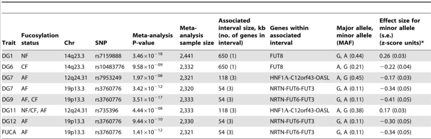
Table 1.Genetic markers associated with plasma N-glycan levels.
Trait
Fucosylation
status Chr SNP
Meta-analysis P-value Meta-analysis sample size Associated interval size, kb (no. of genes in interval) Genes within associated interval Major allele, minor allele (MAF)
Effect size for minor allele (s.e.)
(z-score units)*
DG1 NF 14q23.3 rs7159888 3.46610218 2,441 650 (1) FUT8 G, A (0.44) 0.26 (0.03)
DG6 CF 14q23.3 rs10483776 9.58610209 2,332 650 (1) FUT8 A, G (0.21) 20.22 (0.04) DG7 AF 12q24.31 rs7953249 1.97610208 2,321 118 (3) HNF1A-C12orf43-OASL A, G (0.45) 20.17 (0.03)
DG7 AF 19p13.3 rs3760776 3.42610212 2,320 54 (3) NRTN-FUT6-FUT3 G, A (0.11) 20.34 (0.05)
DG9 AF, CF 19p13.3 rs3760776 3.51610217 2,333 54 (3) NRTN-FUT6-FUT3 G, A (0.11) 20.41 (0.05) DG11 NF/CF, AF 12q24.31 rs735396 4.44610208 2,333 118 (3) HNF1A-C12orf43-OASL A, G (0.38) 0.17 (0.03)
DG12 AF 19p13.3 rs3760776 9.44610210 2,330 54 (3) NRTN-FUT6-FUT3 G, A (0.11) 20.30 (0.05)
FUCA AF 19p13.3 rs3760776 1.41610212 2,321 54 (3) NRTN-FUT6-FUT3 G, A (0.11) 20.34 (0.05)
*Effect size shown is theb-coefficient, which represents change in glycan levels measured in standard deviation units (after adjustment for covariates) per copy of the allele modelled. NF, non-fucosylated; CF, core fucosylated; AF, antennary fucosylated; See also Tables S1 and S2.
HNF1aIs a Master Regulator of Fucosylation
HNF1aregulates multiple stages in protein fucosylation: (1) regulation of GDP-fucose biosynthesis
The shared characteristic of all glycan groups that showed association with HNF1a SNPs was the presence or absence of
antennary fucose (Table S2). We hypothesised that HNF1a transcriptionally regulates the expression of genes involved in the separates steps of fucosylation. This is supported by the fact that a functionally related transcription factor, HNF4a was previously shown to bind the regulatory elements of the GDP-mannose-4,6-dehydratase (GMDS) gene in a genome-wide ChIP-ChIP. GMDS is involved in thede novopathway of L-fucose synthesis to produce GDP-fucose, the substrate used by both core and antennary fucosyltransferases to N-glycosylated proteins [24]. Moreover, HNF4a directly regulates the expression of the hepatic fucosyl-transferase VI gene (FUT6) [36]. Therefore, we tested whether HNF1a and/or HNF4a might regulate other genes involved in GDP-fucose biosynthesis. To this end,HNF1aandHNF4awere
transiently knocked-down in liver and pancreatic cell lines using RNA interference. Both HNF1a and HNF4a expression levels
decreased upon knockdown of either of them in hepatocytes (Figure 3A). In pancreatic cells, HNF1aknockdown up-regulates HNF4a expression but the reverse is not true (Figure S1). This
confirms the differential regulation of gene expression downstream of HNFs in liver vis-a-vis pancreas [28,37]. It also corroborated recent findings in murineHnf1ahetrozygote pancreas, where the
levels ofHnf4amRNA increase [38].
As a positive control, the expression ofFUT6, a known target of HNF4a in hepatocytes, was first analysed. The ablation of the
HNF4a transcript abolished the expression of FUT6 in HepG2
cells confirming that the knockdown was effective. Surprisingly, knockdown of HNF1a resulted in 50% reduction in FUT6
transcript levels suggesting that HNF1a also regulate FUT6
expression in HepG2. This experiment suggested that our hypothesis may potentially explain and provide a direct link between HNF1a and the fucosylation genes. Therefore, we focused on the genes responsible for fucose biosynthesis, a rate limiting step in protein fucosylation. To this end, we analysed the expression ofGMDSand L-Fucokinasewhich regulate de novoand salvage pathways of fucose synthesis, respectively. In HepG2 liver cells,HNF1aandHNF4aknockdown resulted in dramatic
down-regulation in the expression ofGMDS(91 and 77%, respectively) and L-Fucokinase (92 and 98%, respectively) (Figure 3B). In the pancreatic Panc1 cell lines, HNF4a RNAi resulted in a 70%
decrease in GMDS and L-Fucokinase transcript levels (Figure 1). However, HNF1a RNAi led to a 90% reduction in GMDS
transcript levels but did not affectL-FucokinasemRNA abundance (Figure 1). This suggests that HNF1aregulatesde novosynthesis of d-fucose in both cell lines tested (liver and pancreas), but only the salvage pathway in the liver cell line tested. HNF4a, on the other hand, regulates both pathways in both cell types tested.
We therefore focused on HNF1a direct transcriptional regulation ofHNF4a,GMDSand L-Fucokinasein HepG2 cells. In
order to investigate the latter, we performed a bioinformatics analysis to delineate in silico HNF1a and HNF4a binding sites. First, we assessed the conservation of regulatory elements (at the 59
and 39 end) between human and other primates as described previously [39]. It was recently shown the sites are not conserved
between primates and rodents [40]. Second, the conserved regions were then mined for potential sites using ECR browser and the TRANSFAC database [41]. Finally, the potential sites were analysed manually to ascertain the likely binding sites based on homology to HNF1aand HNF4aconsensus binding sites mined using genome-wide ChIP analyses [40,42]. This limited our analysis to 5 sites (primer pairs P16 to P20, Figure 3C) in the
GMDSpromoter, 3 sites in the promoter (primer pairs P21 to P23, Figure 3D) as well as 2 sites at the 39end (primer pairs P24 and P25, Figure 3D) of the L-Fucokinase gene and 3 sites in the promoter (primer pairs P34 to P36, Figure 3D) as well as a single site at the 39end (primer pair P37) of theHNF4agene. The primer
pairs are less than 1Kbps away from each other and some contained both HNF1a and HNF4a binding sites (or half sites) within the 200bps amplifiable regions.
Using these primer pairs, we performed chromatin immuno-precipitation (ChIP) assays to delineate the occupancy of these sites by HNF1a, HNF4aor both proteins as described earlier [43,44]. In HepG2, both HNF1a and HNF4a bind the promoters of
GMDS(P17, Figure 3C), L-Fucokinase(P22 although the two factors cannot be re-precipitated, Figure 3D) andHNF4a(P36, Figure 3E).
Also, we show binding of HNF1aand HNF4aat the 39UTR of
L-Fucokinase as well as HNF4a binds the 39UTR of HNF4a
(Figure 3D and 3E, respectively). The interactions of these proteins is not affected by shearing as the primers acts as genomic controls for each other and no signal above background was apparent in the IgG isotype control antibody. Together, the data suggests a complex network of interactions between HNF4aand HNF1ato regulate fucose biosynthesis gene expression and point to a novel and an unappreciated role for HNF1ain regulating the two genes studied (GMDSand L-fucokinase). We further investigated the role of HNF1a in regulating the activity of the promoter regions bound by HNF factors (i.e. regions amplified by primer pairs P17, P22 and P37). We cloned these fragments into luciferase expressing vector (Promega’s pGL4-basic) and assayed for reporter activity in two systems to delineate whether HNF1ais necessary to drive reporter expression (RNAi in HepG2 cells) and sufficient (expression of HNF1a in HEK293 cells that do not express endogenous HNF1a). Knockdown ofHNF1aleads to a
downreg-ulation in the activity of both GMDS(5 fold reduction) and
L-Fucokinase(2 fold reduction) promoter regions. Conversely, HNF1a
overexpression leads to the induction of the luciferase activity in reporters driven by the two promoter regions. Put together, the expression data combined with the ChIP analysis and the reporter activity results strongly support a direct role for HNF1a in regulating the two key genes GMDS and L-Fucokinase that are responsible forde novoand salvage pathway of fucose synthesis.
HNF1aregulates multiple stages in protein fucosylation: (2) transcriptional regulation of core and antennary fucosyltransferases
After confirming the role of HNF1ain the biosynthesis of GDP-fucose, we analysed the role of HNF1a and HNF4a in the regulation of the expression of fucosyltransferase (FUT) genes (FUT3-11) in HepG2 and Panc1 cell lines to assess whether these hepatic factors regulate other stages of protein fucosylation. In HepG2 cells,HNF1a knockdown down-regulated the expression
Figure 1. Significance plots.Significance plots for regions of interest from the meta-analysis of (a) DG1, (b) DG7 and (c) DG9. A region around the
most significant genotyped SNP (represented by a red diamond) is displayed with the –log10of the association p-values plotted against chromosome
HNF1aIs a Master Regulator of Fucosylation
of all FUT genes (Figure 4A and 4B), except FUT8 whish was induced upon the loss of HNF1a(Figure 4C).HNF4aknockdown
led to a statistically significant downregulation of FUT3, FUT5,
FUT6,FUT10,FUT11but notFUT7orFUT9(Figure 4A and 4B). Conversely,FUT8expression levels increased 10 fold upon the loss of HNF4a((Figure 4C).FUT4was not expressed in HepG2 cells
confirming earlier studies [45]. In the pancreas, allFUTgenes were down-regulated (Figure 2) pointing to a key role for HNF1ain the regulation of fucosylation in the pancreas. Knockdown ofHNF4ain
liver cells reduced the expression of allFUTgenes analysed except
FUT7 or FUT9, but to a lesser extent thanHNF1aknockdown
(Figure 4A and 4B), however, FUT8 was again up-regulated (Figure 4A and 4B). The data supports a wider effect of HNF1aon the expression of the 8 fucosyltransferase genes compared to HNF4a. The data also suggests that HNF1a and HNF4a downregulate FUT8, which adds fucose to the core glycan, in contrast to all other FUTs that add fucose to the antennary arms of glycans [33]. We observed a rather high correlation between concentrations of antennary and core fucose in our population samples (r = 0.574, p = 4.01610285), indicating that the availability of the common substrate of both core and antennary FUTs, GDP-fucose, is a rate-limiting factor in protein fucosylation. It therefore appears that HNF1anot only enhances the activity of antennary FUTs but also, by down-regulating FUT8, increases the amount of GDP-fucose available for antennary fucosylation.
FUT3, FUT6 and FUT5 were the only FUTs to be highly repressed (more than 3-fold) upon the loss of both HNF1a and HNF4ain liver cells (Figure 4A), suggesting a co-regulation of the three genes. In pancreatic cells,FUT3andFUT6, but notFUT5
followed the same dynamics (Figure 2). FUT3 and FUT6
expression was not repressed upon HNF4a loss (Figure 2). This
could be explained by a differential role for HNF4ain regulating
FUT5but notFUT3orFUT6. These data suggest that HNF1ais the major regulator of the fucosylation pathway in both liver and pancreatic cell lines. While HNF4aalso regulates the expression of these genes, its role is probably secondary to HNF1a. However, none of the genes studied here have previously been shown to be regulated in vivo by HNFs. Only the GMDS promoter has previously been shown to be chromatin immunoprecipitated with HNF4aantibody [28].
Bioinformatic analysis showed thatFUT3,FUT5andFUT6are clustered in one locus in the human genome (see and Figure S3) [39]. This also corroborated our findings thatFUT3, FUT5and
FUT6 are co-regulated downstream of HNF4a and HNF1a (Figure 4A). However, theFUT3/5/6cluster was neither syntenic nor conserved in the mouse genome. We therefore focused on primate conservation only.
The promoter, intergenic and 39regulatory element conserved regions were analysed for HNF binding sites as detailed above for
GMDSandL-Fucokinase. This analysis identified a limited number of sites in regulatory regions ofFUT3,FUT5,FUT6, andFUT10. It did not identify any binding sitesin silicoin theFUT11promoter, but a highly conserved long range enhancer was found within the
ADKgene, that is 650 kb upstream and rich in HNF binding sites. We were unable to detect any HNF binding sequences within the
FUT8regulatory elements analysed.
Using ChIP, the binding of HNF1aand HNF4ato the putative response elements identifiedin silicowas analysed. ChIP analysis showed that HNF1a and HNF4a bound multiple sequences within the predicted regulatory regions of multiple FUT genes, including FUT3, FUT5, FUT6, FUT10, FUT11 (Figure 4D–4I). HNF4a, and not HNF1a, bound the promoter ofFUT5(P13 and P15, Figure 4G). The unique binding of HNF4ato the promoter ofFUT5corroborated our findings that knockdown of HNF4ain pancreatic PANC1cells abolished the expression ofFUT5but not
FUT3orFUT6(Figure 2).
Using re-precipitation (reChIP), we confirmed that both HNF transcription factors bound (i) the promoters ofFUT3,FUT6and
FUT10(Figure 4E, 4F, 4D, and 4I respectively); (ii) 39UTRs of
FUT6 (Figure 4D); and (iii) the long range enhancer 650 kb upstream of FUT11 (Figure 4H). This shows that HNF1a and HNF4aare potential regulators of the expression of these genes
in vivo.
Discussion
By performing the first genome-wide association analysis (GWAS) of protein glycosylation we have taken the first steps towards the mapping of the complex network of genes that regulate protein N-glycosylation. We also identified common variants in three genes which exert a relatively strong influence on N-glycans in plasma (1–6% of variance explained). Importantly, all of the identified genes (FUT6, FUT8 and HNF1a)are involved in
fucosylation, indicating that the addition of this unusual sugar may be a rate-limiting step in N-glycan synthesis. A gene encoding the transcription factor HNF1a, with previously unknown biological links to glycosylation, is shown to be strongly associated with the relative proportions of plasma N-glycans. The possible function(s) of HNF1a are a focus of intense current interest following its recently reported associations in GWAS with plasma C-reactive protein (CRP) [27], gamma-glutamyl transferase (GGT) [28], LDL cholesterol and apolipoprotein [29,31] and coronary artery disease [29,46]. Our analysis of gene knockdowns (RNAi) showed that HNF1ais an upstream regulator of several key genes involved in different stages of the fucosylation pathway. We have demonstrated that HNF1a binds the promoters in vivo, and is necessary and sufficient for thein vitro expression, of two genes, fucokinase andGMDS, required forde novoand salvage pathways of fucose synthesis, respectively (Figure 5C). Fucose synthesis is the rate limiting step for fucosylation in eukaryotes and prokaryotes [35] and, by up-regulating its synthesis, HNF1a increases the availability of fucose to the glycosylation machinery. In addition, HNF1adirectly regulates the expression of several fucosyltransfer-ase (FUT) genes (Figure 5D). Our results also demonstrate that HNF1a reciprocally regulates coreversusantennary fucosylation; while activating FUTs involved in antennary fucosylation, HNF1a represses FUT8, which adds fucose to the core-GlcNAc. In this way, HNF1a decreases the consumption of GDP-fucose for core-fucosylation, and further increases the pool of fucose available for antennary fucosylation.
Having shown this novel regulation of fucosylation genes, we scanned earlier genome wide studies for HNF factors to identify whether these genes were picked up. In fact, other genome wide
Figure 2. Quantile–quantile plots and forest plots.Quantile-quantile plots for test statistics (a–c) and forest plots of the most significant SNPs
(d–f) from the meta-analysis of DG1, DG7, and DG9.2Log10of the association p-values are plotted against2log10of the expected p-value under the
null hypothesis of no association for the meta-analysis. The effect size estimates for each individual population and the meta-analysis are shown
along with their standard errors. The effect size presented is theb-coefficient, which represents a change in glycan levels measured in standard
deviation units (adjusted for covariates) per copy of the allele modelled. The mean effect size estimates are represented by a square for individual cohorts where the size is proportional to its contribution to the pooled effect size.
HNF1aIs a Master Regulator of Fucosylation
studies support our findings. Boyd et al (2009) mapped HNF4a binding to both FUT2 and FUT5 in intestinal epithelial cells [42]. A genome wide prediction study for HNF4a functional binding sites identified FUT6, FUT5, FUT9, GMDS and FUT12 as functional targets [47].
We hypothesize that the role of HNF1aand its transcriptional co-factor HNF4a in the regulation of fucosylation is an essential part of mounting an acute phase response to infection in humans. Antennary fucosylation of their glycoprotein ligands is needed for binding of E-, L- and P-selectins to their target cells and the initiation of inflammation [48]. The decrease in fucosylation in the rare Leukocyte Adhesion Deficiency II (LAD II) impairs neutrophil function, which can be restored by oral administration of fucose [49]. Recently, we have reported moderate correlations between fucosylated plasma N-glycans and components of the acute phase response [15], which are also highly glycosylated and have high content of antennary-fucose [50]. Mounting a successful acute-phase response requires a rapid increase in the concentra-tion of acute-phase proteins and this in turn is dependent on their efficient fucosylation. Our results indicate that fucosylation is a rate-limiting step in plasma protein glycosylation, and by both increasing de novo and salvage synthesis of GDP-fucose, up-regulation of antennary fucosyltransferases and down-up-regulation of core-fucosyltransferase, HNF1aappears to be a master regulator of this process. Variants in HNF1a and HNF4a genes were
previously reported to be associated with concentrations of acute phase proteins in human plasma [24,27]. Plasma protein fucosylation plays an important role in inflammation [22] and the central role of HNF1a in the regulation of multiple genes involved in fucosylation may be the molecular mechanism behind the reported association between common variants in HNF1aand inflammatory markers (such as CRP) as well as several diseases in which inflammation plays a key pathogenic role (such as coronary artery disease, inflammatory bowel disease and cancer).
Materials and Methods
Study populations and genotyping
All three populations recruited adult individuals within a community irrespective of any specific phenotype. The CROA-TIA-VIS and CROATIA-KORCULA studies are both cohorts from the Croatian Dalmatian islands recruited in 2003–2004 and 2007 respectively. The ORCADES study is ongoing with participants recruited from the Orkney islands in Scotland. Fasting blood samples were collected, biochemical and physiolog-ical measurements taken and questionnaires of medphysiolog-ical history as well as lifestyle and environmental exposures collected following similar protocols.
The CROATIA-VIS study includes 1008 Croatians, aged 18– 93 years, who were recruited from the villages of Vis and Komiza on the Dalmatian island of Vis during 2003 and 2004 within a larger genetic epidemiology program [32].
The CROATIA-KORCULA study includes 969 Croatians between the ages of 18 and 98 [32]. The field work was performed
in 2007 in the eastern part of the island, targeting healthy volunteers from the town of Korcˇula and the villages of Lumbarda, Zˇ rnovo and Racˇisˇc´e.
The Orkney Complex Disease Study (ORCADES) is an ongoing study in the isolated Scottish archipelago of Orkney [32]. Data for participants aged 18 to 100 years, from a subgroup of ten islands, were used for this analysis.
DNA samples were genotyped according to the manufacturer’s instructions on Illumina Infinium SNP bead microarrays (Hu-manHap300v1 for the CROATIA-VIS cohort, HumanHap300v2 for the ORCADES cohort and HumanCNV370v1 for the CROATIA-KORCULA cohort). Genotypes were determined using Illumina BeadStudio software. Genotyping was successfully completed on 991 individuals from CROATIA-VIS, 953 from CROATIA-KORCULA and 761 from ORCADES.
Ethics statement
All studies conformed to the ethical guidelines of the 1975 Declaration of Helsinki and were approved by appropriate ethics boards with all respondents signing informed consent prior to participation.
Glycan release and labelling
The N-glycans from plasma sample (5ml) proteins were released and labelled with 2-aminobenzamide (LudgerTag 2-AB labelling kit Ludger Ltd., Abingdon, UK) as described previously [13]. Labelled glycans were dried in a vacuum centrifuge and redissolved in known volume of water for further analysis.
Sialidase digestion
After initial HPLC quantification sialidase digestion was performed to improve measurement precision. Aliquots of the 2-AB-labeled glycan pool were dried down in 200-ml microcentrifuge tubes. To these, the following was added: 1ml of 500 mM sodium acetate incubation buffer (pH 5.5), 1ml (0.005 units) of ABS, Arthrobacter ureafaciens sialidase (releases a2–3, 6, 8 sialic acid, Prozyme) and H2O to make up to 10ml. This was incubated overnight (16–18 h) at 37uC and then passed through a Micropure-EZ enzyme remover (Millipore, Billerica, MA, USA) before applying to the HPLC.
Hydrophilic interaction high-performance liquid chromatography (HILIC)
Released glycans were subjected to hydrophilic interaction high performance liquid chromatography (HILIC) on a 25064.6 mm i.d. 5mm particle packed TSKgel Amide 80 column (Tosoh Bioscience, Stuttgart, Germany) at 30uC with 50 mM formic acid adjusted to pH 4.4 with ammonia solution as solvent A and acetonitrile as solvent B. 60 min runs were on a 2795 Alliance separations module (Waters, Milford, MA). HPLCs were equipped with a Waters temperature control module and a Waters 2475 fluorescence detector set with excitation and emission wavelengths of 330 and 420 nm, respectively. The system was calibrated using
Figure 3. HNF1ais major regulator of fucose synthesis pathway genes.A). Real time PCR forHNF1a,HNF4aandFUT6RNAs afterHNF1aand
HNF4aRNA interference (RNAi) in HepG2 cells. B). Real time PCR forGMDSandL-Fucokinasetranscripts afterHNF1aandHNF4aRNA interference
(RNAi) in HepG2 cells. Expression levels were compared to GAPDH transcripts. All experiments were performed in triplicate. C). Chromatin
immunoprecipitation (ChIP) analysis of regions as determined by the indicated primer sets for theGMDSpromoter. D). ChIP analysis of regions as
determined by the indicated primer sets for theL-Fucokinasepromoter and 39UTRs. E). ChIP analysis of regions as determined by the indicated
primer sets for theHNF4apromoter and 39UTRs. F). Reporter activity of the controlFUT6promoter uponHNF1aRNAi in HepG2 cells or
pcDNA3-HNF1aexpression in HEK293 cells. G). Reporter activity of theGMDSandL-Fucokinasepromoters uponHNF1aRNAi in HepG2 cells or pcDNA3-HNF1a
expression in HEK293 cells. Abbreviations: HNF: Hepatocyte nuclear factor; RNAi: RNA interference, GMDS: GDP-mannose 4,6-dehydratase; Fuc:
L-Fucokinase; FUT: Fucosyltranferase. 39UTR: 39un-translated region. reChIP: re-precipitation (or sequential) ChIP. See also Figure S2 for gene expression
results in PANC1 cell line.
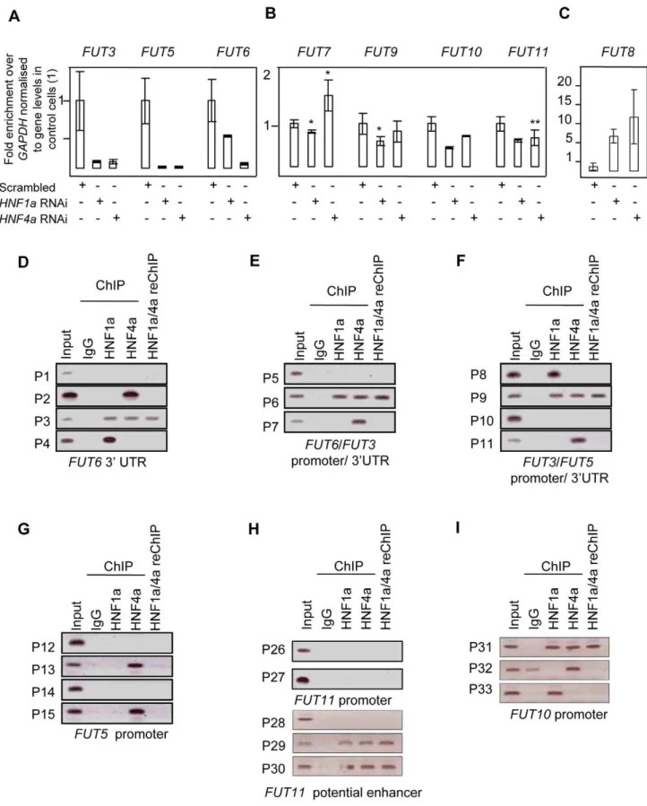
Figure 4. HNF1ais major regulator of the fucosyltransferase genes.Real time PCR forFUT3,FUT5andFUT6RNAs (A);FUT7,FUT9,FUT10and FUT11(B);FUT8(C) afterHNF1aandHNF4aRNA interference (RNAi) in HepG2 cells. H) analyses show that Hnf1a(HNF1) and Hnf4a(HNF4) bind
predicted regulatory elements for key fucosylation genes in liver cells. D), E), F), G), H) and I) Binding of HNF1aand HNF4ato predicted regulatory
elements of theFUT3,FUT5, FUT6, FUT10andFUT11.Abbreviations: the gene names are detailed in Figure 1; P1–P37: Primer sets 1 to 37; 39UTR: 39
un-translated region. reChIP: re-precipitation (or sequential) ChIP. doi:10.1371/journal.pgen.1001256.g004
HNF1aIs a Master Regulator of Fucosylation
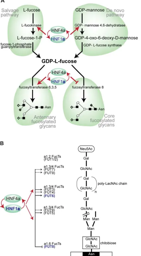
Figure 5. HNF1ais at the heart of fucosylation regulation.A). Diagram showing HNF1aand HNF4aregulation of pathways involved in fucose
synthesis. B). Pathways of core (tri-mannosyl-chitobiose) and antennary fucosylation and their proposed regulation by the transcription factors Hnf1a
and Hnf4a, encoded by theHNF1aandHNF4agenes respectively. Red arrows indicate genes that are targets of Hnf1a/Hnf4a. Solid lines show direct
regulation and dashed lines show indirect regulation of the FUT genes shown. Genes shown in blue depict GWAS identified genes (HNF1a,FUT6and
FUT8). Abbreviations: Neu5Ac, N-Acetylneuraminic (sialic) acid; GlcNAc, N-acetylglucosamine; Man, mannose; Gal, galactose; poly-LacNAc,
Galb1,4GlcNAc (LacNAc) antennary chains; Asn, asparagine residue on glycosylated protein.
an external standard of hydrolyzed and 2-AB-labeled glucose oligomerase from which the retention times for the individual glycans were converted to glucose units (GU) [51]. Glycans were analyzed on the basis of their elution positions and measured in glucose units then compared to reference values in NIBRT’s ‘‘GlycoBase v3.0 ’’ database available at http://glycobase.nibrt.ie) for structure assignment [52].
HPLC analysis was performed partly in the National Institute for Biotechnology and Training (NIBRT) in Dublin, Ireland, and partly in the Glycobiology laboratory of Genos Ltd in Zagreb, Croatia. Both laboratories used the same columns and separation conditions. Duplicate analysis of a number of samples was performed and confirmed full reproducibility of the analytical results both within and between laboratories.
Glycan structural features
Levels of glycans sharing the same structural features were approximated by adding the structures having same characteris-tics: Core fucosylated glycans (FUC-C) = DG6/(DG5+DG6)*100; Antennary fucosylated glycans (FUC-A) = DG7/(DG5+DG7)*100.
Genotype and phenotype quality control
Genotyping quality control was performed using the same procedures for all cohorts. Individuals with a call rate less than 97% were removed as well as SNPs with a call rate less than 98% (95% for CROATIA-VIS), minor allele frequency less than 0.02% or Hardy-Weinburg equilibrium p-value less than 1610210. Differences in SNP call rate threshold were used to account for observed differences between genotyping arrays. 924 individuals passed all quality control thresholds from CROATIA-VIS, 898 from CROATIA-KORCULA and 737 from ORCADES.
Extreme outliers were removed for each glycan measure to account for errors in quantification and to remove individuals not representative of normal variation within the population. An individual was classified to be an extreme outlier if their measure for the trait was more than 3 interquartile distances away from the mean.
Genome-wide association analysis
Each trait was tested for normality within each cohort then the transformation that performed best for all cohorts was used. Models including sex, age and fibrinogen as covariates were tested for each cohort separately. Any covariate that was significant within any cohort was included as a covariate in the final model. Genome-wide associations were performed for all glycan measures using the same transformation to normality and covariates for each cohort separately then combined in a meta-analysis. The ‘‘mmscore’’ function of the GenABEL package for R statistical software [53] was used for the association test under an additive model. This score test for family based association takes into account pedigree structure and allowed unbiased estimations of SNP allelic effect when relatedness is present between examinees [54]. The relationship matrix used in this analysis was generated by the ‘‘ibs’’ function of GenABEL which used IBS genotype sharing to determine the realised pairwise kinship coefficient. Meta-analysis was performed using the MetABEL package for R [53]. An association was considered statistically significant at the genome-wide level if the p-value for an individual SNP was less that 561028 (based on Bonferroni correction to account for multiple testing). All identified SNPs that reached significance or seemed to be suggestive of significance were visualised using Haploview software [55].
Association interval
An associated interval for a region of interest was defined by determining the HapMap SNPs in linkage disequilibrium of r2 .0.5 with the most significantly associated SNP in the region using the web-based program SNAP [56]. The bounds of the associated interval were determined by the flanking HapMap recombination hotspots.
Haplotype analysis
Haplotype analysis was performed on ‘‘unrelated’’ individuals in each population separately to account for possible allele frequency and haplotype differences between populations. Individuals were considered to be unrelated with a kinship coefficient of less than 0.05 (first cousins once removed). This left 525 individuals in the CROATIA-VIS cohort, 568 in CROATIA-KORCULA and 263 in ORCADES. An EM based algorithm was used to infer haplotypes from genotypic data. The ‘‘scan.haplo’’ function of the GenABEL package for R [53], which calls the ‘‘haplo.score.slide’’ function of the haplo.stats package for R [57], was used to test a sliding window of 3- and 5-SNP haplotypes across the associated interval. These results were compared to a single SNP model across the same region obtained using the ‘‘qtscore’’ function of the GenABEL package for R. A significant difference between haplotype and single-SNP analysis was determined using the Akaike information criterion [58].
ChIP identification of HNF binding sites in FUT genes
To establish whether HNF1aand HNF4abind the regulatory elements of the fucosylation genes, their genomic loci were analysed using bioinformatics to identify HNF response elements. Conserved elements between human and mouse genomes [39] were analysed initially to delineate the binding sites of HNF1aand HNF4a using the TRANSFAC database and the ECR browser (http://ecrbrowser.dcode.org/). Primers for ChIP, reChIP and real-time PCR are listed in Text S1.
RNA interference
Production of the RNA duplexes for RNA interference was described in details earlier (Kittler et al., 2005). The target sequences (see Text S1) againstHNF1aandHNF4awere designed
using the siDESIGN Center (Dharmacon). The Trasnfection of HepG2 and PANC1 cells was carried out as described by (Yu et al., 2002).
Luciferase assays
The PCR products of ChIP primers (Sequences are detailed in the Text S1) were cloned into pGEM-T easy vector (Promega) and subcloned into pGL4 vectors (Promega) as described earlier (Essafi et al., 2005). pGL4-lucconstructs (100 ng) and internal control of pRLTK (20 ng) renilla plasmid were transiently co-transfected into HepG2 and PANC1 cells (105) using the calcium phosphate co-precipitation. Cells were harvested 48 hr post-transfection for luciferase reporter assay using the Dual-Luciferase reporter assay system (Promega). The luciferase activity was normalized byRenilla
luciferase activity. All assays were performed in three separate experiments done in triplicate.
ChIP
ChIP was carried out on HepG2 cells essentially as detailed earlier (Essafi et al., 2005). The antibodies used were HNF1a (sc-6547) and HNF4a(sc-6556) from Santa Cruz Biotechnology. The corresponding control IgG antibodies were from Sigma-Aldrich.
HNF1aIs a Master Regulator of Fucosylation
Real-time PCR
RNA isolation, cDNA synthesis and Real time PCR were performed as described earlier (Birkenkamp et al., 2007). PCR primer sequences are listed in Text S1.
Supporting Information
Figure S1 PANC1 cells were treated with RNAi againstHNF1a
and HNF4a. The expression levels of the indicated genes were
analysed by real time SYBR Green PCR.
Found at: doi:10.1371/journal.pgen.1001256.s001 (0.28 MB TIF)
Figure S2 PANC1 cells were treated with RNAi againstHNF1a
and HNF4a. The expression levels of the indicated genes were
analysed by real time SYBR Green PCR.
Found at: doi:10.1371/journal.pgen.1001256.s002 (0.41 MB TIF)
Figure S3 The clustering of FUT3, FUT5 and FUT6 on chromosome 19. The position of primer pairs used for ChIP are indicated at the bottom of the figure.
Found at: doi:10.1371/journal.pgen.1001256.s003 (0.39 MB TIF)
Table S1 (A) Main structures present in glycan groups for which Genome-wide significant associations were found (SNPs inFUT8,
FUT6andHNF1agenes). (B) Glycan structures present in different HPLC peaks.
Found at: doi:10.1371/journal.pgen.1001256.s004 (0.10 MB DOC)
Table S2 Desialylated glycan traits (DG1-13, FUC-A, FUC-C) and their associations with FUT6, FUT8 and HNF1a SNPs
showing their effect sizes (Beta) in standard deviation units (with standard errors) and p-values. The fucosylation status of each trait is also shown.
Found at: doi:10.1371/journal.pgen.1001256.s005 (0.06 MB DOC)
Text S1 Supplemental Materials and Methods.
Found at: doi:10.1371/journal.pgen.1001256.s006 (0.05 MB DOC)
Acknowledgments
The authors collectively thank a large number of individuals for their help in organizing, planning, and carrying out the field work related to the project:
Professor Pavao Rudan and staff of the Institute for Anthropological Research in Zagreb, Croatia; Professor Stipan Jankovic and staff at the University of Split Medical School; Professor Ariana Vorko-Jovic and staff and medical students of the Andrija Stampar School of Public Health of the Faculty of Medicine, University of Zagreb, Croatia; Dr. Branka Salzer from the biochemistry lab ‘‘Salzer’’, Croatia; local general practitioners and nurses; and the employees of several other Croatian institutions who participated in the field work, including but not limited to the University of Rijeka, Croatia; Croatian Institute of Public Health; Institutes of Public Health in Split and Dubrovnik, Croatia. SNP Genotyping of the Vis samples was carried out by the Genetics Core Laboratory at the Wellcome Trust Clinical Research Facility, WGH, Edinburgh, Scotland, and for Korcula by Helmholtz Zentrum Mu¨nchen, GmbH, Neuherberg, Germanycompany. DNA extractions were performed at the Wellcome Trust Clinical Research Facility in Edinburgh. We would like to acknowledge the invaluable contributions of Lorraine Anderson, the research nurses in Orkney, and the administrative team in Edinburgh.
Author Contributions
Conceived and designed the experiments: G Lauc, A Essafi, C Hayward, A Knezevic, O Gornik, U Gyllensten, JF Wilson, AF Wright, ND Hastie, H Campbell, PM Rudd, I Rudan. Performed the experiments: A Essafi, JE Huffman, A Knezevic, JJ Kattla, O Polasek, V Vitart, JL Abrahams, M Pucic, M Novokmet, I Redzic, SH Wild, F Borovecki, I Kolcic, L Zgaga. Analyzed the data: G Lauc, A Essafi, JE Huffman, C Hayward, A Knezevic, JJ Kattla, O Polasek, O Gornik, V Vitart, JL Abrahams, M Pucic, M Novokmet, I Redzic, S Campbell, SH Wild, F Borovecki, W Wang, I Kolcic, L Zgaga, U Gyllensten, JF Wilson, AF Wright, ND Hastie, H Campbell, PM Rudd, I Rudan. Contributed reagents/materials/ analysis tools: G Lauc, C Hayward, O Polasek, V Vitart, S Campbell, F Borovecki, W Wang, I Kolcic, L Zgaga, U Gyllensten, JF Wilson, AF Wright, ND Hastie, H Campbell, PM Rudd, I Rudan. Wrote the paper: G Lauc, A Essafi, JE Huffman, C Hayward, O Gornik, V Vitart, U Gyllensten, JF Wilson, AF Wright, ND Hastie, H Campbell, PM Rudd, I Rudan. Led the writing of the paper: I Rudan, G Lauc. Performed functional genomic studies: A Essafi. Carried out the genotyping and performed statistical analyses: JE Huffman, C Hayward, V Vitart, S Campbell, SH Wild, F Borovecki. Performed laboratory analyses of glycans: A Knezevic, JJ Kattla, O Gornik, JL Abrahams, M Pucic, M Novokmet, I Rudan. Performed field work and constructed genealogies in Croatia: O Polasek, I Kolcic, L Zgaga. Assisted in writing of the paper and checked the manuscript for important intellectual content: W Wang, U Gyllensten, JF Wilson, AF Wright, ND Hastie, H Campbell, PM Rudd. Designed the study: U Gyllensten, JF Wilson, AF Wright, ND Hastie, H Campbell, PM Rudd, I Rudan, G Lauc.
References
1. Marth JD, Grewal PK (2008) Mammalian glycosylation in immunity. Nature Reviews Immunology 8: 874–887.
2. Dennis JW, Lau KS, Demetriou M, Nabi IR (2009) Adaptive Regulation at the Cell Surface by N-Glycosylation. Traffic 10: 1569–1578.
3. Ohtsubo K, Marth JD (2006) Glycosylation in cellular mechanisms of health and disease. Cell 126: 855–867.
4. Apweiler R, Hermjakob H, Sharon N (1999) On the frequency of protein glycosylation, as deduced from analysis of the SWISS-PROT database. Biochimica et Biophysica Acta 1473: 4–8.
5. Skropeta D (2009) The effect of individual N-glycans on enzyme activity. Bioorganic & Medicinal Chemistry 17: 2645–2653.
6. Hart GW, Housley MP, Slawson C (2007) Cycling of O-linked beta-N-acetylglucosamine on nucleocytoplasmic proteins. Nature 446: 1017–1022. 7. Crocker PR, Paulson JC, Varki A (2007) Siglecs and their roles in the immune
system. Nature Reviews Immunology 7: 255–266.
8. Brown JR, Crawford BE, Esko JD (2007) Glycan antagonists and inhibitors: a fount for drug discovery. Critical reviews in biochemistry and molecular biology 42: 481–515.
9. Jaeken J (2003) Komrower Lecture. Congenital disorders of glycosylation (CDG): it’s all in it! J Inherit Metab Dis 26: 99–118.
10. Freeze HH (2006) Genetic defects in the human glycome. Nature Reviews Genetics 7: 537–551.
11. Freeze HH (2002) Human disorders in N-glycosylation and animal models. Biochimica et Biophysica Acta 1573: 388–393.
12. Lauc G, Rudan I, Campbell H, Rudd PM (2010) Complex Genetic Regulation of Protein Glycosylation. Molecular Biosystems 6: 329–335.
13. Royle L, Campbell MP, Radcliffe CM, White DM, Harvey DJ, et al. (2008) HPLC-based analysis of serum N-glycans on a 96-well plate platform with dedicated database software. Analytical Biochemistry 376: 1–12.
14. Rudd PM, Rudan I, Wright AF (2009) High-Throughput Glycome Analysis Is Set To Join High-Throughput Genomics. Journal of Proteome Research 8: 1105– 1105.
15. Knezˇevic´ A, Polasˇek O, Gornik O, Rudan I, Campbell H, et al. (2009) Variability, Heritability and Environmental Determinants of Human Plasma N-Glycome. Journal of Proteome Research 8: 694–701.
16. Gornik O, Wagner J, Pucˇic´ M, Knezˇevic´ A, Redzˇic´ I, et al. (2009) Stability of N-glycan profiles in human plasma. Glycobiology 19: 1547–1553.
17. Knezˇevic´ A, Gornik O, Polasˇek O, Pucˇic´ M, Novokmet M, et al. (2010) Effects of aging, body mass index, plasma lipid profiles, and smoking on human plasma N-glycans. Glycobiology 20: 959–969.
18. Pucˇic´ M, Pinto S, Novokmet M, Knezˇevic´ A, Gornik O, et al. (2010) Common aberrations from normal human N-glycan plasma profile. Glycobiology 20: 970–975.
19. Becker DJ, Lowe JB (2003) Fucose: biosynthesis and biological function in mammals. Glycobiology 13: 41R–53R.
21. Javaud C, Dupuy F, Maftah A, Julien R, Petit JM (2003) The fucosyltransferase gene family: an amazing summary of the underlying mechanisms of gene evolution. Genetica 118: 157–170.
22. Gornik O, Lauc G (2008) Glycosylation of serum proteins in inflammatory diseases. Disease Markers 25: 267–278.
23. Miyoshi E, Moriwaki K, Nakagawa T (2008) Biological function of fucosylation in cancer biology. Journal of Biochemistry 143: 725–729.
24. Odom DT, Zizlsperger N, Gordon DB, Bell GW, Rinaldi NJ, et al. (2004) Control of pancreas and liver gene expression by HNF transcription factors. Science 303: 1378–1381.
25. Yamagata K, Furuta H, Oda N, Kaisaki PJ, Menzel S, et al. (1996) Mutations in the hepatocyte nuclear factor-4alpha gene in maturity-onset diabetes of the young (MODY1). Nature 384: 458–460.
26. Yamagata K, Oda N, Kaisaki PJ, Menzel S, Furuta H, et al. (1996) Mutations in the hepatocyte nuclear factor-1alpha gene in maturity-onset diabetes of the young (MODY3). Nature 384: 455–458.
27. Elliott P, Chambers JC, Zhang W, Clarke R, Hopewell JC, et al. (2009) Genetic Loci associated with C-reactive protein levels and risk of coronary heart disease. JAMA 302: 37–48.
28. Yuan X, Waterworth D, Perry JR, Lim N, Song K, et al. (2008) Population-based genome-wide association studies reveal six loci influencing plasma levels of liver enzymes. American Journal of Human Genetics 83: 520–528.
29. Reiner AP, Gross MD, Carlson CS, Bielinski SJ, Lange LA, et al. (2009) Common coding variants of the HNF1A gene are associated with multiple cardiovascular risk phenotypes in community-based samples of younger and older European-American adults: the Coronary Artery Risk Development in Young Adults Study and The Cardiovascular Health Study. Circ Cardiovasc Genet 2: 244–254.
30. Barrett JC, Lee JC, Lees CW, Prescott NJ, Anderson CA, et al. (2009) Genome-wide association study of ulcerative colitis identifies three new susceptibility loci, including the HNF4A region. Nature Genetics 41: 1330–1334.
31. Kathiresan S, Willer CJ, Peloso GM, Demissie S, Musunuru K, et al. (2009) Common variants at 30 loci contribute to polygenic dyslipidemia. Nature Genetics 41: 56–65.
32. Vitart V, Rudan I, Hayward C, Gray NK, Floyd J, et al. (2008) SLC2A9 is a newly identified urate transporter influencing serum urate concentration, urate excretion and gout. Nature Genetics 40: 437–442.
33. Taniguchi N, Honke K, Fukuda M, eds (2002) Handbook of glycosyltransferases and related genes. Tokyo: Springer Verlag.
34. Brinkman-Van der Linden EC, Mollicone R, Oriol R, Larson G, Van den Eijnden DH, et al. (1996) A missense mutation in the FUT6 gene results in total absence of alpha3-fucosylation of human alpha1-acid glycoprotein. J Biol Chem 271: 14492–14495.
35. Ma B, Simala-Grant JL, Taylor DE (2006) Fucosylation in prokaryotes and eukaryotes. Glycobiology 16: 158R–184R.
36. Higai K, Miyazaki N, Azuma Y, Matsumoto K (2008) Transcriptional regulation of the fucosyltransferase VI gene in hepatocellular carcinoma cells. Glycoconjugate Journal 25: 225–235.
37. Servitja JM, Pignatelli M, Maestro MA, Cardalda C, Boj SF, et al. (2009) Hnf1alpha (MODY3) controls tissue-specific transcriptional programs and exerts opposed effects on cell growth in pancreatic islets and liver. Molecular & Cellular Biology 29: 2945–2959.
38. Boj SF, Petrov D, Ferrer J (2010) Epistasis of transcriptomes reveals synergism between transcriptional activators Hnf1alpha and Hnf4alpha. PLoS Genet 6: e1000970. doi:10.1371/journal.pgen.1000970.
39. Bejerano G, Siepel AC, Kent WJ, Haussler D (2005) Computational screening of conserved genomic DNA in search of functional noncoding elements. Nature Methods 2: 535–545.
40. Odom DT, Dowell RD, Jacobsen ES, Gordon W, Danford TW, et al. (2007) Tissue-specific transcriptional regulation has diverged significantly between human and mouse. Nat Genet 39: 730–732.
41. Loots G, Ovcharenko I (2007) ECRbase: database of evolutionary conserved regions, promoters, and transcription factor binding sites in vertebrate genomes. Bioinformatics 23: 122–124.
42. Boyd M, Bressendorff S, Moller J, Olsen J, Troelsen JT (2009) Mapping of HNF4alpha target genes in intestinal epithelial cells. BMC Gastroenterol 9: 68. 43. Benko S, Fantes JA, Amiel J, Kleinjan DJ, Thomas S, et al. (2009) Highly conserved non-coding elements on either side of SOX9 associated with Pierre Robin sequence. Nat Genet 41: 359–364.
44. Essafi A, Fernandez de Mattos S, Hassen YA, Soeiro I, Mufti GJ, et al. (2005) Direct transcriptional regulation of Bim by FoxO3a mediates STI571-induced apoptosis in Bcr-Abl-expressing cells. Oncogene 24: 2317–2329.
45. Withers DA, Hakomori SI (2000) Human alpha (1,3)-fucosyltransferase IV (FUTIV) gene expression is regulated by elk-1 in the U937 cell line. Journal of Biological Chemistry 275: 40588–40593.
46. Erdmann J, Grosshennig A, Braund PS, Konig IR, Hengstenberg C, et al. (2009) New susceptibility locus for coronary artery disease on chromosome 3q22.3. Nature Genetics 41: 280–282.
47. Kel AE, Niehof M, Matys V, Zemlin R, Borlak J (2008) Genome wide prediction of HNF4alpha functional binding sites by the use of local and global sequence context. Genome Biol 9: R36.
48. Brandley BK, Kiso M, Abbas S, Nikrad P, Srivasatava O, et al. (1993) Structure-function studies on selectin carbohydrate ligands. Modifications to fucose, sialic acid and sulphate as a sialic acid replacement. Glycobiology 3: 633–641. 49. Luhn K, Marquardt T, Harms E, Vestweber D (2001) Discontinuation of fucose
therapy in LADII causes rapid loss of selectin ligands and rise of leukocyte counts. Blood 97: 330–332.
50. Klein A (2008) Human total serum N-glycome. Advances in Clinical Chemistry 46: 51–85.
51. Royle L, Radcliffe CM, Dwek RA, Rudd PM (2006) Detailed structural analysis of N-glycans released from glycoproteins in SDS-PAGE gel bands using HPLC combined with exoglycosidase array digestions. Methods Mol Biol 347: 125–143.
52. Campbell MP, Royle L, Radcliffe CM, Dwek RA, Rudd PM (2008) GlycoBase and autoGU: tools for HPLC-based glycan analysis. Bioinformatics 24: 1214–1216.
53. Aulchenko YS, Ripke S, Isaacs A, van Duijn CM (2007) GenABEL: an R library for genome-wide association analysis. Bioinformatics 23: 1294–1296. 54. Chen WM, Abecasis GR (2007) Family-based association tests for genomewide
association scans. Am J Hum Genet 81: 913–926.
55. Barrett JC, Fry B, Maller J, Daly MJ (2005) Haploview: analysis and visualization of LD and haplotype maps. Bioinformatics 21: 263–265. 56. Johnson AD, Handsaker RE, Pulit SL, Nizzari MM, O’Donnell CJ, et al. (2008)
SNAP: a web-based tool for identification and annotation of proxy SNPs using HapMap. Bioinformatics 24: 2938–2939.
57. Schaid DJ, Rowland CM, Tines DE, Jacobson RM, Poland GA (2002) Score tests for association between traits and haplotypes when linkage phase is ambiguous. Am J Hum Genet 70: 425–434.
58. Akaike H (1974) New Look at Statistical-Model Identification. Ieee Transactions on Automatic Control AC19: 716–723.
HNF1aIs a Master Regulator of Fucosylation