Advanced Pharmaceutical Bulletin, 2013, 3(2), 425-428 doi: http://dx.doi.org/10.5681/apb.2013.068
http://apb.tbzmed.ac.ir/
*Corresponding author: Hadi Valizadeh, Department of Pharmaceutics, Faculty of Pharmacy, Tabriz University of Medical Sciences, Tabriz, Iran. Tel: +98 (411) 339-2649, Fax: +98 (411) 334-4798, Email: [email protected]
Copyright © 2013 by Tabriz University of Medical Sciences
Preparation, Physicochemical Characterization and Performance
Evaluation of Gold Nanoparticles in Radiotherapy
Ali Kamiar1, Reza Ghotaslou2, Hadi Valizadeh3*
1 Faculty of Pharmacy, Student Research Committee, Tabriz University of Medical Sciences, Tabriz, Iran.
2
Department of Microbiology, School of Medicine, Tabriz University of Medical Sciences, Tabriz, Iran.
3
Research Center for Pharmaceutical Nanotechnology and Faculty of Pharmacy, Tabriz University of Medical Sciences, Tabriz, Iran.
Introduction
Nanotechnology is beginning to show its impact on the way the health care is administered. These include new interventions in disease detection, treatment and prevention, which are collectively termed as nanomedicine.1 The most well-studied nanoparticles include quantum dots, carbon nanotubes, paramagnetic nanoparticles, liposomes, gold nanoparticles (GNPs), and many others.2 In recent years, gold nanoparticles have attracted much attention. They are agents with numerous applications in biomedicine like cancer research, diagnostic assay,3-5 thermal ablation, gene and drug delivery,6-8 etc. Nano gold have several unique properties, For example they are inert and nontoxic9 and have good anti-bacterial,10 anti-angiogenesis properties,11 etc. GNPs have been prepared by both “physical” and “chemical” methods. For the “physical” preparation method, Au bulk is broken down by a strong attack force, for example, ion irradiation in air or arc discharge in water, to generate GNPs. Chemical method including chemical reduction of Au salts, electrochemical pathways and decomposition of organometallic compounds. Among them, the chemical reduction method is simple and controllable to prepare various sizes and shapes of GNPs.12,13
Today Cancer is the third leading cause of death in developed countries and the second leading cause of death in the United States.2 Treatment of Cancer includes chemotherapy, surgery and radiotherapy. Although radiation therapy is one of the most preferred cure and has been practiced for about 100 years in cancer treatment, but this treatment has a lot of side effects. So scientists are looking for new ways to enhance effect of radiotherapy and lower damage to the normal cell.14 The concept of using high-Z materials as dose enhancement in cancer radiotherapy has long been investigated. Several studies have focused on the potential application of GNPs in conjunction with radiation therapy.15 The aim of this project was preparation, characterization of GNPs with the intention of absorbed dose enhancement in tumor cells. Anti-bacterial effect of prepared GNPs against clinical strains of E. coli was also investigated.
Materials and Methods Materials
HAuCl4 was purchased from Alfa Aesar (Great Britain). Tri sodium citrate was obtained from Scharlau (Spania). N,N'-methylenebis-acrylamide (bis) acrylamide (AA), Tetrakis (Hydroxymethyl) A R T I C L E I N F O A B S T R A C T
Article Type: Research Article
Article History: Received: 23 April 2013 Revised: 30 May 2013 Accepted: 2 June 2013 ePublished: 20 August 2013
Keywords: Gold Nano particle Dose enhancement Radiation therapy Gel dosimetry Anti-bacterial
426 | Advanced Pharmaceutical Bulletin, 2013, 3(2), 425-428 Copyright © 2013 by Tabriz University of Medical Sciences Kamiar et al.
Phosphonium Chloride and Gelatinwere obtained from Sigma chemical company. Mueller Hinton agar was purchased from Liofilchem. De-ionized water was used to prepare aqueous solutions.
Gold nanoparticles preparation
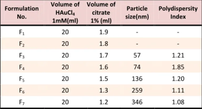
Gold nanoparticles were prepared by the classical citrate reduction (frens method). Briefly, 20ml of HAuCl4 water solution (1 mM) was kept boiling. Various volume of 1% sodium citrate water solution was then added to the solution and stirred for about 10 min, until the formation of a colored gold nanoparticle suspension. Table 1 shows different citrate volume that use for preparation of GNPs.
Table 1. Different formulation for preparation of gold nanoparticle and their particle size and polydispersity index
Formulation No.
Volume of HAuCl4
1mM(ml)
Volume of citrate 1% (ml)
Particle size(nm)
Polydispersity Index
F1 20 1.9 - -
F2 20 1.8 - -
F3 20 1.7 57 1.21
F4 20 1.6 74 1.85
F5 20 1.5 136 1.20
F6 20 1.3 259 1.11
F7 20 1.2 346 1.08
Characterization of GNPs
The mean particle-size values of GNPs were measured by using a laser diffraction particle-size analyzer (Sald 2101, Shimadzu, Japan) equipped with Wing software (version 1.20). The morphology of the nanoparticles was investigated by Transmission electron microscopy (TEM) (LEO906, Germany). Drops of the gold suspension (formulation F6) were deposited and dried onto a Formvar-coated copper grid. The UV–visible absorption spectra of the one of the prepared colloidal solutions recorded using a spectrophotometer (Shimadzu, Japan), from 400 to 900 nm.
Gel dosimetry
Gel fabrication
The gel solution consists of water (89 % of total mass), acrylamide (3 %), N,N-methylene-bisacrylamide (3 %) and gelatin (5 %). The gel components were mixed together at 35-40 °C in a 500 ml beaker. An oxygen scavenger, Tetrakis (hydroxyl methyl) phosphonium chloride (THPC), was added to the gel mixture at a concentration of 10 mM as anti-oxidant. Nano gold (formulation F3) was used as a part of water in gel preparation to fabricate gel with GNPs batch. The GNPs were observed to mix homogeneously in the gel. Another batch of gel without GNPs served as a control. The gel was then quickly poured into separate tubes.
Irradiation
The tubes of the both batches were irradiated with CT scanner after put them in a head and neck phantom.
The gel samples were exposed to radiation doses of 40, 80 and 120 Gy. Irradiation of the gel samples was carried out at Day CT scanner center with following parameters: slice thickness=1 cm, t=0.8 s/turn, mA=200, kVp =140.
Magnetic Resonance Imaging (MRI)
Irradiated and non-irradiated gel samples were scanned using a 1.5 T MRI scanner (GE Sigma, Milwaukee, USA), to measure spin–spin relaxation time of the free protons using a head coil. A fast-spin echo sequence was used with following parameters: field of view =105 × 120mm2, slice thickness = 5 mm (kV X-ray beams), effective echo time TE = 22 ms, turbo factor = 14, repetition time (TR) = 5,710 ms, the field of view = 128 ×128 matrix, total imaging time = 20 min. At least 24 h elapsed after irradiation prior to imaging to allow for polymerization. All the samples were scanned at room temperature.
Data analysis
Analysis of the image was performed using MATLAB software (version 3.5.7) (The Math Works Inc, Natick, Massachusetts, USA). The program examined the data before analyzing it to determine the region of interest. T2 values were calculated and formed T2 maps on a pixel-by-pixel basis. The levels of the polymerization of the irradiated gels with and without GNPs were compared by calculating the R2 (1/T2).
Anti-bacterial test
Antibacterial activity was studied by the agar-well-diffusion method, wherein 100 l bacterial suspension was added to 20-mL sterile nutrient Mueller Hinton agar at 45 °C and the mixture was solidified on a Petri dish. After the medium had solidified, 7-mm-diameter wells were made in the agar (three wells per dish) that were equidistant from one another and from the dish edge. The wells received 150 L of different concentration of GNPs from formulation F2 (400 ppm, 200 ppm, 100 ppm). The petri dishes were incubated in a thermostat at 37 °C for 24 h. After incubation, the diameter of the zone of bacterial-growth inhibition was measured. All experiments were done for five clinical strains of E. coli and repeated thrice.
Results and Discussion
Physicochemical properties of GNPs
| 427 Advanced Pharmaceutical Bulletin, 2013, 3(2), 425-428
Copyright © 2013 by Tabriz University of Medical Sciences
Enhanced radiotherapy dose absorption by gold nanoparticles
Figure 1. TEM image of gold nanoparticle (formulation F6)
Figure 2. Particle size distribution of gold nanoparticles (formulation F1)
Figure 3. UV–Vis spectra of 250 nM gold nanoparticles (formulation F3)
Gel dosimetry
The relationship between the delivered X-ray dose and the R2 (spin–spin relaxation rate) was investigated to characterize the effect of GNPs using polymer gel. R2 is equal to 1/T2 (spin–spin relaxation time) and is a function of dose (dose delivered to water). A linear relationship is found between delivered dose and R2 (Figure 4). The dose–response slopes for R2 versus delivered X-ray dose for gel–GNP and pure gel were calculated. The ratio of these slopes was taken as the dose enhancement factor (DEF). The DEF of 1.21 was obtained for the dose–response relationship. Dose enhancement by high Z material is believed to be caused predominantly by enhancing the likelihood of the photoelectric interaction. When GNPs are added to the gel prior to irradiation and bombarded with kilovoltage X-rays, the photoelectric interaction cross section will increase. This can be clearly inferred from the interaction probability of these X-ray photons with gold atoms compared to their interaction with the tissue equivalent medium such as water.
Figure 4. dose–response curve for pure gel and gel–GNPs
Antimicrobial activity of GNPs against E. coli clinical strains
428 | Advanced Pharmaceutical Bulletin, 2013, 3(2), 425-428 Copyright © 2013 by Tabriz University of Medical Sciences Kamiar et al.
CTAB is a potent anti-microbial agent, so this difference may be related to use of this material.
Table 2. Inhibition zone diameter (mm) of different
concentration of nano gold loaded in plates with E. coli clinical
strainsinoculums
Strain 100 ppm 200 ppm 400 ppm
1 0 8.33 10
2 5.5 9 11
3 0 8.66 11.33
4 0 5.33 11.33
5 0 2.66 8.16
Conclusion
We reported here the measurement of radiation dose enhancement generated by GNPs using polymer gel dosimeters as a phantom. This study has found a significant dose enhancement from the inclusion of the GNPs within polymer gels irradiated with kilovoltage X-rays beams from a therapy machine. Besides, GNPs exhibited a good anti-microbial effect against E. coli clinical strains at 400 ppm concentration.
Acknowledgements
The authors would like to thank the authority of student research committee, Tabriz university medical sciences for their support.
Conflict of Interest
The authors report no conflicts of interest.
References
1. Rees M, Moghimi SM. Nanotechnology: From fundamental concepts to clinical applications for healthy aging. Nanomed Nanotechnol Biol Med
2012;8(Suppl 1):S1-4.
2. Cai W, Gao T, Hong H, Sun J. Applications of gold nanoparticles in cancer nanotechnology.
Nanotechnol Sci Appl 2008;1(1):17-32.
3. Sun IC, Eun DK, Na JH, Lee S, Kim IJ, Youn IC, et al. Heparin-coated gold nanoparticles for
liver-specific CT imaging. Chemistry
2009;15(48):13341-7.
4. Fournelle M, Bost W, Tarner IH, Lehmberg T, WeiB E, Lemor R, et al. Antitumor necrosis factor- antibody-coupled gold nanorods as nanoprobes for molecular optoacoustic imaging in arthritis.
Nanomed 2012;8(3):346-54.
5. Lee H, Lee K, Kim IK, Park TG. Synthesis, characterization, and in vivo diagnostic applications of hyaluronic acid immobilized gold nanoprobes.
Biomaterials 2008;29(35):4709-18.
6. Lee SH, Bae KH, Kim SH, Lee KR, Park TG. Amine-functionalized gold nanoparticles as
non-cytotoxic and efficient intracellular siRNA delivery carriers. Int J Pharm 2008;364(1):94-101.
7. Ryou SM, Kim S, Jang HH, Kim JH, Yeom JH, Eom MS, et al. Delivery of shRNA using gold nanoparticle-DNA oligonucleotide conjugates as a universal carrier. Biochem Biophys Res Commun
2010;398(3):542-6.
8. Rivera Gil P, Huhn D, Del Mercato LL, Sasse D, Parak WJ. Nanopharmacy: Inorganic nanoscale devices as vectors and active compounds.
Pharmacol Res 2010;62(2):115-25.
9. Patra CR, Bhattacharya R, Mukhopadhyay D, Mukherjee P. Fabrication of gold nanoparticles for targeted therapy in pancreatic cancer. Adv Drug Deliv Rev 2010;62(3):346-61.
10. Dastjerdi R, Montazer M. A review on the application of inorganic nano-structured materials in the modification of textiles: focus on anti-microbial properties. Colloids Surf B Biointerfaces
2010;79(1):5-18.
11. Mukherjee P, Bhattacharya R, Wang P, Wang L, Basu S, Nagy JA, et al. Antiangiogenic properties of gold nanoparticles. Clin Cancer Res
2005;11(9):3530-4.
12. Schmid G, Corain B. Nanoparticulated gold: Syntheses, structures, electronics, and reactivities.
Eur J Inorg Chem 2003;2003(17):3081-98.
13. Nguyen DT, Kim DJ, Kim KS. Controlled synthesis and biomolecular probe application of gold nanoparticles. Micron 2011;42(3):207-27. 14. Cho SH, Jones BL, Krishnan S. The dosimetric
feasibility of gold nanoparticle-aided radiation therapy (gnrt) via brachytherapy using low-energy gamma-/x-ray sources. Phys Med Biol
2009;54(16):4889-905.
15. Chang MY, Shiau AL, Chen YH, Chang CJ, Chen HH, Wu CL. Increased apoptotic potential and dose-enhancing effect of gold nanoparticles in combination with single-dose clinical electron beams on tumor-bearing mice. Cancer Sci
2008;99(7):1479-84.
16. Lasagna-Reeves C, Gonzalez-Romero D, Barria MA, Olmedo I, Clos A, Sadagopa Ramanujam VM, et al. Bioaccumulation and toxicity of gold nanoparticles after repeated administration in mice.
Biochem Biophys Res Commun 2010;393(4):649-55.
17. Cui Y, Zhao Y, Tian Y, Zhang W, Lu X, Jiang X. The molecular mechanism of action of bactericidal gold nanoparticles on Escherichia coli. Biomaterials
2012;33(7):2327-33.
18. Arshi N, Ahmed F, Kumar S, Anwar MS, Lu J, Koo BH, et al. Microwave assisted synthesis of gold nanoparticles and their antibacterial activity against escherichia coli (E. Coli). Curr Appl Phys