Int J Anat Res 2014, 2(1):287-88. ISSN 2321-4287 288 Case Report
THE RARE CASE OF LEFT RETRO AORTIC RENAL VEIN: SURGICO
ANATOM IC RAM IFICATIONS OF THE ABNORM ALITY
Ashfaq Ul Hassan *
1, Zahida Rasool
2, Nissar Chaudhary
3, M uneeb Ul Hassan
4.
ABSTRACT
Address for Correspondence: Dr. Ashfaq Ul Hassan,M BBS M D, Lect urer SKIM S M edical College Bemina Srinagar, Kashmir, India. E-M ail: [email protected]
Access this Article online
Quick Response code Web site:
*1 M BBS M D, Lect urer, SKIM S M edical College Bemina Srinagar, Kashmir, India.
2 M BBS, M edical Consultant , Islamic Universit y of Science and Technology, Awant ipora, Kashmir
India.
3 Professor, General Surgery, SKIM S, Soura.
4 Assistant Surgeon, Direct orate of Healt h Services, Srinagar, Kashmir, India.
The Art icle present s a rare Anom oly of Course of Renal Vein w here it lies behind t he Aorta . The Con sequence of failure t o Recognize t his clinical ent it y can be dangerous if left Undet ect ed. This Vein is locat ed bet w een t he Aor ta and t he Vert ebrae. The Pat ient present ed w it h left Flank Pain and on Evaluat ion w as found t o have Left Ret roAort ic Renal Vein.
KEYW ORDS:Kidney; Renin; Transplant ; Ret ro Aor t ic; Abberrat ion.
INTRODUCTION
Int ernat ional Journal of Anatomy and Research,
Int J Anat Res 2014, Vol 2(1):287-88. ISSN 2321- 4287
Received: 08 M arch 2014
Peer Review : 05 M arch 2014 Published (O):30 M arch 2014 Accepted: 18 M arch2014 Published (P):30 M arch 2014
Internat ional Journal of Anat omy and Research ISSN 2321-4287
w w w.ijmhr.org/ ijar.ht m
The kidneys are sit uated ret roperit onealy on t he posterior abdominal wall on each side of t he ver-tebral column. The right kidney is slight ly low er t han left and t he left kidney is a litt le nearer t o t he median plane. The kidneys vert ically extend from upper border of T12 vertebra t o cent re of body of L3 Vertebra. The Real Veins are t he main Venous drainage t o t he Kidney. The Renal veins are know n for t heir Anat om ic Variat ion and hence for a Surgeon Operat ing in t his region. A Proper Know ledge of t he Vasculat ure of Kidneys is of Ut most Importance.
DISCUSSION
The Normal course of t he left and right Renal v ein sho u l d b e w el l k n ow n t o a su r geo n especially in case of vascular inj ur y t o t he Kidneys . For t he blunt abdominal t rauma or t he Penet rat ing t rauma especially in pat ient w it h
hem at ur ia, it is helpful if t he preoperat ive evaluat ion includes an assessm ent of renal f unct ion [1]. In case a surgical Approach is n eed ed , Th e l ef t r en al v essel s can b e approached t hrough a direct exposure at t he base of t he mesocolon. During Renal Surgeries t his Anat om ic Variat ion severly effect s t he technical feasibilit y of t he Surgery [2]. Failure t o recognize can lead to a profound Hemorrage and severe renal failure. The Posterior Nut cracker Phenomenon can occur as a result of decreased space left and cause renal venous hypertension w it h subsequent renal vein compression and obst ruct ion and m ore chances of Varicocele format ion [3].
Int J Anat Res 2014, 2(1):287-88. ISSN 2321-4287 28930
Ashfaq Ul Hassan et al., THE RARE CASE OF LEFT RETRO AORTIC RENAL VEIN: SURGICO ANATOM IC RAM IFICATIONS OF THE ABNORM ALITY.
Evaluat ing t he various causes of failure of Renal t ran spl an t p ro cedu r es a cau se can b e t hrombosis of renal vein as a result of w hich nor m al Urinar y f low m ay be obst r uct ed by edema, clot , stenosis, or kinking of t he ureter w hich in t urn can impair glomerular filt rat ion.
In cases w here Early reexploration t o cont rol t he profound bleeding, relieve renal compression, and rest ore blood volume usually causes prompt r et u r n o f r en al f un ct io n . M o der n Day Radionuclide renal scans and arteriography are helpful in t he different ial diagnosis of early postt ransplantat ion of w hich one cause can be an aberrant course of renal veins [5].
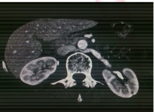
Fig. 1: CT Scan Dem onst rat ing t he Rare Left Ret roAort ic Renal Vein.
Fig. 2: CT Scan Dem onst rat ing t he Rare Left Ret roAort ic Renal Vein.
The kidney may also be affected adversely by by renal vein t hrom bosis, w hich may also cause renal arterial t hrombosis. The Chain of event s w hich follow can be very dangerous leading t o Renal Dysfunct ion or renal failure.
In case of Renal Transplant s, usually The left kidney is chosen as it has t he longer renal vein
w h ich faci li t at es t h e reci pi en t o p er at io n . Ho w ever, if t he veno gr am sh ow s such an aberrat ion ext ra care is needed.
For t he The convent ional splenorenal shunt w hich consist s of anast omosis of t he proximal part of t he splenic vein t o t he renal vein. W it h such an abbert ion in t he course of Renal vein A non convent ional splenorenal shunt w ould be required.
M odern day assessment of Hypertension and t he use of renal vein renin measurement have considerable importance especially in pat ient s w it h significant renovascular hypertension [6]. Pat ient s w it h hypertension and abnormal renal arteries only rarely exhibit a renal vein renin ratio greater t han 1.5 t o 2.0:1, w hereas in pat ient s w it h funct ionally significant renal stenosis, renal vein renin concent rat ions are at least one and one-half t o t w o t im es higher in renal venous blood of t he involved or affected kidney t han in t he renal effluent of t he cont ralateral venous drainage. The Outcomes of Ret ro Aort ic Renal Vein are t hus of great significance.
CONCLUSION
As a Conclusion t he Det ailed Know ledge of Anomalies of Renal Veins is important t o make a dist inct ive diagnosis of ret roperit oneal nodes, pathologies, masses and renal vascular diseases and t o impede complicat ions w hich can occur during ret roperit oineal surgical procedures.
Conflicts of Interests: None
REFERENCES
[1] . Hayashi M , Kum e T, Nihira H. Abnorm alit ies of r e n al ven o u s syst e m an d u n exp l ai n ed r en al
hematuria. J Urol. 1980;124:12–16.
[2] . Thom as TV. Surgical im plicat ions of ret roaor t ic left renal vein. Arch Surg. 1970;100:738–740.
[3]. M at hew s R, Sm it h PA, Fishm an EK, M arshall FF. Anom alies of t he inferior vena cava and renal veins: e m b r y o l o gi c an d su r gi cal
considerations. Urology. 1999;53:873–880.
[4] . Shindo S, Kubot a K, Kojim a A, Iyori K, Ishim ot o T, Kobayashi M , et al. Anom alies of infer ior vena cava and left renal vein: r isks in aort ic surgery. Ann Vasc
Surg. 2000;14:393–396.
[5]. Hoeltl W, Hruby W, Aharinejad S. Renal vein anat omy and it s im plicat ions for ret roperit oneal surgery. J
Urol. 1990;143:1108–1114.
[6] . Gi bo M , Onit suka H. Ret roaort ic left renal vein w i t h r e n al v ei n h y p er t en si o n cau si n g hem at uria.Clin Im aging. 1998;22:422–424.