Genomic Analysis Reveals the Molecular
Basis for Capsule Loss in the Group B
Streptococcus
Population
Roberto Rosini1, Edmondo Campisi1, Matteo De Chiara1, Hervé Tettelin2, Daniela Rinaudo1, Chiara Toniolo1, Matteo Metruccio1, Silvia Guidotti1, Uffe B.
Skov Sørensen3, Mogens Kilian3, DEVANI Consortium¶, Mario Ramirez4,
Robert Janulczyk1, Claudio Donati5, Guido Grandi1, Immaculada Margarit1
*
1Novartis Vaccines and Diagnostics Srl, Siena, Italy,2Institute for Genome Sciences, University of Maryland School of Medicine, Baltimore, Maryland, United States of America,3Department of Biomedicine, Aarhus University, Aarhus, Denmark,4Instituto de Microbiologia, Instituto de Medicina Molecular,
Faculdade de Medicina, Universidade de Lisboa, Lisbon, Portugal,5Department of Computational Biology, Research and Innovation Centre, Fondazione Edmund Mach, Via E. Mach 1, San Michele all'Adige, Italy
¶ Membership of the Devani Consortium is listed in the Acknowledgments. *[email protected]
Abstract
The human and bovine bacterial pathogenStreptococcus agalactiae(Group B Streptococ-cus, GBS) expresses a thick polysaccharide capsule that constitutes a major virulence factor and vaccine target. GBS can be classified into ten distinct serotypes differing in the chemical composition of their capsular polysaccharide. However, non-typeable strains that do not react with anti-capsular sera are frequently isolated from colonized and infected humans and cattle. To gain a comprehensive insight into the molecular basis for the loss of capsule ex-pression in GBS, a collection of well-characterized non-typeable strains was investigated by genome sequencing. Genome based phylogenetic analysis extended to a wide population of sequenced strains confirmed the recently observed high clonality among GBS lineages mainly containing human strains, and revealed a much higher degree of diversity in the bo-vine population. Remarkably, non-typeable strains were equally distributed in all lineages. A number of distinct mutations in thecpsoperon were identified that were apparently responsi-ble for inactivation of capsule synthesis. The most frequent genetic alterations were point mutations leading to stop codons in thecpsgenes, and the main target was found to becpsE encoding the portal glycosyl trasferase of capsule biosynthesis. Complementation of strains carrying missense mutations incpsEwith a wild-type gene restored capsule expression al-lowing the identification of amino acid residues essential for enzyme activity.
Introduction
Streptococcus agalactiae(Group B Streptococcus, GBS) colonizes the gastrointestinal and/or genital tract of about 20% of women and can cause neonatal sepsis and meningitis with high
a11111
OPEN ACCESS
Citation:Rosini R, Campisi E, De Chiara M, Tettelin H, Rinaudo D, Toniolo C, et al. (2015) Genomic Analysis Reveals the Molecular Basis for Capsule Loss in the Group BStreptococcusPopulation. PLoS ONE 10(5): e0125985. doi:10.1371/journal. pone.0125985
Academic Editor:Eliane Namie Miyaji, Instituto Butantan, BRAZIL
Received:January 14, 2015
Accepted:March 27, 2015
Published:May 6, 2015
Copyright:© 2015 Rosini et al. This is an open access article distributed under the terms of the
Creative Commons Attribution License, which permits unrestricted use, distribution, and reproduction in any medium, provided the original author and source are credited.
Data Availability Statement:The newly sequenced genomes were deposited in the NCBI database under the Bioproject ID PRJNA278931.
Funding:The authors confirm that the Novartis Vaccines funder provided support in the form of salaries for authors RR DR SG RJ GG IM, but did not have any additional role in the study design, data collection and analysis, decision to publish, or preparation of the manuscript. The specific roles of all authors are articulated in the‘author contributions’
mortality rates. It is also an important cause of morbidity in the elderly and immunocompro-mised adults, and of bovine mastitis [1]. The polysaccharide capsule of GBS is an important virulence factor conferring anti-phagocytic properties. Low anti-capsular antibody titers corre-late with increased risk of disease, and capsular polysaccharide-based vaccines are currently under development [2]. The synthesis of the GBS capsular polysaccharide (CPS) requires mul-tiple enzymes encoded in thecpsoperon. This gene cluster shares homologies with operons from other encapsulated Gram-positive bacteria [3] and with those responsible for the synthe-sis of the lipopolysaccharide O-antigen in Gram-negatives [4]. In all these bacteria, the polysac-charide repeating unit is synthesized on the membrane acceptor undecaprenyl phosphate by multiple glycosyl transferases, transported outside the cell membrane by a translocase and as-sembled into high molecular weight polymers [3].
GBS can be classified into 10 distinct serotypes varying in polysaccharide composition, based on reactivity with specific antisera by using Lancefield precipitation or latex agglutina-tion tests [5]. Yet, some isolates are considered as non-typeable (NT) because they do not react with any anti-capsular serum. The frequencies of detected NTs in GBS epidemiological studies have recently decreased with the development of serotyping methods of improved sensitivity [6,7]. However, they still account for 5–20% of the isolates colonizing or infecting human adults [8] and for 30–77% of those obtained from bovine mastitis [6,9–11].
The GBS NT phenotype could in principle be due to low or complete lack of capsule expres-sion, or to the presence of yet unknown polysaccharide variants that do not react with the avail-able typing antisera [6]. The molecular basis for the absence of capsule expression, and the genetic relationship between NT and encapsulated GBS strains have been poorly investigated to date. Newly developed molecular typing methods based on PCR amplification of the differ-ent GBScpsalleles have allowed assigning capsular genotypes to most NT strains [6,12,13]. Ramaswamyet al., [14] identified an IS1381 insertion in thecpsEgene as possibly responsible for the NT phenotype of a capsular genotype V strain. Furthermore, a single strain lacking the entire capsule locus was recently described [15].
Here we undertook a comprehensive study of the GBS non-encapsulated population by geno-mic analysis of a large collection of colonizing and invasive strains obtained both from humans and cattle. To achieve a high-resolution characterization of the GBS population structure and to map the distribution of NT isolates among the different lineages, we constructed phylogenetic trees based on genome Single Nucleotide Polymorphisms (SNPs) or Multiple Locus Sequence Typing (MLST), including in the analysis all publicly available sequenced strains. Furthermore, the different types of genetic alterations leading to capsule loss and the main target genes of these mutations were identified by DNA sequence comparison with encapsulated reference strains.
Materials and Methods
Ethics Statement
All animal studies were carried out in compliance with current Italian legislation on the care and use of animals in experimentation (Legislative Decree 116/92) and with the Novartis Ani-mal Welfare Policy and Standards. Protocols were approved by the internal "Novartis AniAni-mal Ethical Committee" (approval number: AEC 200825) and authorized by the "Italian Ministry of Health" (authorization number: 21/2009-B).
Bacterial strains and culture conditions
A total of 206 strains that were non-typeable by latex agglutination methods (Group B kit, no 73259, SSI diagnostic, Copenhagen, Denmark; GBS serotyping kit, Essum, Umeå, Sweden) were included in the study. Fifty strains from healthy pregnant women were collected,
identified and serotyped during the European DEVANI study (2009–2010, Italy, Germany, Spain, Denmark, Belgium, Bulgaria, Czech Republic, UK) [16]. Thirty-six strains were obtained in Portugal from adult carriers or from patients with invasive GBS disease [17]. Ten strains were obtained from bovine mastitis in Denmark. For 96 of these strains the lack of capsule was confirmed by flow-cytometry, and they were subjected to deep genome sequencing. The final dataset of sequenced strains also included 32 encapsulated reference strains of serotypes Ia (n = 7), Ib (n = 5), II (n = 6), III (n = 10), V (n = 4) from both colonized and infected neonates (S2 Table). Bacteria were grown at 37°C in Todd Hewitt broth (THB, Difco Laboratories), or in trypticase soy agar supplemented with 5% sheep blood.
Isolation of genomic DNA
For genomic DNA isolation and purification, bacteria were grown overnight at 37°C in THB. Chromosomal DNA was prepared from 2 mL of culture using the GenElute Bacterial Genomic DNA Kit (Sigma) according to the manufacturer’s instructions. DNA concentration was esti-mated by optical density determination at 260nm.
Deep sequencing of genomic DNA
Whole genome sequencing was performed using either of the two methods described below.
Method A: 5μg of bacterial DNA randomly sheared by means of Covaris S2 focused
ultraso-nicator into fragments of 300 bp on average. Genomic libraries were automatically generated starting from 2.5μg of sheared DNA by means of SPRIworks Fragment Library Kit I cartridges
(Beckman Coulter) and barcoded with TruSeq indexes (Illumina), with a 300–600 bp size selec-tion. Library enrichment was performed using a TruSeq Sample Preparation Kit (Illumina) and amplified products were purified by Ampure XP Magnetic Beads (Beckman Coulter) ac-cording to manufacturer’s protocol. Adapted libraries were quantified by qPCR using the
“KAPA SYBR FAST (ABI Prism) Kit”(KAPA) and the 7900HT Fast RT-PCR System (Life Technologies), to allow optimal cluster density of reads and equimolar multiplexing of librar-ies. Libraries were then pooled, denatured and diluted to a final concentration of 6 pM, and loaded on the cBot clonal amplification system (Illumina) for cluster generation. A deep se-quencing run was later performed on a HiSeq2500 platform using TruSeq SBS chemistry (Illu-mina) to generate 2x100 bp paired-end reads.
Method B: DNA libraries were constructed for sequencing on the Illumina platform using the NEBNext DNA Sample Prep Master Mix Set 1 (New England Biolabs, Ipswich, MA). First, DNA was fragmented with the Covaris E210. Then libraries were prepared using a modified version of manufacturer’s protocol. The DNA was purified between enzymatic reactions and the size selection of the library was performed with AMPure XT beads (Beckman Coulter Ge-nomics, Danvers, MA). The PCR amplification step was performed with primers containing an index sequence seven nt in length.
Adapted libraries were quantified by qPCR using the“KAPA Library Quantification Kit”
(KAPA) and the ViiA 7 RT-PCR System (Life Technologies), to allow optimal cluster density of reads and equimolar multiplexing of libraries. Libraries were then pooled, denatured and di-luted to a final concentration of 10.5 pM, and loaded on the cBot clonal amplificationsystem (Illumina) for cluster generation. A deep sequencing run was later performed on a HiSeq2000 platform using TruSeq SBS chemistry (Illumina) to generate 2x100 bp paired-end reads.
Flow cytometry analysis
grown in THB to exponential phase were harvested and fixed in PBS containing 0.1% (w/v) paraformaldehyde (PFA). The fixed cells were washed with PBS and incubated for 1 h at room temperature with secondary antibody alone or immune mouse monoclonal antibodies raised against purified polysaccharides, diluted 1:200 in PBS containing 0.1% BSA. The cells were in-cubated for 1 h at room temperature with R-phycoerythrin-conjugated F(ab)2 goat anti-mouse immunoglobulin G, diluted 1:100 in PBS containing 0.1% BSA. All data were collected using a BD FACS CANTO II (BD Bioscience) by acquiring 10,000 events, and data were analyzed using the Flow-Jo software (v.8.6, TreeStar Inc.).
Genome Assembly and Annotation
The draft genome sequences of 96 NT isolates and 32 typeable isolates of GBS (S2 Table) were assembled using Celera Assembler 7.0 [19]. Each genome was assembled using raw read age ranging from 40 to 150x. Criteria for selection of the best assembly were the highest cover-age possible resulting in the lowest number of contigs/scaffolds with a genome size (cumulative size of contigs) closest to the average GBS genome size. The draft genomes were annotated using the Institute for Genome Sciences CloVR-Microbe pipeline [20]. The newly sequenced genomes were deposited in the NCBI database under the Bioproject ID PRJNA278931.
Genome alignments and SNPs selection
All the sequenced genomes were aligned to the reference complete genome of the strain 2603V/R using the program Nucmer from the Mummer suite [21]. From these, the SNPs in the pairwise alignment were identified using the command“show-snps”(parameters–ClrHI) of the MUMmer software suite. Using custom scripts, the list of polymorphic sites was then fil-tered to include only sites in the core genome of GBS,i.e. those regions of the sequence of the reference strain 2603V/R that could be aligned against all other strains, including 0.42Mbp. The alleles of all strains in the polymorphic sites were determined using pair-wise sequence alignments against the reference 2603V/R strain. Using this procedure, we identified 14,092 SNPs.
Generation of a MST tree
Sequence Types (STs) of our GBS strain collection were assigned on the basis of FASTA analy-sis against the 7 reference housekeeping genes present in the GBS MLST database (http:// pubmlst.org/sagalactiae/) (S2 Table). A minimum spanning tree (MST) based on the goe-BURST algorithm was generated by integrating these data with the ST-types present in the GBS MLST database using the PHYLOVIZ program [22].
Sequence alignments and phylogenetic analysis
Thecpsloci were extracted from sequenced genomes and analyzed by sequence alignment using MUSCLE algorithm implemented on Geneious suite (Geneious R7, created by Biomat-ters, available fromhttp://www.geneious.com/). The BLAST algorithm [23] was used to assign capsular type using referencecpsloci. To search for mutations events thecpslocus from each of the sequenced NT isolates was aligned to that extracted from its serotype-specific encapsulated reference strain genome (Serotype Ia (A909, NC_007432), Ib (H36B, NZ_AAJS00000000.1), II (ES-PW-160, this study), III (COH1, HG939456) and V (2603 V/R, NC_004116).
using the Maximum Composite Likelihood method [25]. All positions containing gaps and missing data were eliminated.
For analysis of the GBS pan-genome, the coding sequences of internally sequenced and fully annotated genomes were clustered by the CD-HIT algorithm ( http://weizhongli-lab.org/cd-hit/) using a 70% cut-off for protein identity over 70% of the length.
Construction of plasmid pAM-
cpsE
vector and complementation
analysis
The complementation vector pAM-cpsEwas constructed using the primerscpsE-f
CCTGTCATGCGGCCGCGAAAAAGGAAGTAAGGGGCTCTTGTATTGandcpsE-r
CTCTCTCTGAGATCTCATTATATTCCTTTCAAACCTTACCTTTACfrom GBS 515 Ia strain
ge-nomic DNA to amplify a 1,442 bp fragment. The PCR product was digested with NotI/BglII and ligated in pAM80 plasmid under the promoter of the GBS pilus 1 operon [26]. The resulting pAM-cpsEplasmid was transformed into GBS NT strains DK-PW-097, 325662, ES-PW-195, IT-PW-0094 and BE-PW-101. Transformations were performed essentially as de-scribed [27]. DNA plasmid concentration was optimized for each strain: ranging between 1μg
and 3μg. Transformants were selected on TSA plates with 10μg/ml of chloramphenicol at
37°C.
RNA extraction and qRT-PCR
Bacteria were grown in triplicate in 10 ml THB at 37°C and harvested at OD600= 0.4 (log
phase). To rapidly arrest transcription, the cultures were cooled on ice and added to 10 ml of frozen THB medium in a 50 ml conical tube. Bacteria were collected by centrifugation for 15 min at 4000 rpm at 4°C, washed in 400μl PBS added with 800μl RNAprotect (Qiagen) and
finally incubated in 100μl TE buffer with 30μg/ml lysozyme (Sigma) and 200 units
mutanoly-sin (Sigma), for 40 min at 37°C with gentle agitation.
RNA was extracted using the Qiagen RNeasy Mini Kit (Qiagen) according to the manufac-turer’s instructions. RNA samples were treated with DNase (Roche) for 2 h at 37°C and further purified using the RNeasy Mini Kit (Qiagen), including a second DNase treatment on the col-umn for 30 min at room temperature, according to the manufacturer’s instructions. The cDNA was prepared by the Reverse Transcription System (Promega) by using 500 ng of RNA per re-action. Real time quantitative PCR (qRT-PCR) was performed on 50 ng of cDNA that was am-plified using LightCycler 480 DNA SYBR Green I Master (Roche). Reactions were monitored using a LightCycler 480 instrument and software (Roche). For each biological replicate, three technical replicates were performed. To quantify thecpsoperon transcription levels, we used primers annealing oncpsA(cpsA-F/R and cpsAup-F/R) andcpsE(cpsE-F/R,). Primer se-quences are reported inS1 Table. The transcript amounts were standardized to the housekeep-ing gene (gyrA) and compared with standardized expression in the wild-type strain (ΔΔCT
method).
Results
Identification of GBS non-encapsulated isolates for molecular analysis
The first step in the investigation of the molecular basis for the loss of capsule expression inpurified CRM-conjugated polysaccharides of the five most frequent capsular genotypes,i.e. Ia, Ib, II, III, V, [8,30]. In a control experiment, the obtained monoclonals specifically stained ref-erence encapsulated strains of the five corresponding serotypes (S1A Fig); by contrast, none of the antibodies reacted with non-encapsulated strains of the same capsular genotypes (S1B Fig).
The developed flow-cytometry-based assay was then used to re-examine a collection of Eu-ropean strains previously reported as NT according to conventional serological methods. These strains were selected among those NT belonging to genotypes Ia, Ib, II, III, V, as deter-mined by PCR analysis [13].
Among the 206 re-examined strains, 96 showed completely negative fluorescence signals and were chosen for the intended molecular analysis. These included 10 isolates from bovine mastitis, 59 from human adult carriers and 27 from adult invasive disease (S2 Table). The 110 remaining strains showed comparable levels of fluorescence to reference encapsulated bacteria, confirming a higher sensitivity of the used flow-cytometry assay compared to classical serotyp-ing methods [28,29].
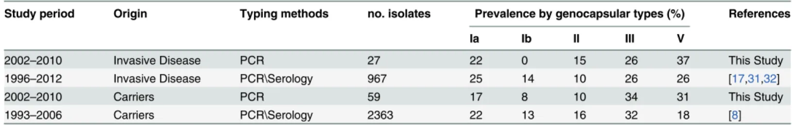
The relative distribution of confirmed non-encapsulated isolates among the five selected capsular genotypes reflected the relative prevalence of the corresponding serotypes in GBS col-onizing and invasive strains from European epidemiological studies, with Ia, III and V being the most frequent genotypes, followed by II and Ib (Table 1). Concerning the ten examined NT strains obtained from cattle, 6 belonged to capsular genotype II while the other four genotypes were represented by a single isolate (S2 Table).
Phylogenetic structure of the GBS population and evidence of random
distribution of human NT isolates among the different lineages
To undertake a population-scale molecular analysis of non-encapsulated GBS, the 96 selected NT strains along with 32 reference encapsulated isolates belonging to serotypes Ia (n = 7), Ib (n = 5), II (n = 6), III (n = 10) and V (n = 4) were subjected to whole genome next generation sequencing.De novoassembly produced DNA sequences ranging from 2.09 to 2.38 Mb and au-tomatic annotation yielded 1940 to 2625 ORFs per genome.
SNP-based comparative analysis was conducted on the 128 obtained genome sequences from NT and encapsulated strains, plus 245 genomes downloaded from the NCBI database. The analysis was based on the alignment of 0.42 Mbp core sequences to the reference complete genome of 2603V/R, the first fully sequenced GBS strain, belonging to the Clonal Complex 19 (CC19) [33].
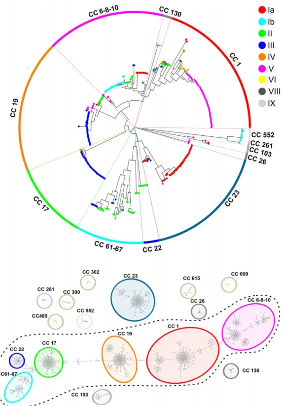
A neighbor joining phylogenetic tree constructed on the basis of the 14,092 detected SNPs is shown inFig 1A. The tree appeared constituted by 6 major clades containing from 34 to 69 iso-lates, plus 6 smaller lineages comprising 2 to 9 strains. Two random strains belonging to sepa-rate clades differed on average by 2,208 SNPs, while the average number of SNPs within each Table 1. Relative frequency of capsular genotypes Ia, Ib, II, III and V among our collection of NT isolates and in encapsulated GBS strains obtained from adults in Europe.
Study period Origin Typing methods no. isolates Prevalence by genocapsular types (%) References
Ia Ib II III V
2002–2010 Invasive Disease PCR 27 22 0 15 26 37 This Study
1996–2012 Invasive Disease PCR\Serology 967 25 14 10 26 26 [17,31,32]
2002–2010 Carriers PCR 59 17 8 10 34 31 This Study
1993–2006 Carriers PCR\Serology 2363 22 13 16 32 18 [8]
Fig 1. GBS SNP-based and MST-based phylogenetic trees.(A) SNP-based Neighbor-joining phylogenetic tree. The tree was generated using 14,092 polymorphic sites extracted from the alignment of 0.42 Mbp non-duplicated core regions shared by all 373 strains aligned to the reference strain 2603 V/R. CCs assigned to the 12 major clusters are indicated in colored ribbons. Dots represent single strains and are colored according to their capsular genotype. Asterisks indicate strains where the CC assigned by MLST (CC-1 or CC-6-8-10) differs from that assigned to strains belonging to the same SNP clade. (B) Minimum Multilocus Sequence Typing spanning tree of GBS strains. Each node represents one ST and STs differing by only one allele are connected by a line. Node dimensions refer to the relative number of strains belonging to each ST. Colored dots represent the assigned 17 CCs after refinement based on the SNP analysis. CCs included in the dotted line circle are linked by transition STs.
clade was 471. The capsular genotypes of the 373 strains included in this analysis are indicated by dots in different colors inFig 1A. As shown, strains belonging to the same capsular genotype were mostly grouped in monophyletic branches. Yet there were few exceptions of“misplaced”
strains indicative of capsular switching (at least 10 strains, of which 5 NT).
We next assigned Sequence Types (STs) to the 128 internally sequenced isolates (S2 Table) and generated a Minimum Spanning Tree (MST) by integrating these data with the STs already present in the GBS MLST database (http://pubmlst.org/sagalactiae). The obtained MST tree (Fig 1B) contained 706 previously described STs, plus 12 STs extracted from the newly se-quenced genomes. Similar to what was previously observed by Martinset al., [34], a clear sepa-ration between CCs,i.e. clusters of related STs differing by only one allele and associated by a founder ST, was not achievable due to the presence of a number of transition STs linking more than one previously described CC. As a result, strictly speaking most STs constituted a single large group (dotted line inFig 1B).
A better discrimination between CCs belonging to different population lineages was achieved by taking advantage of the conducted genome-wide SNP analysis. This integrated MLST/SNP approach allowed assigning most GBS STs to 17 CCs (Fig 1BandS2 Table), 6 of which interconnected by transition STs and 11 independent clusters. Twelve of these 17 CCs correspond to the 12 clades defined by the SNP-based tree (Fig 1A).
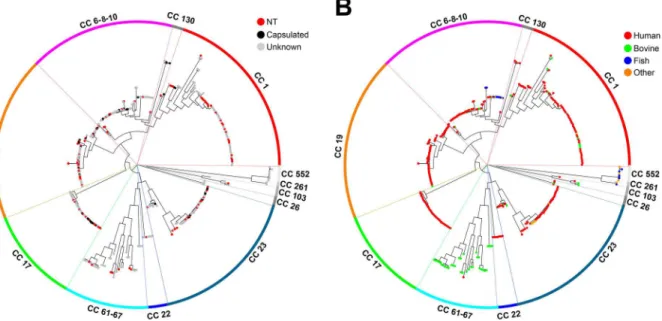
Concerning the distribution of non-encapsulated strains in the GBS population, they were found in all the main lineages of the obtained SNP phylogenetic tree (Fig 2A). This random dis-tribution suggests that genetic mutation events leading to capsule loss occur randomly in the GBS population. Interestingly, we found 11 capsular genotype III human NTs belonging to CC17, a hypervirulent CC frequently associated to late-onset neonatal disease [35].
Fig 2Bdisplays the same SNP-based tree as in Figs1Aand2A, but highlighting each GBS strain according to its host species. As shown, human strains were found in all clades, while strains isolated from frog, dog, dolphin and fish species clustered in the clades corresponding to clonal complexes CC23, CC6-8-10, CC552 and CC261. Few of the bovine strains were found in clades mainly containing human isolates, while most of them belonged to a highly divergent
Fig 2. GBS SNP-based Neighbor-joining phylogenetic trees highlighting GBS hosts and NT strains.(A) Phylogenetic tree where non-typeable or capsulated phenotypes are indicated by colored dots. (B) Phylogenetic tree where strain host origin is indicated by colored dots.
separate clade (average of 1,304 SNPs) corresponding to the CC61–67. This bovine CC ap-peared phylogenetically related to the human CC22, but not to the neonatal hypervirulent CC17 as previously predicted on the basis of MLST analysis only [35].
Taking advantage of 123 fully annotated internally sequenced genomes, we sought to com-pare the diversity between GBS isolates of bovine (n = 10) and human (n = 113) origin and be-tween encapsulated (n = 32 strains) and NT (n = 91) strains at the level of the full pan-genome, i.e including both core and accessory genes. To this aim, we first extracted all the coding se-quences from these genomes and the corresponding proteins were clustered by the CD-HIT al-gorithm (http://weizhong-lab.ucsd.edu/cd-hit/) using 70% amino acid identity and 70% query coverage as cut-off. The analysis yielded 6790 clusters each containing orthologous proteins, of which 1435 were found in at least 95% of the analyzed genomes while the remaining 5355 were considered accessory clusters.
To assess diversity in the bovine CC61–67 and in the five main human CCs 1, 6-8-10, 17, 19 and 23, we performed pairwise comparisons for every unique combination of two strains with-in each of these CCs. The distance between pairwise compared strawith-ins was defwith-ined as the semi-sum of the number of protein clusters exclusively identified in either of the two strains. As shown inS3A Fig, the average distance between pairwise strains was about two fold higher within the CC61–67 than within each of the five human CCs (P<0.0001), consistent with the higher variability of the bovine CC already revealed by SNP analysis of the core genome (Fig 2B).
We next compared the diversity between NT and encapsulated strains within each of the human CCs 17, 23, 19, 6-8-10 (at least 9 NT and 7 encapsulated isolates in each CC). The cor-responding pairwise distances are, shown inS3B Fig, and indicated a slightly higher diversity for the NT strains compared to their encapsulated counterparts of the same CC (1.2–1.4 fold, P<0.01).
Identification of genetic events leading to loss of capsule expression
To identify the type of genetic alterations that most often lead to capsule loss in GBS, we ex-tracted the DNA sequences corresponding to thecpslocus and its 5’flanking region (20 kbp in total) from the genomes of the 96 NT sequenced strains.The extracted DNA sequences were aligned with the internally derivedcpsreference se-quences from encapsulated strains and with those present in public databases. The analysis confirmed the capsular genotypes of all 96 NT strains predicted by PCR analysis, and allowed detecting genetic alterations potentially responsible for the NT phenotype.
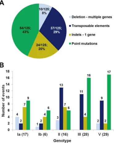
We found that all 96 NT strains differed from their reference counterparts by at least one nucleotide, either in the promoter region or in thecpsORFs. More specifically, 89 strains con-tained single or multiple genetic alterations representing a total of 125 different mutations that are likely to prevent capsule expression. These included deletions affecting multiplecpsgenes (range 80–9,059 bp), insertions of large transposable elements, point mutations in the -10 pro-moter region, and insertions/deletions (indels, range 2–253 bp) or point mutations resulting in frame-shifts and/or in premature stop codons in one of thecpsgenes. The remaining 7 strains contained only non-synonymous single nucleotide polymorphisms that could not be unambig-uously associated to gene inactivation or impaired transcription. These missense mutations are described in detail in a separate section below.
different types of genetic alterations among the five investigated capsular genotypes are shown inFig 3B.
As reported inS2 Table, 62 isolates showed only one single alteration, while more than one inactivating mutation was present simultaneously in 27 isolates.
Of note, 30 strains contained non-unique mutations that were found in more than one iso-late, either alone or in combination with other mutations. In particular, 10 single point muta-tions were encountered in 2 to 8 different strains. Additional genomic comparison confirmed that strains that carried the samecpsmutation were not identical in the DNA regions outside the capsular locus.
Target genes of
cps
inactivation mutations
We next investigated the frequency of genetic alterations possibly causing inactivation of cap-sule expression in each of the genes and the promoter of thecpsoperon.
This DNA region is composed by 16–18 genes in the different GBS serotypes [36]. The 5’ cpsABCDgenes are predicted to be involved in the regulation of capsule synthesis; the central region fromcpsEtocpsLencodes the enzymes responsible for the synthesis, transport, and po-lymerization of the polysaccharide repeating units; finally,neuBCDAgenes are responsible for the synthesis of the activated sialic acid, a sugar component present in all GBS capsular poly-saccharides [37].
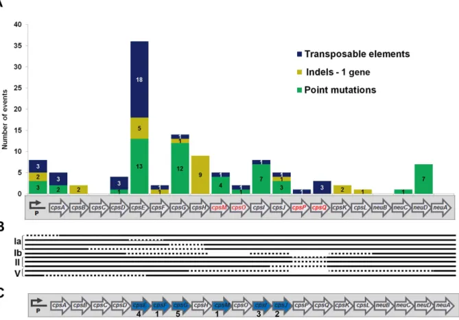
Fig 3. Relative frequency of the different types of mutations possibly responsible for the lack of capsule expression in GBS.(A) Distribution of 126 genetic alteration events detected in 89 isolates. (B) Number of strains bearing each of thecpsmutation types among to the five selected capsular genotypes.
The number of point mutations, indels, and transposable elements detected along the oper-on are indicated inFig 4A. As shown, thecpspromoter region and allcpsgenes exceptneuA
andcpsC, were targets of at least one kind of genetic alteration.
Overall, we found 55 point mutation events across 10cpsgenes or the promoter, and the most frequent targets werecpsEandcpsG, encoding the first and second enzymes in the synthe-sis pathway of the polysaccharide repeating unit. Remarkably, we did not find any NT strains harboring capsule inactivating point mutations incpsA,cpsB,cpsCorcpsD.
Twenty-four indels were detected in 8cpsgenes or the promoter, and most of these were found in thecpsH. This gene is homologous to wzy fromE.coliand fromS.pneumoniae, pre-sumably encoding the polymerases that link repeating units via glycosidic linkages to form the long polysaccharide chains [37,38].
Ten isolates contained deletions affecting more than one gene, while 37 transposable ele-ments were present in 32 isolates (Fig 4B). The transposable elements present in thecpsregion of the different strains were classified according to the IS finder database nomenclature (http:// www-is.biotoul.fr) [39]. In total, 9 different IS types were identified, 8 of which belonging to known IS families (Table 2). As shown, most IS were found in thecpsEgene, and the most fre-quently encountered IS type was IS1381 as previously reported for another NT GBS strain [14]. Fig 4. Different kinds of genetic alterations detected in the GBScpsoperon.(A) Distribution of 37 transposable elements (blue), 24 insertion or deletion (indels) targeting a single gene (light green) and 54 point mutations (dark green) scattered across thecpsoperon. Genes that are missing in one or more capsular types are shown with red font color in the operon diagram. (B) Deletions comprising more than a single gene are indicated with dotted lines; empty spaces represent the absence ofcpsgenes in a specific capsular genotype. (C) Gene targets of capsule inactivation mutations that appeared as single events (blue arrows); the figures below the target genes indicate the number of strains presenting the individual mutation.
IS type IS family Promoter cpsA cpsB cpsC cpsD cpsE cpsF cpsG cpsH cpsM cpsO cpsI cpsJ cpsP cpsQ cpsK cpsL neuB neuC neuD neuA
IS1381 IS5 3 10 1 1 1
ISSa4 IS982 3 1 1 1
IS1062 like IS30 1
ISSag11 IS256 2 1 1
IS1548 ISAs1 3 3 1
Unknown Unknown 1
ISSpo8 IS1595 1
ISSpo8- like IS1595 1
doi:10.1371/journal.pone.0125985.t002
Genomics
of
Non-Type
able
|DOI:10.137
1/journal.p
one.0125985
May
6,
Finally, sixteen of the 96 NT strains harbored only one single mutation resulting in a prema-ture stop codon in thecpslocus, and the target genes of these single mutations werecpsE,F,G,
I,JandcpsM(Fig 4C).
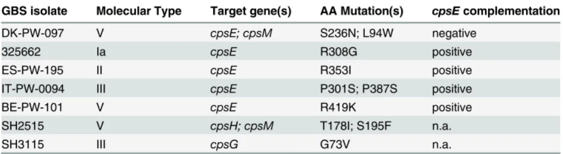
Mutations in amino acid residues essential for functional activity of CpsE
As shown above, 7 NT strains belonging to capsular genotypes Ia, II, III and V presented mis-sense mutations for which the effect on capsule synthesis was unpredictable (Table 3). The genes carrying these mutations were compared with the corresponding genes from the 32 en-capsulated reference strains and loci from genome draft sequences present in the NCBI data-base. All the missense mutations were unique to these seven NT isolates, and five of them targeted thecpsEgene. The CpsE portal enzyme of GBS capsule biosynthesis is highly con-served among the nine serotypes, with an amino acid identity ranging between 98.2% and 99.8%. BLAST analysis indicated that 4 of the detected mutations (R308G, R353I, P387S, R419K) were located in gene positions that were conserved in more than 95% of 500 examined CpsE homologs from a wide range of bacterial species.We investigated whether the CpsE amino acid substitutions were sufficient to account for capsule loss. The plasmid pAM-cpsEcontaining a wild typecpsEIa gene sequence was generat-ed and transformgenerat-ed into the five different NT strains harboring mutations incpsE. The trans-formed bacteria were tested for their capacity to synthesize a capsular polysaccharide by latex agglutination and by flow-cytometry assays. As shown inFig 5, CpsE overexpression restored capsular synthesis in the 4 strains carrying exclusively one missense mutation in thecpsEgene. The results show that the highly conserved amino acid residues R308, R353, P387, R419 are es-sential for enzyme activity. One of the five mutations could not be complemented, possibly due to a concomitant missense mutation in thecpsMgene.
Genetic events that abolish transcription of the
cps
operon
The transcription of the cps operon is a prerequisite for capsule production. Consequently, mutations leading to insufficient or abolished transcription were expected to result in a NT phenotype. We used qRT-PCR to compare the transcription of the cps operon in a subset of 48 NT and 6 encapsulated reference isolates (strains 1–48 inS2 Fig). The operon is transcribed from the promoter region upstream of its first genecpsA[40], therefore, two primers annealing to the 3’region ofcpsAwere used (seeMethodssection). As shown inS2 Fig, thecpsA tran-script amount for most strains was normally distributed between 0.4–1.42 fold compared to Table 3. NT GBS strains presenting missense mutations and the results ofcpsEcomplementation in same strains.
GBS isolate Molecular Type Target gene(s) AA Mutation(s) cpsEcomplementation
DK-PW-097 V cpsE; cpsM S236N; L94W negative
325662 Ia cpsE R308G positive
ES-PW-195 II cpsE R353I positive
IT-PW-0094 III cpsE P301S; P387S positive
BE-PW-101 V cpsE R419K positive
SH2515 V cpsH; cpsM T178I; S195F n.a.
SH3115 III cpsG G73V n.a.
The nucleotide and amino acid changes in the isolates were based on the encapsulated 2603 V/R reference genome sequence. AA, single letter amino acid designation; del, deletion of the nucleotide; n.a. not applicable.
the encapsulated strain GBS515. However, 9 NTs were devoid of any detectablecpsAtranscript and, notably, all of them presented point mutations in the promoter region or IS either in the promoter or the first four genes of the operon (Fig 6A). On the basis of the above observations, we subsequently selected for further transcription analysis all the NT isolates presenting genetic alterations in thecpspromoter region or thecpsA,B,C,Dgenes. Eighteen out of 96 NT isolates, including the 9 above described strains lacking thecpsAtranscript, contained different kinds of Fig 5. Complementation ofcpsEmissense mutations in selected NT GBS strains.Flow cytometry analysis of NT GBS strains carrying missense mutations incpsEand of their counterpart after transformation with pAM-cpsE. Bacteria were incubated with mouse monoclonal antibodies specific for the five capsular polysaccharides, and then treated with labeled secondary antibodies. Fluorescence after incubation with the secondary antibody alone is indicated by empty histograms, while staining after treatment with type the specific antibodies is shown by the colored histograms.
doi:10.1371/journal.pone.0125985.g005
Fig 6. Analysis of the influence of genetic alterations on the transcription of thecpsoperon in NT isolates.(A) Mutations in thecpspromoter or
cpsA-Ddetected in 18 NT strains. Their positions are indicated as +/- numbers in relation to the first base pair of thecpsAcoding sequence. ISs are
represented by colored triangles; point mutations in the -10 sequence are marked in bold; deleted regions are represented by dotted lines and stop codons by crosses. Numbers within parentheses identify the strains reported in the x-axis of panel B. (B) Transcription of thecpsoperon in the 18 NT strains measured with primers cpsAup-F/R (filled bars) and to cpsE-F/R (hatched bars). The relative fold expression for each strain was estimated in comparison to the expression ofcpsAin strain 515. For strains 25, 26, 52 and 53 the transcript incpsEgene was not determined.
mutations in this region. These alterations comprised IS elements inserted in the vicinity of the Shine-Dalgarno sequence, point mutations in the -10 box, large deletions comprising thecps
promoter, IS elements in thecpsAandcpsDcoding regions, and point mutations or indels lead-ing to stop codons incpsAandcpsB(Fig 6A).
Therefore, all 18 isolates were then analyzed by qRT-PCR using primers annealing to the
cpsEgene, and to the 5’region of thecpsAgene (upstream the different IS elements identified in this gene). As shown inFig 6B, all the mutations present in the promoter region completely abolished transcription. In the strains containing IS elements incpsAor incpsDwe found tran-scripts upstream the transposable element, but not downstream of this insertion. Finally, genet-ic alterations leading to premature stop codons incpsAorin cpsBappeared to have limited or no effect, indicating that these genes are not essential forcpstranscriptional regulation.
Discussion
The sialic acid containing capsular polysaccharide that surrounds GBS is essential for bacterial survival in animal models of infection. Furthermore, vaccines targeting this antigen are in de-velopment phase. The occurrence of non-encapsulated GBS strains devoid of this important protective“shield”is therefore difficult to explain and could be of concern in GBS vaccinology.
In the present study we confirmed the complete absence of capsule expression in 96 well-characterized GBS isolates, and 27 of these strains were obtained from patients suffering from invasive disease.
In principle one cannot exclude that in some of the cases the NT phenotype could have aris-en in an already infected patiaris-ent, or after isolation of the microorganism duringin vitro cultiva-tion. However, 11 of the 27 NT strains causing invasive disease harbored more than one mutation possibly leading to capsule inactivation, and it appears highly unlikely that in all these strains more than one independent mutation event could occur during or after infection. We show that the NT phenotype occurs randomly in all lineages of the GBS population and that it is mainly associated with genetic alterations in thecpsoperon. Capsule loss can also occur among hypervirulent CC17 strains, and some of the resulting non-encapsulated isolates are still able to cause invasive disease in humans.
The observed random distribution of non-encapsulated isolates in the GBS population dif-fers from the distribution observed forS.pneumoniae, where the large majority of NTs cluster in few CCs that consist almost exclusively of non-encapsulated strains (http://pubmlst.org/ spneumoniae/). Indeed, a comprehensive epidemiological study on pneumococcal carriage re-vealed a high frequency of NT strains mostly belonging to one single phylogenetic lineage [41]. This NT lineage showed the highest rates of receipt and donation of recombined DNA frag-ments among pneumococcal clades, and the authors proposed that carriage NT strains could be a major reservoir of genetic diversity in this species [42].
The NT phenotype is much less frequent among GBS strains obtained from human carriers compared to the pneumococci, suggesting the absence of selective advantage for human coloni-zation. However, the high frequency (up to 77%) of NTs among isolates obtained from bovine mastitis could reflect a high degree of fitness for colonization of the udder epithelia. Alterna-tively, less efficient complement deposition on the surface of GBS present in bovine milk com-pared to human blood [43] could make the capsule shield less necessary.
results confirm the essential role of the corresponding enzymes in the synthesis of the GBS capsule.
The CpsE enzyme catalyzes the addition of glucose-1-phosphate to undecaprenyl carrier of the bacterial membrane as first reaction in the synthesis of the polysaccharide repeating unit [38]. CpsE overexpression restored capsular synthesis in the 4 strains carrying exclusively one missense mutation in thecpsEgene, allowing the identification of amino acid residues that are essential for enzyme activity. Moreover, expression of a single enzyme variant rescued biosyn-thesis of different capsular types, confirming that serotype specificity does not depend on CpsE.
On the basis of its homology with a well-characterized galactosyl transferase fromS. pneu-moniae, CpsG was predicted to attach galactose to undecaprenyl-glucose during the second step of polysaccharide repeating unit biosynthesis [37]. It is tempting to speculate that inacti-vating mutations in the enzymes responsible for the early steps of capsule biosynthesis could prevent unnecessary energy expenditure and potential sequestration of monosaccharides.
We also detected non-encapsulated strains that contained only one single mutation result-ing in a premature stop codon incpsF,G,I,JandcpsM. The data suggest that, in addition to the above mentionedcpsE,GandH, these genes are essential for GBS capsule expression.
In agreement with findings from Cieslewiczet al., we did not find any NT strains that har-bored single capsule inactivation mutations incpsA,cpsB,cpsCorcpsD[36]. In fact, these au-thors demonstrated that out of five genes examined by in-frame deletion mutations (cpsA,
cpsB,cpsC,cpsDandcpsE), only a disruption ofcpsEresulted in the non-encapsulated pheno-type. It was therefore suggested thatcpsA-Dare not required for biosynthesis of the repeating unit, but rather direct the coordinated polymerization and export of the polysaccharide [36]. The authors also observed a decreased transcription in thecpsAknockout mutant. According to our observations, genetic alterations leading to premature stop codons incpsAor incpsB ap-peared to have limited or no effect, indicating that these genes are not essential forcps
transcriptional regulation.
Genome wide analysis conducted on both NT and encapsulated strains provided new in-sights into the evolution of the GBS species and its adaptation to cause human disease. The phylogenetic tree obtained by integrated SNP-based analysis of the 128 sequenced isolates plus the 245 publicly available genomes appeared constituted by 6 major clades, plus 6 smaller line-ages comprising a low number of strains. The tree was consistent with the one recently re-ported by Da Cunhaet al. [44]. The authors noted that human strains belonging to the same phylogenetic clade contained a limited number of polymorphisms, and proposed that this clon-al pattern could be the result of recent globclon-al spreading of few successful GBS clones having ac-quired a conjugative element conferring resistance to tetracycline after the introduction of this antibiotic in 1948. Our analysis confirmed very low variability within clades containing mainly human strains (from 50 SNPs for CC17 to 601 SNPs for CC6-8-10). However, it revealed a higher degree of diversity in the bovine population (average of 1,304 SNPs within the CC61–
conferring Tetracycline resistance was found in 216 out of 291 human strains and only in 6 out of 53 strains from bovine origin (none of which belonging to CC61–67), consistent with the above mentioned hypothesis according to which most human GBS strains would have arisen from few clones having acquired a conjugative element bearingtetM.
Finally, the slightly higher diversity detected in the NT accessory genomes compared to the encapsulated strains might suggest a higher uptake of foreign genetic elements in the first group, although this will need to be confirmed with more in depth analysis. Further mining into the now available large number of genomes is indeed expected to provide new important hints on GBS host adaptation and pathogenesis mechanisms.
The strategy used by non-encapsulated GBS strains to evade the innate immune system re-mains unknown. Further research in this field will be crucial for the development of new pre-ventive and therapeutic initiatives against this relevant human and animal pathogen.
Supporting Information
S1 Fig. Flow cytometry analysis of representative encapsulated and non-encapsulated GBS isolates.Encapsulated (A) or non-encapsulated bacteria (B) were stained with mouse mono-clonal antibodies anti-Ia, Ib, II, III and V, and labeled with R-Phycoerythrin conjugated sec-ondary antibodies. Bacterial staining obtained with pre-immune mouse serum is indicated by black histograms, and staining with type specific antibodies is shown by colored histograms. Empty histograms indicate the signals obtained with antibodies against heterologous capsular types.
(TIF)
S2 Fig. Relative transcription ofcpsAin NT and encapsulated GBS strains.Transcript levels in NT (grey) and encapsulated strains (black) obtained using primers cpsA-F/R were compared to those of the housekeeping genegyrAby qRT-PCR. The relative fold expression for each strain was in comparison with strain 515. Columns show results from three independent growth replicates, each analyzed performed in technical triplicates. Error bars represent standard deviations.
(TIF)
S3 Fig. Diversity within major human and cattle GBS CCs.Pairwise comparisons were per-formed for every unique combination of two strains within the indicated CCs (x-axis). The dis-tances between strain pairs (y-axis) were defined as the semi-sum of the number of unique protein classes identified in the two compared strains. In the box-and-whiskers plots, the box represents the interquartile range and the whiskers extend from the minimum to the maxi-mum distances; the median distances are represented by a horizontal line and the average dis-tances by a cross; statistical differences between groups were calculated by the two-tailed Mann Whitney test. (A)Pairwise distances between CCs. Significant differences were detected be-tween the bovine CC61–67 and the human CCs1, 6-8-10, 17, 19 and 23 (P<0,0001), as well as between CCs 6-8-10 or 19 and CCs 1, 17 or 23 (P<0.01). (B)Pairwise distances between NT and encapsulated strains belonging to CCs 6-8-10, 17, 19 and 23. Significant differences be-tween NT and encapsulated strains were detected in all four CCs (P<0.01)
(TIF)
S1 Table. Primers used for qRT-PCR analysis ofcpsoperon transcription. (PDF)
leading to stop codons; IND: Insertions/Deletions involving 1 gene and leading to stop codons; MIS: Missense point mutations; LOD: Late Onset Disease; EOD: Early Onset Disease; ID: Inva-sive Disease; ST: Sequence type; CC: Clonal Complex.
(PDF)
Acknowledgments
We would like to acknowledge Nicola Pacchiani, Stefano Censini, Alessandro Muzzi, Paola Parisi, Maddalena Lazzarin, Qi Su, Ankit Maroo, Sean C. Daugherty, Sonia Agrawal, Naomi Sengamalay, Lisa Sadzcewicz, and Luke J. Tallon for their contribution to genome sequencing and analysis. The DEVANI Consortium (Uffe B. Skov Sørensen, Mogens Kilian, P. Melin, A. Decheva and B. Petrunov, P. Kriz, R. Berner, M. Hufnagel, M. Kunze, R. Creti, L. Baldassarri, A. Berardi, G. Orefici, J. R. Granger, M. De La Rosa Fraile, B. Afshar, A. Efstratiou, D. Maione, J.L. Telford) for the collection, identification and serotyping of the GBS NT isolates.
Author Contributions
Conceived and designed the experiments: RR IM GG. Performed the experiments: RR EC HT DR CT MM SG. Analyzed the data: RR EC MD RJ CD IM. Contributed reagents/materials/ analysis tools: US MK MR. Wrote the paper: IM RR.
References
1. Schuchat A. Group B streptococcus. Lancet. 1999; 353(9146):51–6. doi:10.1016/S0140-6736(98) 07128-1PMID:10023965.
2. Baker CJ, Rench MA, Edwards MS, Carpenter RJ, Hays BM, Kasper DL. Immunization of pregnant women with a polysaccharide vaccine of group B streptococcus. N Engl J Med. 1988; 319(18):1180–5. doi:10.1056/NEJM198811033191802PMID:3050524.
3. Yother J. Capsules of Streptococcus pneumoniae and other bacteria: paradigms for polysaccharide biosynthesis and regulation. Annu Rev Microbiol. 2011; 65:563–81. doi:10.1146/annurev.micro.62. 081307.162944PMID:21721938.
4. Whitfield C. Biosynthesis and assembly of capsular polysaccharides in Escherichia coli. Annu Rev Bio-chem. 2006; 75:39–68. doi:10.1146/annurev.biochem.75.103004.142545PMID:16756484.
5. Afshar B, Broughton K, Creti R, Decheva A, Hufnagel M, Kriz P, et al. International external quality as-surance for laboratory identification and typing of Streptococcus agalactiae (Group B streptococci). J Clin Microbiol. 2011; 49(4):1475–82. doi:10.1128/JCM.02365-10PMID:21325542; PubMed Central PMCID: PMC3122801.
6. Kong F, Lambertsen LM, Slotved HC, Ko D, Wang H, Gilbert GL. Use of phenotypic and molecular se-rotype identification methods to characterize previously nonsese-rotypeable group B streptococci. J Clin Microbiol. 2008; 46(8):2745–50. doi:10.1128/JCM.00189-08PMID:18562579; PubMed Central PMCID: PMC2519501.
7. Gherardi G, Imperi M, Baldassarri L, Pataracchia M, Alfarone G, Recchia S, et al. Molecular epidemiol-ogy and distribution of serotypes, surface proteins, and antibiotic resistance among group B streptococ-ci in Italy. J Clin Microbiol. 2007; 45(9):2909–16. doi:10.1128/JCM.00999-07PMID:17634303; PubMed Central PMCID: PMC2045288.
8. Ippolito DL, James WA, Tinnemore D, Huang RR, Dehart MJ, Williams J, et al. Group B streptococcus serotype prevalence in reproductive-age women at a tertiary care military medical center relative to global serotype distribution. BMC Infect Dis. 2010; 10:336. doi:10.1186/1471-2334-10-336PMID: 21106080; PubMed Central PMCID: PMC3004907.
9. Rato MG, Bexiga R, Florindo C, Cavaco LM, Vilela CL, Santos-Sanches I. Antimicrobial resistance and molecular epidemiology of streptococci from bovine mastitis. Vet Microbiol. 2013; 161(3–4):286–94. doi:10.1016/j.vetmic.2012.07.043PMID:22964008.
11. Martinez G, Harel J, Higgins R, Lacouture S, Daignault D, Gottschalk M. Characterization of Strepto-coccus agalactiae isolates of bovine and human origin by randomly amplified polymorphic DNA analy-sis. J Clin Microbiol. 2000; 38(1):71–8. PMID:10618066; PubMed Central PMCID: PMC86023. 12. Imperi M, Pataracchia M, Alfarone G, Baldassarri L, Orefici G, Creti R. A multiplex PCR assay for the
di-rect identification of the capsular type (Ia to IX) of Streptococcus agalactiae. J Microbiol Methods. 2010; 80(2):212–4. doi:10.1016/j.mimet.2009.11.010PMID:19958797.
13. Poyart C, Tazi A, Reglier-Poupet H, Billoet A, Tavares N, Raymond J, et al. Multiplex PCR assay for rapid and accurate capsular typing of group B streptococci. J Clin Microbiol. 2007; 45(6):1985–8. doi: 10.1128/JCM.00159-07PMID:17376884; PubMed Central PMCID: PMC1933079.
14. Ramaswamy SV, Ferrieri P, Madoff LC, Flores AE, Kumar N, Tettelin H, et al. Identification of novel cps locus polymorphisms in nontypable group B Streptococcus. J Med Microbiol. 2006; 55(Pt 6):775–83. doi:10.1099/jmm.0.46253-0PMID:16687599.
15. Creti R, Imperi M, Pataracchia M, Alfarone G, Recchia S, Baldassarri L. Identification and molecular characterization of a S. agalactiae strain lacking the capsular locus. Eur J Clin Microbiol Infect Dis. 2012; 31(3):233–5. doi:10.1007/s10096-011-1298-7PMID:21614482.
16. Rodriguez-Granger J, Alvargonzalez JC, Berardi A, Berner R, Kunze M, Hufnagel M, et al. Prevention of group B streptococcal neonatal disease revisited. The DEVANI European project. Eur J Clin Micro-biol Infect Dis. 2012; 31(9):2097–104. doi:10.1007/s10096-012-1559-0PMID:22314410.
17. Martins ER, Pessanha MA, Ramirez M, Melo-Cristino J, Portuguese Group for the Study of Streptococ-cal I. Analysis of group B streptococStreptococ-cal isolates from infants and pregnant women in Portugal revealing two lineages with enhanced invasiveness. J Clin Microbiol. 2007; 45(10):3224–9. doi:10.1128/JCM. 01182-07PMID:17699641; PubMed Central PMCID: PMC2045366.
18. Maione D, Margarit I, Rinaudo CD, Masignani V, Mora M, Scarselli M, et al. Identification of a universal Group B streptococcus vaccine by multiple genome screen. Science. 2005; 309(5731):148–50. doi:10. 1126/science.1109869PMID:15994562; PubMed Central PMCID: PMC1351092.
19. Myers EW, Sutton GG, Delcher AL, Dew IM, Fasulo DP, Flanigan MJ, et al. A whole-genome assembly of Drosophila. Science. 2000; 287(5461):2196–204. PMID:10731133.
20. Angiuoli SV, Matalka M, Gussman A, Galens K, Vangala M, Riley DR, et al. CloVR: a virtual machine for automated and portable sequence analysis from the desktop using cloud computing. BMC Bioinfor-matics. 2011; 12:356. doi:10.1186/1471-2105-12-356PMID:21878105; PubMed Central PMCID: PMC3228541.
21. Kurtz S, Phillippy A, Delcher AL, Smoot M, Shumway M, Antonescu C, et al. Versatile and open soft-ware for comparing large genomes. Genome Biol. 2004; 5(2):R12. doi:10.1186/gb-2004-5-2-r12 PMID:14759262; PubMed Central PMCID: PMC395750.
22. Francisco AP, Vaz C, Monteiro PT, Melo-Cristino J, Ramirez M, Carrico JA. PHYLOViZ: phylogenetic inference and data visualization for sequence based typing methods. BMC Bioinformatics. 2012; 13:87. doi:10.1186/1471-2105-13-87PMID:22568821; PubMed Central PMCID: PMC3403920. 23. Altschul SF, Gish W, Miller W, Myers EW, Lipman DJ. Basic local alignment search tool. J Mol Biol.
1990; 215(3):403–10. doi:10.1016/S0022-2836(05)80360-2PMID:2231712.
24. Tamura K, Stecher G, Peterson D, Filipski A, Kumar S. MEGA6: Molecular Evolutionary Genetics Anal-ysis version 6.0. Mol Biol Evol. 2013; 30(12):2725–9. doi:10.1093/molbev/mst197PMID:24132122; PubMed Central PMCID: PMC3840312.
25. Stamatakis A. RAxML-VI-HPC: maximum likelihood-based phylogenetic analyses with thousands of taxa and mixed models. Bioinformatics. 2006; 22(21):2688–90. doi:10.1093/bioinformatics/btl446 PMID:16928733.
26. Lauer P, Rinaudo CD, Soriani M, Margarit I, Maione D, Rosini R, et al. Genome analysis reveals pili in Group B Streptococcus. Science. 2005; 309(5731):105. doi:10.1126/science.1111563PMID: 15994549.
27. Framson PE, Nittayajarn A, Merry J, Youngman P, Rubens CE. New genetic techniques for group B streptococci: high-efficiency transformation, maintenance of temperature-sensitive pWV01 plasmids, and mutagenesis with Tn917. Appl Environ Microbiol. 1997; 63(9):3539–47. PMID:9293004; PubMed Central PMCID: PMC168659.
28. Yao K, Poulsen K, Maione D, Rinaudo CD, Baldassarri L, Telford JL, et al. Capsular gene typing of Streptococcus agalactiae compared to serotyping by latex agglutination. J Clin Microbiol. 2013; 51 (2):503–7. doi:10.1128/JCM.02417-12PMID:23196363; PubMed Central PMCID: PMC3553911. 29. Sellin M, Olofsson C, Hakansson S, Norgren M. Genotyping of the capsule gene cluster (cps) in
30. Phares CR, Lynfield R, Farley MM, Mohle-Boetani J, Harrison LH, Petit S, et al. Epidemiology of inva-sive group B streptococcal disease in the United States, 1999–2005. JAMA. 2008; 299(17):2056–65. doi:10.1001/jama.299.17.2056PMID:18460666.
31. Tazi A, Morand PC, Reglier-Poupet H, Dmytruk N, Billoet A, Antona D, et al. Invasive group B strepto-coccal infections in adults, France (2007–2010). Clin Microbiol Infect. 2011; 17(10):1587–9. doi:10. 1111/j.1469-0691.2011.03628.xPMID:21883671.
32. Bergseng H, Rygg M, Bevanger L, Bergh K. Invasive group B streptococcus (GBS) disease in Norway 1996–2006. Eur J Clin Microbiol Infect Dis. 2008; 27(12):1193–9. doi:10.1007/s10096-008-0565-8 PMID:18560908.
33. Tettelin H, Masignani V, Cieslewicz MJ, Eisen JA, Peterson S, Wessels MR, et al. Complete genome sequence and comparative genomic analysis of an emerging human pathogen, serotype V Streptococ-cus agalactiae. Proc Natl Acad Sci U S A. 2002; 99(19):12391–6. doi:10.1073/pnas.182380799PMID: 12200547; PubMed Central PMCID: PMC129455.
34. Martins ER, Andreu A, Melo-Cristino J, Ramirez M. Distribution of pilus islands in Streptococcus aga-lactiae that cause human infections: insights into evolution and implication for vaccine development. Clin Vaccine Immunol. 2013; 20(2):313–6. doi:10.1128/CVI.00529-12PMID:23269415; PubMed Cen-tral PMCID: PMC3571264.
35. Bisharat N, Crook DW, Leigh J, Harding RM, Ward PN, Coffey TJ, et al. Hyperinvasive neonatal group B streptococcus has arisen from a bovine ancestor. J Clin Microbiol. 2004; 42(5):2161–7. PMID: 15131184; PubMed Central PMCID: PMC404684.
36. Cieslewicz MJ, Kasper DL, Wang Y, Wessels MR. Functional analysis in type Ia group B Streptococcus of a cluster of genes involved in extracellular polysaccharide production by diverse species of strepto-cocci. J Biol Chem. 2001; 276(1):139–46. doi:10.1074/jbc.M005702200PMID:11027683.
37. Cieslewicz MJ, Chaffin D, Glusman G, Kasper D, Madan A, Rodrigues S, et al. Structural and genetic diversity of group B streptococcus capsular polysaccharides. Infect Immun. 2005; 73(5):3096–103. doi: 10.1128/IAI.73.5.3096-3103.2005PMID:15845517; PubMed Central PMCID: PMC1087335. 38. Yamamoto S, Miyake K, Koike Y, Watanabe M, Machida Y, Ohta M, et al. Molecular characterization of
type-specific capsular polysaccharide biosynthesis genes of Streptococcus agalactiae type Ia. J Bac-teriol. 1999; 181(17):5176–84. PMID:10464185; PubMed Central PMCID: PMC94020.
39. Flechard M, Gilot P. Physiological impact of transposable elements encoding DDE transposases in the environmental adaptation of Streptococcus agalactiae. Microbiology. 2014; 160(Pt 7):1298–315. doi: 10.1099/mic.0.077628-0PMID:24760965.
40. Chaffin DO, Beres SB, Yim HH, Rubens CE. The serotype of type Ia and III group B streptococci is de-termined by the polymerase gene within the polycistronic capsule operon. J Bacteriol. 2000; 182 (16):4466–77. PMID:10913080; PubMed Central PMCID: PMC94618.
41. Chewapreecha C, Harris SR, Croucher NJ, Turner C, Marttinen P, Cheng L, et al. Dense genomic sam-pling identifies highways of pneumococcal recombination. Nat Genet. 2014; 46(3):305–9. doi:10.1038/ ng.2895PMID:24509479; PubMed Central PMCID: PMC3970364.
42. Turner P, Turner C, Jankhot A, Helen N, Lee SJ, Day NP, et al. A longitudinal study of Streptococcus pneumoniae carriage in a cohort of infants and their mothers on the Thailand-Myanmar border. PLoS One. 2012; 7(5):e38271. doi:10.1371/journal.pone.0038271PMID:22693610; PubMed Central PMCID: PMC3365031.
43. Rainard P. The complement in milk and defense of the bovine mammary gland against infections. Vet Res. 2003; 34(5):647–70. doi:10.1051/vetres:2003025PMID:14556699.
44. Da Cunha V, Davies MR, Douarre PE, Rosinski-Chupin I, Margarit I, Spinali S, et al. Streptococcus agalactiae clones infecting humans were selected and fixed through the extensive use of tetracycline. Nat Commun. 2014; 5:4544. doi:10.1038/ncomms5544PMID:25088811.
45. Sorensen UB, Poulsen K, Ghezzo C, Margarit I, Kilian M. Emergence and global dissemination of host-specific Streptococcus agalactiae clones. MBio. 2010; 1(3). doi:10.1128/mBio.00178-10PMID: 20824105; PubMed Central PMCID: PMC2932510.
46. Richards VP, Choi SC, Pavinski Bitar PD, Gurjar AA, Stanhope MJ. Transcriptomic and genomic evi-dence for Streptococcus agalactiae adaptation to the bovine environment. BMC Genomics. 2013; 14:920. doi:10.1186/1471-2164-14-920PMID:24369756; PubMed Central PMCID: PMC3890567. 47. Broker G, Spellerberg B. Surface proteins of Streptococcus agalactiae and horizontal gene transfer. Int