Differential MicroRNA Expression in Human
Macrophages with
Mycobacterium
tuberculosis
Infection of Beijing/W and
Non-Beijing/W Strain Types
Lin Zheng
1, Eric Leung
1, Nelson Lee
2, Grace Lui
2, Ka-Fai To
3, Raphael C. Y. Chan
1,
Margaret Ip
1*
1Department of Microbiology, The Chinese University of Hong Kong, Hong Kong SAR, China,
2Department of Medicine & Therapeutics, The Chinese University of Hong Kong, Hong Kong SAR, China,
3Department of Anatomical & Cellular Pathology, The Chinese University of Hong Kong, Hong Kong SAR, China
Abstract
Objectives
The role of microRNAs in association with
Mycobacterium tuberculosis
(MTB) infection and
the immunology regulated by microRNAs upon MTB infection have not been fully
unrav-elled. We examined the microRNA profiles of THP-1 macrophages upon the MTB infection
of Beijing/W and non-Beijing/W clinical strains. We also studied the microRNA profiles of
the host macrophages by microarray in a small cohort with active MTB disease, latent
infec-tion (LTBI), and from healthy controls.
Results
The results revealed that 14 microRNAs differentiated infections of Beijing/W from
non-Bei-jing/W strains (P
<
0.05). A unique signature of 11 microRNAs in human macrophages was
identified to differentiate active MTB disease from LTBI and healthy controls. Pathway
anal-yses of these differentially expressed miRNAs suggest that the immune-regulatory
interac-tions involving TGF-
β
signalling pathway take part in the dysregulation of critical TB
processes in the macrophages, resulting in active expression of both cell communication
and signalling transduction systems.
Conclusion
We showed for the first time that the Beijing/W TB strains repressed a number of miRNAs
expressions which may reflect their virulence characteristics in altering the host response.
The unique signatures of 11 microRNAs may deserve further evaluation as candidates for
biomarkers in the diagnosis of MTB and Beijing/W infections.
a11111
OPEN ACCESS
Citation:Zheng L, Leung E, Lee N, Lui G, To K-F, Chan RCY, et al. (2015) Differential MicroRNA Expression in Human Macrophages with
Mycobacterium tuberculosisInfection of Beijing/W and Non-Beijing/W Strain Types. PLoS ONE 10(6): e0126018. doi:10.1371/journal.pone.0126018
Academic Editor:Selvakumar Subbian, Public Health Research Institute at RBHS, UNITED STATES
Received:December 12, 2014
Accepted:March 27, 2015
Published:June 8, 2015
Copyright:© 2015 Zheng et al. This is an open access article distributed under the terms of the Creative Commons Attribution License, which permits unrestricted use, distribution, and reproduction in any medium, provided the original author and source are credited.
Introduction
Tuberculosis is one of the most common causes of death from infectious diseases. Studies have
shown that one-third of the world
’
s population is infected with
M
.
tuberculosis
(MTB). The
people who are infected with MTB but who do not have active tuberculosis have latent
infec-tion (LTBI), and they have a 10% lifetime chance that they will progress to having the active
disease.
Macrophages play a key role in the immune defence, and in particular, early clearance of
MTB. MTB invade and replicate within alveolar macrophages. They evade the host defence
system by blocking the formation of the apoptotic envelope [1] or inhibiting plasma membrane
repair [2], which lead to macrophage necrosis and dissemination of infection in the lung.
MicroRNAs (miRNAs) are small, non-coding RNAs that have an important regulatory role
in gene expression programs [3]. Each miRNA has the potential to repress the expression of
hundreds of genes [4]. Disease-associated miRNAs represent a new class of diagnostic marker
or therapeutic targets [5]. Several of these have recently been demonstrated to regulate the
components of important inflammation signalling pathways under the challenge of specific
MTB antigens [6
–
12]. For example, miR-144
were over-expressed in the T cells of active TB
patients [6], miR-146a regulating IL-6 production in dendritic cells [7]. High miR-125b
expres-sion and low miR-155 expresexpres-sion with correspondingly low TNF production regulate the
mac-rophage inflammatory response [9
–
10], while the miR-155/miR-155
ratio was increased in
PBMCs of MTB patients [12].
The effect of miRNA expression on the infection of various MTB strain types is as yet
un-known. While most studies used laboratory strains, clinical strains such as that of the Beijing/
W family have been associated with outbreaks and multidrug resistance, and may harbour a
genetic advantage for disease. We hypothesized that miRNAs have a role in regulating the
unique gene expression of macrophages in a strain- and host-dependent way. In this study, we
examined the expression of 384 unique human-specific and widely expressed miRNAs from
PMA-treated THP-1 derived macrophages infected with different clinical MTB strains. The
results revealed unique signatures that differentiated infections of Beijing/W from
non-Bei-jing/W strains. In addition, we also revealed that differentially expressed miRNA profiles of
macrophages of patients with active MTB infection differed from those of LTBI patients and
healthy controls. Pathway analyses suggested that cell membrane and extracellular matrix
metabolite involve glycosaminoglycan biosynthesis and fatty acid biosynthesis; and that
im-mune-regulatory interactions involving TGF-β
signalling pathway take part in the
dysregula-tion of critical TB processes in the macrophages. These miRNAs profiles may serve as
disease-associated markers and enhance our understanding in the host-bacterial interactions in
MTB infections.
Materials and Methods
Bacterial Strains
Twelve clinical isolates of MTB, including six Beijing/W, six non-Beijing/W strains previously
isolated from patients at the Prince of Wales Hospital, Hong Kong were examined. The
pheno-types and genopheno-types of these strains were respectively confirmed by MIC and DTM-PCR
meth-ods, as described by Chen
et al
. [13]. Briefly, DTM-PCR used three primers in a multiplex PCR
to target the RD105 deletion in Beijing/W genotypes and produced a 1,466 bp product for the
non-Beijing genotype and a 761 bp for the Beijing/W genotype.
Funding:The study was supported by the Health and Medical Research Fund (previously Research Fund for the Control of Infectious Diseases), Food and Health Bureau, HKSAR government (RFCID No. 09080392). The funders had no role in study design, data collection and analysis, decision to publish, or preparation of the manuscript.
Patient recruitment and characteristics
Participants were recruited from the Prince of Wales Hospital, Hong Kong. All participants
were older than 18 years and gave written informed consent. Patients who were pregnant,
im-mune-suppressed, or who had diabetes or autoimmune disease were excluded. From each
indi-vidual in the three cohorts: the healthy (n = 3), the latent (n = 4), and the active TB patients
(n = 3), whole blood specimens were collected for monocytes isolation. Patients with active TB
were confirmed by a positive acid-fast smear and culture. Active TB patients were prospectively
recruited and sampled before any anti-mycobacterial treatment was started. LTBI cases were
identified to be positive in the IFN-γ
release assay (IGRA) but without their having signs and
symptoms of active disease. Healthy controls were volunteers who were excluded from any
known acute or chronic infections and who were negative by IGRA. Ethics approval was
ob-tained from the Joint Chinese University of Hong Kong, New Territories East Cluster Clinical
Research Ethics Committee. All participants were older than 18 years and gave written
in-formed consent.
IFN-
γ
release assay (IGRA) testing
The QuantiFERON TB-Gold Test (Cellestis) was performed in accordance with the
manufacturer
’
s instructions.
PBMC isolation from whole blood
PBMCs were freshly harvested from the patients
’
whole blood by using the Ficoll-Hypaque
col-umn (GE healthcare) in accordance with manufacturer
’
s instructions. The supernatant
con-taining the autologous donor-specific plasma was saved and heat inactivated at 56°C for 30
min. The PBMC was resuspended in ice-cold monocyte adhesion medium (RPMI1640 + 7.5%
autologous plasma, 1% penicillin-streptomycin) and incubated in a petri dish for 90minutes at
37°C. The adherent monocytes were washed with warm RPMI medium several times to remove
loosely attached cells. The monocytes were detached by incubation with PBS containing 5 mM
EDTA for 10
–
20 minutes at room temperature and were collected by centrifugation. The
dif-ferentiation into macrophages was according to protocol previously described [14]. The
mono-cytes were refed by fresh medium every 2 days and allowed to differentiate into macrophages
for 10 days in vitroRNA of macrophages was harvested and kept for downstream TaqMan
miRNA array experiments.
Infection of macrophages
THP-1 cells were maintained in RPMI 1640 (Gibco,Carlsbad, CA) supplemented with 10%
fetal bovine serum (Gibco). Cells were incubated with phorbolmyristate acetate (5ng/ml PMA;
Sigma-Aldrich, St Louis, MO) for 48 hours to induce differentiation into a macrophage
pheno-type [15]. MTB isolates were cultured in Middlebrook7H9 (BD Biosciences) at 37°C, 5% CO
2until the cultures reached McFarland 1 (about 10
7CFU/mL). The MTB cells were harvested by
centrifugation and the pellet was resuspended in RPMI medium and added to the
macro-phages. Macrophages were infected at a multiplicity of infection (MOI) of 3 bacilli/cell for 2
hours, and the excess free-floating bacilli were removed by washing the culture with fresh
RPMI containing 10
μ
g/ml gentamicin. The culture was incubated in a fresh RPMI medium
RNA isolation and Quantification
RNA was isolated from macrophages with the mirVana miRNA Isolation Kit (Ambion, Austin,
TX, USA) in accordance with the manufacturer
’
s instructions. The purity and quantity of RNA
were measured by NanoDrop (ND-1000 spectrophotometer, Thermo Scientific, Wilmington,
DE, USA). The samples were used immediately or stored at -80°C.
TaqMan microRNA Array Quantitative PCR
The TaqMan MicroRNA Reverse Transcription Kit (Applied Biosystems, Foster City, CA,
USA) was used for preparation of cDNA. RT reactions were performed on a GeneAmp PCR
System 9600 (Applied Biosystems) with the following conditions: 40 cycles of 16°C, 2 min;
42°C, 1 min; 50°C, 1 sec; and 1 cycle of holding at 85°C, 5 minutes. All samples were analysed
with the Human TaqMan low density miRNA array (TLDA, Applied Biosystems) which
cov-ered 384 different miRNAs simultaneously and performed using a fast real-time PCR system
(ABI Prism 7900HT). The cycle threshold (Ct) raw data was analyzed by two manufacturer
’
s
softwares; SDS 2.4 and RQ Manager 1.2.1. The uninfected control results were set as the
base-line against the infected in the analyses.
Analysis of potential mRNAs targeted by differentially expressed
microRNAs
Family names were specified by miRBase release 19, while clustered microRNA described in
miRBase release 19 were assumed to be polycistronic pri-miRNAs. Possible mRNA targets of
the differentially expressed miRNA were identified by using the miRwalk databases [16],
through an integrative evaluation with different algorithms: DIANA-mT (http://diana.cslab.
ece.ntua.gr/), miRanda (http://www.microrna.org/microrna/home.do), miRDB (http://mirdb.
org/miRDB/), RNA22(http://cbcsrv.watson.ibm.com/rna22.html), and TargetScan v 6.2
(http://www.targetscan.org/). Only mRNA predicted by at least three of these algorithms were
considered as potential targets. Cellular pathway analysis of the differentially expressed
miR-NAs was performedby using the DIANA-miRPath v2.0 [17], based on information from
DIANA-microT-CDS (http://diana.cslab.ece.ntua.gr/micro-CDS/?r = search) and the KEGG
pathway database (http://www.genome.jp/kegg/pathway.html).
Statistical analysis
Results
1. MicroRNA expression profiling of THP-1macrophages infected with
MTB strains
PMA-induced THP-1 macrophages were infected separately with six Beijing/W and six
non-Beijing/W strains. MicroRNAs were quantitated by RT-PCR using the Human TaqMan Low
Density Array (TLDA). Of the miRNAs that were fully expressed in all samples, statistically
significant differential expression (p
<
0.05) of 14 miRNAs in macrophages of Beijing/W MTB
infection were identified when compared with that of non-Beijing/W strains (Table 1). Of
these, 13 miRNAs (hsa-let-7e, hsa-let-7f, 10a, 21, 26a,
99a, 140-3p, 150, 181a, 320, 339-5p,
hsa-miR-425, and hsa-miR-582-5p) were repressed in the Beijing/W TB infected group (Fig 1). The
clus-ter analysis is shown in
S1 Fig.
Based on these significantly altered miRNA profiles, a number of biological processes were
highlighted in infection with different MTB strains. The pathway analyses of miRNA profiles
induced by Beijing/W versus non-Beijing/W strains (Table 2) showed that immune-regulatory
interactions of the TGF-
β
signalling pathway were involved. In particular, a change of
path-ways leading to cell communication (Gap junction, focal adhesion, and adherens junction) and
cellular process (endocytosis and apoptosis), as well as signal transduction through MAP
ki-nases, mTOR, ECM receptor, and Wnt were implicated.
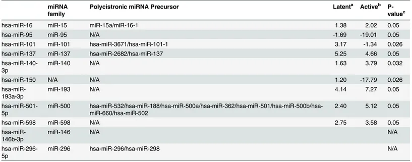
2. MicroRNA Expression in host macrophages of active MTB, latent
infection and healthy controls
The miRNA expression in macrophages of active MTB (n = 3), LTBI infection (n = 4), and
healthy controls (n = 3) were examined. Details of the subjects are listed (S1 Table). Eleven
miRNAs was found to be differentially expressed in the active MTB versus the latent/healthy
controls (p
<
0.05) (Table 3). Among these 11 miRNAs, no differences were observed between
the latent and healthy controls groups. Seven miRNAs had different expression levels between
active TB and healthy controls: six miRNAs (miR-16, miR-137, miR-140-3p,
hsa-miR-193a-3p, hsa-miR-501-5p, and hsa-miR-598) were upregulated while hsa-miR-95 was
down-regulated. Two miRNAs (hsa-miR-101 and hsa-miR-150) were found to differentiate
the LTBI group from the MTB active disease group (S2 Fig). Interestingly, hsa-miR-146b-3p
and hsa-miR-296-5p were expressed in all of LTBI group but not in the active MTB and
healthy controls.
Fig 2
shows a tendency for these 11 differentially expressed miRNAs to cluster
independently the groups of active MTB disease and the LTBI or healthy controls.
The biological pathways potentially implicated by these differentially expressed miRNAs are
listed in
Table 4. Pathway analyses identified that the change of cell membrane and
extracellu-lar matrix metabolite involving glycosaminoglycan biosynthesis-HS and fatty acid biosynthesis
might play a role in MTB infection. This might result in signal transduction through MAP
ki-nases, mTOR, ECM receptor, and Wnt, and finally activate the immune-regulatory
interac-tions involving the TGF-β
signalling pathway and the T cell receptor signalling pathway.
Discussion
In our studies, miRNA profiles in the macrophages were found to be altered in MTB
infec-tion in a strain- and host-dependent way. The Beijing genotype strain is the most predominant
M
.
tuberculosis
strain in south China, and it has caused large outbreaks of MDR-TB. The
Bei-jing strains showed increased transmission fitness when they acquired streptomycin resistance
[21]. Beijing genotype strains were also found to induce the STAT1 activation and
interferon-related immune response [22
–
23]. We showed for the first time that the Beijing/W strains
pressed a number of miRNAs as compared to the non-Beijing/W TB strains, which might
re-flect their virulence characteristics in altering the host response. Hsa-miR485-3p was found to
be upregulated in Beijing/W infected macrophages. Hsa-miR-485-3p has been shown to be
in-volved in cell survival [24] and knockdown of this miRNA in hepatic cells increased apoptosis
[25]. Previous report indicated that miR-485-3p post-transcriptionally targeted NF-YB [24], a
direct transcriptional repressor of Top2α
gene and of MDR1 and CCNB2 genes [26] in
regula-tion of the cell cycle, Our results suggest that high miR-485-3p possibly facilitates survival of
the Beijing/W strains in macrophages and evades apoptosis or alters macrophage lysis and
sub-sequent downstream immune response toward clearance of MTB.
The difficulty in discriminating the spectrum of MTB infections and of latency is prompting
the need to search for new biomarkers for MTB infection. Previous studies that have utilized
such microarrays as diagnostic markers are listed in
Table 5. Studies used whole-genome
tran-scriptional profiling of peripheral blood mononuclear cells (PBMCs) [27] or whole blood cells
Table 1. MicroRNAs differentially expressed in THP-1 macrophages infected with Beijing/W and non-Beijing/W clinical TB strains.
miRNA family
Polycistronic miRNA Precursor Ratioa
P-valueb
hsa-let-7e let-7 hsa-mir-99b/hsa-let-7e/hsa-mir-125a -1.65 0.041
hsa-let-7f let-7 N/A -1.87 0.026
hsa-miR-10a
miR-10 N/A -2.35 0.015
hsa-miR-21 miR-21 N/A -2.65 0.025
hsa-miR-26a
miR-26 N/A -1.83 0.015
hsa-miR-99a
miR-99 hsa-let-7c/hsa-miR-99a -4.34 0.026
hsa-miR-140-3p
miR-140 N/A -2.10 0.015
hsa-miR-150
N/A N/A -8.01 0.002
hsa-miR-181a
miR-181 hsa-miR-181a/hsa-miR-181b -2.85 0.015
hsa-miR-320
miR-320 N/A -1.55 0.026
hsa-miR-339-5p
miR-339 N/A -3.03 0.004
hsa-miR-425
miR-425 hsa-miR-191/hsa-miR-425 -1.70 0.041
hsa-miR-485-3p
miR-485 hsa-miR-381/hsa-miR-487b/hsa-miR-539/hsa-miR-889/hsa-miR-544a/hsa-miR-655/hsa-miR-487a/hsa- miR-382/hsa-miR-134/hsa-miR-668/hsa-miR-485/hsa-miR-323b/hsa-miR-154/hsa-miR-496/hsa-miR-377/hsa-miR-541/hsa-miR-409
14.62 0.041
hsa-miR-582-5p
miR-582 N/A -2.90 0.041
a
Fold difference in miRNA expression in THP-1 cells infected with Beijing/W clinical strains vs non-Beijing/W strains.
b
P-value was calculated by Mann-Whitney test.
[28] found that FcGR1B (CD64) and Fc gamma receptor 1B (FCGRIB) were the most
differen-tially expressed genes in the individuals with active TB. A recent report found a dominant
TNF-a+ MTB
–
specific CD4+ T cell response that discriminated between LTBI and active
dis-ease [29]. The miRNA expression profile of PBMCs [30] and sputum supernatant [31]
exhib-ited a characteristic expression in MTB infection, while the miRNA signatures from serum also
associated to different phases of TB infections [32
–
35]. These data may shed some light to the
roles of miRNAs in MTB infections, but do not yet explain the transition of latency to active
TB disease. We were able to distinguish with the expression of 11-miRNA signature profiles of
the active TB group from that of the LTBI group but not that of latent and healthy groups.
When we carried out the analysis using group-wise comparisons, the variations between
Fig 1. miRNAs expression level in the THP-1 macrophages infected with Beijing/W and non-Beijing/W clinical TB strains.The relative quantity (RQ, 2-ΔΔCt) was used to normalize the relative gene expression data. Statistical analysis between two groups was performed using Mann-Whitney test. Individual values were denoted by black dots/squares from each group of Beijing/W versus non-Beijing strains. The mean RQ and S.D. of each group were represented by the—————bar and short bars—in each
figure, respectively.
individual group members showed that 10
–
25% of the latent patients remained clustered with
the active TB patients, and this corroborated with a previous study which concluded that the
whole-blood transcript dominated by neutrophil-driven interferon (IFN)-inducible genes
cor-related with the radiological extent of active MTB [36].
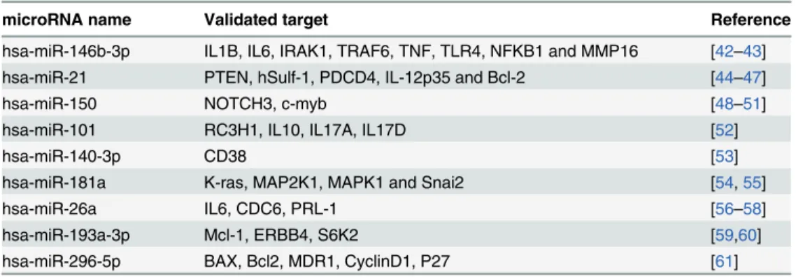
The microRNA profile in the human macrophage was quite different from that of whole
blood, sputum and PBMCs from the literature. In our study, some of the miRNAs were proven
to play key roles in the immune and inflammatory pathways, and their biological targets in
MTB infection have been previously described (Table 6). The miRNA-146 family was found to
play key roles in the anti-inflammatory reaction. miR-146b could be induced by LPS or PG
from bacteria [42]. miR-146a/b was a negative regulator of constitutive NF-kB activity, which
results in the suppression of IL-1 receptor-associated kinase 1 and TNF receptor-associated
factor 6 protein levels [42,43]. In our study, the expression of miR-146b in the LTBI group was
significantly higher than that in active TB infections. We propose that hsa-146b-3p may be
highly related to the LTBI.
miR-21 can be induced after Bacillus Calmette-Guerin (BCG) vaccination by NF-kB
activa-tion. miR-21 suppressed the IL-12 production by targeting IL-12p35, which impaired
anti-my-cobacterial T cell responses both
in vitro
and
in vivo
. Additionally, miR-21 also promoted
Table 2. Biological pathways potentially affected by the differentially expressed microRNAs in THP-1 macrophages infected with Beijing/W and non-Beijing/W TB clinical strains.KEGG pathway p-value #genes Description
TGF-beta signalling pathway 1.60E-05 31 Regulate cell differentiation, proliferation, migration and apoptosis
Wnt signalling pathway 2.07E-05 50 Required for developmental processes: cell-fate specification, cell proliferation and cell division
Lysine degradation 3.25E-05 15 Amino acid metabolism
ECM-receptor interaction 6.05E-05 22 Control of adhesion, migration, differentiation, proliferation, and apoptosis
mTOR signalling pathway 0.000931 22 Signal transduction
T cell receptor signalling pathway 0.002746 37 Activation of T lymphocytes proliferation, cytokine production and differentiation into effector
MAPK signalling pathway 0.003241 74 Involved in various cellular functions: cell proliferation, differentiation and migration Cytokine-cytokine receptor interaction 0.003241 60 Engaged in host defenses, cell growth, differentiation, cell death, angiogenesis,
development and repair processes
Adherens junction 0.004389 25 Important for maintaining tissue architecture and cell polarity and can limit cell movement and proliferation
Protein processing in endoplasmic reticulum 0.004952 46 Newly synthesized peptides glycosylated. Glycosaminoglycan biosynthesis–
heparansulfate
0.005768 11 Cell membrane and extracellular matrix component biosynthesis
Insulin signalling pathway 0.011156 41 Activation of glycogen synthesis and gene transcription Endocrine and other factor-regulated calcium
reabsorption
0.013969 15 Calcium (Ca2+) homeostasis
Apoptosis 0.014 25 Program cell death
Gap junction 0.014 24 Contain intercellular channels that allow communication between the cytosolic compartments of adjacent cells
Adipocytokine signalling pathway 0.017781 22 Positively correlated with leptin production, and negatively correlated with production of adiponectin
Cysteine and methionine metabolism 0.024674 10 Amino acid synthesization Glycosaminoglycan biosynthesis–
keratansulfate
0.036004 5 Glycan biosynthesis and metabolism
Osteoclast differentiation 0.036168 36 Responsible for bone resorption
Fc gamma R-mediated phagocytosis 0.044864 27 An essential role in host-defense mechanisms through the uptake and destruction of infectious pathogens
Table 3. miRNAs differentially expressed in human macrophages with active MTB and latent infections against healthy controls.
miRNA family
Polycistronic miRNA Precursor Latenta Activeb
P-valuec
hsa-miR-16 miR-15 miR-15a/miR-16-1 1.38 2.02 0.05
hsa-miR-95 miR-95 N/A -1.69 -19.01 0.05
hsa-miR-101 miR-101 hsa-miR-3671/hsa-miR-101-1 3.17 -1.34 0.026
hsa-miR-137 miR-137 hsa-miR-2682/hsa-miR-137 5.25 4.66 0.05
hsa-miR-140-3p
miR-140 N/A 1.63 3.79 0.032
hsa-miR-150 N/A N/A 1.20 -17.79 0.026
hsa-miR-193a-3p
miR-193 N/A 4.14 7.27 0.05
hsa-miR-501-5p
miR-500 hsa-miR-532/hsa-miR-188/hsa-miR-500a/hsa-miR-362/hsa-miR-501/hsa-miR-500b/hsa-miR-660/hsa-miR-502
2.40 5.12 0.05
hsa-miR-598 miR-598 N/A 2.75 3.58 0.05
hsa-miR-146b-3p
miR-146 N/A N/A
hsa-miR-296-5p
miR-296 hsa-miR-296/hsa-miR-298 N/A
aIndicates miRNA expression in macrophages of latent group vs healthy controls. bIndicates miRNA expression in macrophages of active group vs healthy controls. c
P-value was obtainedby an independent median test.
doi:10.1371/journal.pone.0126018.t003
Fig 2. Clustering analysis of the 11 miRNAs was performed using DataAssist 3.0v based onΔ Ct-values of the TLDA results.Upregulated miRNAs are designated by various shades of red and down-regulated miRNAs by various shades of green. Clinical phenotypes are labelled in different colours: active MTB infection (red), latent infection (blue), and healthy controls (green).
dendritic cell (DC) apoptosis by targeting Bcl-2. Therefore, miR-21 may potentially be involved
in the fine-tuning of the anti-mycobacterial Th1 response and in regulating the efficacy of BCG
vaccination [44
–
47].
miR-150 has been one of the extensively studied miRNAs, and it has been demonstrated to
be selectively expressed in mature naive B and T cells, being down-regulated in their
progeni-tors or in lymphocyte activation and strongly upregulated as maturation progresses [48
–
51].
The well-known targets for miR-150 are NOTCH3 (a member of the Notch receptor family)
and c-Myb (a transcription factor that plays an essential role in the hematopoietic process that
plays important roles both in T-cell differentiation and leukemogenesis). In our study, the
Bei-jing/W clinical strains suppressed the miR-150 and miR-21expression and they may play a role
in virulence. Lower expression of miR-150 in the active TB patients compared with the latent
and healthy controls may be due to the reduced mature T cells and B cells in patients with
ac-tive TB, as previous studies have shown [36].
Both miR-150 and miR-140-3p were differentially expressed in macrophages infected in
vitro and those from active TB patients. These two miRNAs are related with the secondary
sig-nal transduction pathway, which and likely involved in MTB infection. Four predicted
path-ways, including Wnt signalling pathway, insulin signaling pathway, TGF-β
signalling pathway
and glycosaminoglycan biosynthsis, are involved in Beijing/W & non-Beijing/W (Table 2) and
active MTB& LTBI (Table 4) studies. This reaffirms the involvement of the inflammatory
de-fence and signal transduction and cell communication in the macrophages in in MTB infection
in vitro and in the host.
Four pathways of cell membrane and communication (adherens junction, gap junction,
gly-cosaminoglycan biosynthsis-heparan sulfate/keratin sulfate metabolite), suggesting that
Table 4. Biological pathways potentially affected by the differentially expressed microRNAs of significance from macrophages of active MTB dis-ease, LTBI and healthy controls.
KEGG pathway p-value # of
genes
Description
Glycosaminoglycan biosynthesis–
heparansulfate
6.02E-28 4 Cell membrane and extracellular matrix component biosynthesis
Fatty acid biosynthesis 8.58E-15 1 Lipid Metabolism
MAPK signalling pathway 0.000271 59 Involved in various cellular functions: cell proliferation, differentiation and migration Wnt signalling pathway 0.004258 38 Required for developmental processes: cell-fate specification, cell proliferation and
cell division
Ubiquitin mediated proteolysis 0.005826 33 Functions as a signal for 26S proteasome dependent protein degradation Insulin signalling pathway 0.006315 33 Activation of glycogen synthesis and gene transcription
VEGF signalling pathway 0.008692 21 A crucial signal transducer in both physiologic and pathologic angiogenesis Circadian rhythm—mammal 0.011601 8 An internal biological clock to sustain the absence of environmental cues
Oocyte meiosis 0.011601 24 Involved in cell growth and death
TGF-beta signalling pathway 0.011601 18 Regulate cell differentiation, proliferation, migration and apoptosis ErbB signalling pathway 0.012265 23 Regulate proliferation, differentiation, cell motility and survival
Focal adhesion 0.013426 42 Cell-matrix adhesions
Phosphatidylinositol signalling system 0.015405 16 An important intracellular second-messenger signaling system Notch signalling pathway 0.016696 14 Essential for proper embryonic development in all metazoan organisms
T cell receptor signalling pathway 0.016696 26 Activation of T lymphocytes proliferation, cytokine production and differentiation into effector cells
Endocytosis 0.016696 42 Bring ligands, nutrients, plasma Membrane proteins and lipids from the cell surface into the cell interior
Beijing/W TB strain may affect macrophage survival by altering their cell membrane structure
and limit the downstream host immunological defence reaction.
The inflammatory miRNA miR-146b-3p, miR-101 and the cell survival miRNA
miR-193a-3p and miR-296-5p were only found differentially expressed in macrophages of active TB
group, suggesting response that alters macrophage survival in the infected host.
In addition, compared with whole blood, the microRNA profile revealed from the adherent
human macrophages reflect the molecular changes in the TB-engulfed macrophages, bringing
insights into the immunological defence mechanisms of these macrophages, where the initial
clearance of MTB takes place during infection. On the contrary, the microRNA profiles of
Table 5. Potential biomarkers for latent and active TB infections based on miRNA or whole genome microarray studies.
Test groups Sample Array type Finding Reference
TB active, latent and normal
Whole blood Whole-genome oligonucleotide microarray (Agilent
Technologies)
Fc gamma receptor 1B (FCGRIB) [28]
H37Rv orΔ-mce1 H37Rv bacteria
Murine macrophages Oligo whole-mouse Genome microarrays (Agilent Technologies)
Mce1 protein complex [37]
Active TB, LTBI, and Healthy Control
PBMCs The Agilent Human miRNA
microarray platform
Different pathways [30]
Active TB, LTBI, and Healthy Control
PBMCs Agilent custom designed
oligonucleotide microarrays
CD64 [27]
Active TB and Healthy Control
PBMCs Agilent’s human miRNA
microarray
miR-155 [12]
Active TB and Healthy Control
serum miRCURY LNA array (Exiqon) miR-29a [11]
Active TB and Healthy Control
PBMCs miRCURY LNA microRNA array
(Exiqon)
miR-144* [6]
Healthy donor infected with M. avium subsp.
hominissuis
PBMC derived macrophages
miRCURY LNA microRNA array (Exiqon)
Let-7e, miR-29a, miR-886-5p [38]
Active TB and Healthy Control
Sputum miRCURY LNA microRNA array
(Exiqon)
miR-19b-2*, miR-3179, miR-147 [31]
Beijing strain & latent MTB strain
Rabbit lung Whole genome rabbit microarray (Agilent)
Inflammatory response and STAT1 activation [22]
Beijing MTB strain THP-1 cell HG-U133 Plus 2.0 array (Affymetrix)
Interferon-related immune response [23]
PTB, EPTB, LTBI serum Taqman low density array (TLDA,
Life Technologies)
10 miRNA profile for European group; 12 miRNA profile for African group
[33]
PTB serum Taqman low density array (TLDA,
Life Technologies)
miR-361-5p, miR-889, miR-576-3p [32]
Active TB, LTBI, and Healthy Control
Whole blood Illumina human HT-12 beadchip array
Neutrophil-driven IFN-inducible gene profile [36]
H37Rv Murine dendritic cell miRCURY LNA microRNA array
(Exiqon)
miR-99b, miR-146a, miR-125a-5p [8]
H37Rv RAW264.7 SYBR Green-based miRNA
profiling array (SA Biosciences)
Let-7f [39]
H37Rv PBMC-derived
macrophage from healthy donor
TaqMan Low-Density Array v2.0 (Applied Biosystem, CA, USA)
miR-155,miR-146a, miR-145,miR-222*, miR-27a, miR-27b
[40]
H37Rv and H37Ra THP-1 macrophages Microarray from commercial provider‘LC Sciences’, USA
30a, 30e, 155, 1275, miR-3665, miR-3178, miR-4484, miR-4668-5p and miR-4497
[41]
blood are the orchestrated outcome of all inflammatory cells and their immune mediators in
the host-bacterial interaction, not simply MTB infection of a single immune cell type [11,
28,
32,
33]. Differentially expressed miRNAs and their transcriptional targets might potentially
af-fect the regulation of multiple biological networks. Pathway analysis of our expression profile
determined different transcripts that were modified by these miRNAs. These results provide
clues for the identification of transcriptionally regulated mechanisms of key biological
process-es in TB, enhance our understanding of the fundamental biology of MTB, and offer leads for
new diagnostics in the future.
Supporting Information
S1 Fig. Clustering analysis of the 16 miRNAs was performed using DataAssist 3.0v based
on
Δ
Ct-values of the TLDA results.
Upregulated miRNAs are designated by various shades of
red and down-regulated miRNAs by various shades of green.
(TIF)
S2 Fig. miRNA expression levels in human macrophages with LTBI, active MTB disease
and in healthy controls.
Statistical analysis between two groups was performed using the
un-paired t-test. Individual values were denoted by black dots/squares/triangles from each group.
The mean RQ and S.D. of each group were represented by the
—————
bar and short bars
—
in each figure, respectively.
(TIF)
S1 Table. Characteristics of active TB, latent and healthy controls in this study.
(DOCX)
Acknowledgments
The study was supported by the Health and Medical Research Fund (previously Research Fund
for the Control of Infectious Diseases), Food and Health Bureau, HKSAR government (RFCID
No. 09080392). We are also indebted to the administrators and technicians of the BSL3
Labora-tory, Li Ka Shing Institute of Health Sciences, The Chinese University of Hong Kong for their
invaluable advice and support in this study.
Table 6. Previously reported microRNAs with differential expression related to current study of MTB infections and their validated transcript targets.
microRNA name Validated target Reference
hsa-miR-146b-3p IL1B, IL6, IRAK1, TRAF6, TNF, TLR4, NFKB1 and MMP16 [42–43]
hsa-miR-21 PTEN, hSulf-1, PDCD4, IL-12p35 and Bcl-2 [44–47]
hsa-miR-150 NOTCH3, c-myb [48–51]
hsa-miR-101 RC3H1, IL10, IL17A, IL17D [52]
hsa-miR-140-3p CD38 [53]
hsa-miR-181a K-ras, MAP2K1, MAPK1 and Snai2 [54,55]
hsa-miR-26a IL6, CDC6, PRL-1 [56–58]
hsa-miR-193a-3p Mcl-1, ERBB4, S6K2 [59,60]
hsa-miR-296-5p BAX, Bcl2, MDR1, CyclinD1, P27 [61]
Author Contributions
Conceived and designed the experiments: EL MI KFT RCYC. Performed the experiments: LZ
EL. Analyzed the data: LZ EL MI. Contributed reagents/materials/analysis tools: LZ EL NL GL
KFT RCYC MI. Wrote the paper: LZ MI NL.
References
1. Gan H, Lee J, Ren F, Chen M, Kornfeld H, Remold HG.Mycobacterium tuberculosisblocks crosslinking of annexin-1 and apoptotic envelope formation on infected macrophages to maintain virulence. Nat Immunol. 2008; 9: 1189–1197. doi:10.1038/ni.1654PMID:18794848
2. Divangahi M, Chen M, Gan H, Desjardins D, Hickman TT, Lee DM, et al.Mycobacterium tuberculosis
evades macrophage defenses by inhibiting plasma membrane repair. Nat Immunol. 2009; 10: 899–
906. doi:10.1038/ni.1758PMID:19561612
3. Bartel DP. MicroRNAs: genomics, biogenesis, mechanism, and function. Cell 2004; 116: 281–297. PMID:14744438
4. Lim LP, Lau NC, Garrett-Engele P, Grimson A, Schelter JM, Castle J, et al. Microarray analysis shows that some microRNAs downregulate large numbers of target mRNAs. Nature 2005; 433: 769–773. PMID:15685193
5. Jackson AL, Levin AA. Developing microRNA therapeutics: approaching the unique complexities. Nu-cleic Acid Ther. 2012; 22:213–225. doi:10.1089/nat.2012.0356PMID:22913594
6. Liu YH, Wang XJ, Jiang J, Cao ZH, Yang BF, Cheng X. Modulation of T cell cytokine production by miR-144*with elevated expression in patients with pulmonary tuberculosis. Mol Immunol. 2011; 48: 1084–1090. doi:10.1016/j.molimm.2011.02.001PMID:21367459
7. Chatterjee S, Dwivedi VP, Singh Y, Siddiqui I, Sharma P, Van Kaer L, et al. Early secreted antigen ESAT-6 ofMycobacterium tuberculosispromotes protective T helper 17 cell responses in a Toll-Like receptor-2-dependent manner. PLoS Pathogens 2011; 7:e1002378. doi:10.1371/journal.ppat. 1002378PMID:22102818
8. Singh Y, Kaul V, Mehra A, Chatterjee S, Tousif S, Dwivedi VP, et al.Mycobacterium tuberculosis con-trols miR-99b expression in infected murine dendritic cells to modulate host immunity. J Biol Chem. 2013; 288: 5056–5061. doi:10.1074/jbc.C112.439778PMID:23233675
9. Rajaam MV, Ni B, Morris JD, Brooks MN, Carlson TK, Bakthavachalu B, et al.Mycobacterium tubercu-losislipomannan blocks TNF biosynthesis by regulating macrophage MAPK-activated protein kinase 2 (MK2) and microRNA miR-125b. Proc Natl Acad Sci USA 2011; 108: 17408–17413. doi:10.1073/pnas. 1112660108PMID:21969554
10. O’Connell RM, Taganov KD, Boldin MP, Cheng G, Baltimore D. MicroRNA- 155 is induced during the macrophage inflammatory response. Proc Natl Acad Sci USA 2007; 104: 1604–1609. PMID:17242365
11. Fu Y, Yi Z, Wu X, Li J, Xu F. Circulating microRNAs in patients with active pulmonary tuberculosis. J Clin Microbiol. 2011; 49:4246–4251. doi:10.1128/JCM.05459-11PMID:21998423
12. Wu J, Lu CY, Diao N, Zhang S, Wang S, Wang F, et al. Analysis of microRNA expression profiling iden-tifies miR-155 and miR-155*as potential diagnostic markers for active tuberculosis: a preliminary study. Hum Immunol 2012; 73:31–37. doi:10.1016/j.humimm.2011.10.003PMID:22037148
13. Chen J, Tsolaki AG, Shen X, Jiang X, Mei J, Gao Q, et alDeletion-targeted multiplex PCR (DTM-PCR) for identification of Beijing/W genotypes of Mycobacterium tuberculosis. Tuberculosis (Edinb.) 2007; 87:446–449. PMID:17632035
14. Wong KC, Leong WM, Law HK, Ip KF, Lam JT, Yuen KY, et al. Molecular characterization of clinical iso-lates of Mycobacterium tuberculosis and their association with phenotypic virulence in human macro-phages. Clin Vaccine Immunol. 2007; 10: 1279–84. PMID:17715326
15. Tsuchiya S, Kobayashi Y, Goto Y, Okumura H, Nakae S, Konno T, et al. Induction of maturation in cul-tured human monocytic leukemia cells by a phorboldiester. Cancer Research 1982; 42: 1530–1536. PMID:6949641
16. Dweep H, Sticht C, Pandey P, Gretz N. miRWalk—database: prediction of possible miRNA binding sites by“walking”the genes of 3 genomes. J Biomed Inform. 2011; 44:839–847. doi:10.1016/j.jbi. 2011.05.002PMID:21605702
17. Vlachos IS, Kostoulas N, Vergoulis T, Georgakilas G, Reczko M, Maragkakis M, et al. DIANA miRPath v.2.0: investigating the combinatorial effect of microRNAs in pathways. Nucleic Acids Res 2012; 40: W498–504. doi:10.1093/nar/gks494PMID:22649059
19. Baltimore D, Boldin MP, O’Connell RM, Rao DS, Taganov KD. MicroRNAs: new regulators of immune cell development and function. Nat Immunol. 2008; 9: 839–845. doi:10.1038/ni.f.209PMID:18645592
20. Moschos SA, Williams AE, Perry MM, Birrell MA, Belvisi MG, Lindsay MA. Expression profilingin vivo
demonstrates rapid changes in lung microRNA levels following lipopolysaccharide-induced inflamma-tion but not in the anti-inflammatory acinflamma-tion of glucocorticoids. BMC Genomics 2007; 240:1–12.
21. Buu TN, van Soolingen D, Huyen MN, Lan NT, Quy HT, Tiemersma EW, et al. Increased transmission ofMycobacterium tuberculosisBeijing genotype strains associated with resistance to streptomycin: a population-based study. PLoS One 2012; 7: e42323. doi:10.1371/journal.pone.0042323PMID: 22912700
22. Subbian S, Bandyopadhyay N, Tsenova L, O Brien P, Khetani V, Kushner NL, et al. Early innate immu-nity determines outcome ofMycobacterium tuberculosispulmonary infection in rabbits. Cell Commun Signal 2013; 11:60. doi:10.1186/1478-811X-11-60PMID:23958185
23. Wu K, Dong D, Fang H, Levillain F, Jin W, Mei J, et al. An interferon-related signature in the transcrip-tional core response of human macrophages toMycobacterium tuberculosisinfection. PLoS One 2012; 7: e38367. doi:10.1371/journal.pone.0038367PMID:22675550
24. Lucotti S, Rainaldi G, Evangelista M, Rizzo M. (2013) Fludarabine treatment favors the retention of miR-485-3p by prostate cancer cells: implications for survival. Mol Cancer 12(1):52. doi:10.1186/ 1476-4598-12-52PMID:23734815
25. Yang H, Cho ME, Li TW, Peng H, Ko KS, Mato JM, et al. MicroRNAs regulate methionine adenosyl-transferase 1A expression in hepatocellular carcinoma. J Clin Invest. 2013; 123: 285–298. doi:10. 1172/JCI63861PMID:23241961
26. Chen CF, He XL, Arslan AD, Mo YY, Reinhold WC, Pommier Y, et al. Novel regulation of nuclear factor-γb by miR-485-3p affects the expression of DNA topoisomerase IIαand drug responsiveness. Mol Pharmacol. 2011; 79: 735–741. doi:10.1124/mol.110.069633PMID:21252292
27. Jacobsen M, Repsilber D, Gutschmidt A, Neher A, Feldmann K, Mollenkopf HJ, et al. Candidate bio-markers for discrimination between infection and disease caused byMycobacterium tuberculosis. J Mol Med (Berl.) 2007; 85: 613–621. PMID:17318616
28. Maertzdorf J, Repsilber D, Parida SK, Stanley K, Roberts T, Black G, et al. Human gene expression profiles of susceptibility and resistance in tuberculosis. Genes Immun. 2011; 12: 15–22. doi:10.1038/ gene.2010.51PMID:20861863
29. Harari A, Rozot V, Enders FB, Perreau M, Stalder JM, Nicod LP, et al. Dominant TNF-alpha(+) Myco-bacterium tuberculosis-specific CD4(+) T cell responses discriminate between latent infection and ac-tive disease. Nat Med. 2011; 17:372–U174. doi:10.1038/nm.2299PMID:21336285
30. Wang C, Yang S, Sun G, Tang X, Lu S, Neyrolles O, et al. Comparative miRNA expression profiles in individuals with latent and active tuberculosis. PLoS One 2011; 6:e25832. doi:10.1371/journal.pone. 0025832PMID:22003408
31. Yi Z, Fu Y, Ji R, Li R, Guan Z. Altered microRNA signatures in sputum of patients with active pulmonary tuberculosis. PLoS One 2012; 7: e43184. doi:10.1371/journal.pone.0043184PMID:22900099
32. Qi Y, Cui L, Ge Y, Shi Z, Zhao K, Guo X, et al. Altered serum microRNAs as biomarkers for the early di-agnosis of pulmonary tuberculosis infection. BMC Infect Dis. 2012; 12:384. doi:10.1186/1471 -2334-12-384PMID:23272999
33. Miotto P, Mwangoka G, Valente IC, Norbis L, Sotgiu G, Bosu R, et al. miRNA signatures in sera of pa-tients with active pulmonary tuberculosis. PLoS One 2013; 8: e80149. doi:10.1371/journal.pone. 0080149PMID:24278252
34. Zhang X, Guo J, Fan S, Li Y, Wei L, Yang X, et al. Screening and identification of six serum microRNAs as novel potential combination biomarkers for pulmonary tuberculosis diagnosis. PLoS One 2013; 8: e81076. doi:10.1371/journal.pone.0081076PMID:24349033
35. Zhang H, Sun Z, Wei W, Liu Z, Fleming J, Zhang S, et al. Identification of serum microRNA biomarkers for tuberculosis using RNA-seq. PLoS One 2014; 9: e88909. doi:10.1371/journal.pone.0088909 PMID:24586438
36. Berry MP, Graham CM, McNab FW, Xu Z, Bloch SA, Oni T, et al.An interferon-inducible neutrophil-driv-en blood transcriptional signature in human tuberculosis. Nature 2010; 466: 973–U998. doi:10.1038/ nature09247PMID:20725040
37. Stavrum R, Valvatne H, Stavrum AK, Riley LW, Ulvestad E, Jonassen I, et al.Mycobacterium tubercu-losisMce1 protein complex initiates rapid induction of transcription of genes involved in substrate traf-ficking. Genes Immun. 2012; 13: 496–502. doi:10.1038/gene.2012.24PMID:22695749
39. Kumar M, Kumar Sahu S, Kumar R, Subuddhi A, Kumar Maji R, Jana K, et al. MicroRNA let-7 Modu-lates the Immune Response to Mycobacterium tuberculosis Infection via Control of A20, an Inhibitor of the NF-κB Pathway. Cell Host & Microbe 2015; 17: 345–56.
40. Furci L, Schena E, Miotto P, Cirillo DM. Alteration of human macrophages microRNA expression profile upon infection with Mycobacterium tuberculosis. Int J Mycobacteriol. 2013; 2: 128–34.
41. Dasa K, Saikolappana S, Dhandayuthapani S. Differential expression of miRNAs by macrophages in-fected with virulent and avirulent Mycobacterium tuberculosis. Tuberculosis 2013; 93; Supplement: S47–S50.
42. Taganov KD, Boldin MP, Chang KJ, Baltimore D. NF-kappaB-dependent induction of microRNA miR-146, an inhibitor targeted to signaling proteins of innate immune responses. Proc Natl Acad Sci USA2006; 103: 12481–12486. PMID:16885212
43. Bhaumik D, Scott GK, Schokrpur S, Patil CK, Campisi J, Benz CC, et al. Expression of microRNA-146 suppresses NF-kappaB activity with reduction of metastatic potential in breast cancer cells. Oncogene 2008; 27:5643–5647. doi:10.1038/onc.2008.171PMID:18504431
44. Bao L, Yan Y, Xu C, Ji W, Shen S, Xu G, et al. MicroRNA-21 suppresses PTEN and hSulf-1 expression and promotes hepatocellular carcinoma progression through AKT/ERK pathways. Cancer Lett 2013; 337: 226–236. doi:10.1016/j.canlet.2013.05.007PMID:23684551
45. Zhu S, Wu H, Wu F, Nie D, Sheng S, Mo YY. MicroRNA-21 targets tumor suppressor genes in invasion and metastasis. Cell Res. 2008; 18: 350–359. doi:10.1038/cr.2008.24PMID:18270520
46. Liu C, Yu J, Yu S, Lavker RM, Cai L, Liu W, et al. MicroRNA-21 acts as an oncomir through multiple tar-gets in human hepatocellular carcinoma. J Hepatol. 2010; 53: 98–107. doi:10.1016/j.jhep.2010.02.021 PMID:20447717
47. Wu Z, Lu H, Sheng J, Li L. Inductive microRNA-21 impairs anti-mycobacterial responses by targeting IL-12 and Bcl-2. FEBS Lett. 2012; 586: 2459–2467. doi:10.1016/j.febslet.2012.06.004PMID: 22710123
48. Lin YC, MW Kuo, J Yu, HH Kuo, RJ Lin, WL Lo, et al. c-Myb is an evolutionary conserved miR-150 tar-get and miR-150/c-Myb interaction is important for embryonic development. Mol Biol Evol. 2008; 25: 2189–2198. doi:10.1093/molbev/msn165PMID:18667440
49. Xiao C, Calado DP, Galler G, Thai TH, Patterson HC, Wang J, et al. miR-150 controls B cell differentia-tion by targeting the transcripdifferentia-tion factor c-Myb. Cell 2007; 131: 146–159. PMID:17923094
50. Zhou B, Wang S, Mayr C, Bartel DP, Lodish HF. miR-150, a microRNA expressed in mature B and T cells, blocks early B cell development when expressed prematurely. Proc Natl Acad Sci USA 2007; 104: 7080–7085. PMID:17438277
51. Ghisi M, Corradin A, Basso K, Frasson C, Serafin V, Mukherjee S, et al. Modulation of microRNA ex-pression in human T-cell development: targeting of NOTCH3 by miR-150. Blood 2011; 117:7053–
7062. doi:10.1182/blood-2010-12-326629PMID:21551231
52. Schaefer JS, Montufar-Solis D, Vigneswaran N, Klein JR. Selective upregulation of microRNA expres-sion in peripheral blood leukocytes in IL-10-/- mice precedes expresexpres-sion in the colon. J Immunol. 2011; 187: 5834–5841. doi:10.4049/jimmunol.1100922PMID:22043014
53. Jude JA, Dileepan M, Subramanian S, Solway J, Panettieri RA Jr, Walseth TF, et al. miR-140-3p regu-lation of TNF-α-induced CD38 expression in human airway smooth muscle cells. Am J Physiol Lung Cell Mol Physiol. 2012; 303: L460–468. doi:10.1152/ajplung.00041.2012PMID:22773691
54. He Q, Zhou X, Li S, Jin Y, Chen Z, Chen D, et al. MicroRNA-181a suppresses salivary adenoid cystic carcinoma metastasis by targeting MAPK-Snai2 pathway. Biochim Biophys Acta 2013; 1830: 5258–
5266. doi:10.1016/j.bbagen.2013.07.028PMID:23911747
55. Shin KH, Bae SD, Hong HS, Kim RH, Kang MK, Park NH, et al. miR-181a shows tumor suppressive ef-fect against oral squamous cell carcinoma cells by downregulating K-ras. Biochem Biophys Res Com-mun. 2011; 404:896–902. doi:10.1016/j.bbrc.2010.12.055PMID:21167132
56. Zhang R, Tian A, Wang J, Shen X, Qi G, et al. miR26a Modulates Th17/T reg Balance in the EAE Model of Multiple Sclerosis by Targeting IL6. Neuromolecular Med. 2015; 17:24–34. doi:10.1007/ s12017-014-8335-5PMID:25362566
57. Zhang X, Xiao D, Wang Z, Zou Y, Huang L, Lin W, et al. MicroRNA-26a/b regulate DNA replication li-censing, tumorigenesis, and prognosis by targeting CDC6 in lung cancer. Mol Cancer Res. 2014; 12: 1535–46. doi:10.1158/1541-7786.MCR-13-0641PMID:25100863
58. Dong J, Sui L, Wang Q, Chen M, Sun H. MicroRNA-26a inhibits cell proliferation and invasion of cervi-cal cancer cells by targeting protein tyrosine phosphatase type IVA 1. Mol Med Rep. 2014; 10: 1426–
59. Kwon JE, Kim BY, Kwak SY, Bae IH, Han YH. Ionizing radiation-inducible microRNA miR-193a-3p in-duces apoptosis by directly targeting Mcl-1. Apoptosis 2013; 18: 896–909. doi: 10.1007/s10495-013-0841-7PMID:23546867
60. Yu T, Li J, Yan M, Liu L, Lin H, et al. MicroRNA-193a-3p and -5p suppress the metastasis of human non-small-cell lung cancer by downregulating the ERBB4/PIK3R3/mTOR/S6K2 signaling pathway. On-cogene 2015; 34: 413–23. doi:10.1038/onc.2013.574PMID:24469061