274
Radiol Bras. 2017 Jul/Ago;50(4):266–276 Letters to the Editordecompression and enema use were not considered because of the risk of cecal perforation. Therefore, the pseudo-obstruction was conirmed surgically. Thereafter, the patient was treated with gastric rest and her electrolyte levels were monitored.
Ogilvie’s syndrome was named after William Heneage Ogil -vie, who, in 1948, described a disorder of gastrointestinal motil -ity, with dilation of the cecum and right colon in the absence of mechanical obstruction, that was autonomic in origin, with suppression of parasympathetic activity and activation of sym-pathetic activity(1).
The acute form of Ogilvie’s syndrome arises from an au -tonomic imbalance, with a mismatch between parasympathetic and sympathetic activity, which are downregulated and upregu-lated, respectively. The distal colon is often atonic, whereas the proximal colon can still be functional(2). Some chemotherapeu -tic agents have been implicated, such as those in the rituximab-cyclophosphamide-doxorubicin-vincristine-prednisone regimen, as have factors such as trauma, acute myocardial injury, elec -trolyte disturbances, hypothyroidism, renal failure, and neu-ropathy(2). Lee et al.(3) observed that cancer patients developed Ogilvie’s syndrome two to ten days after infusion of vincristine, the syndrome resolving after its discontinuation. Sandler et al.(4) found that patients treated with vincristine experienced abdomi -nal pain and constipation within the irst 4–72 hours after re -ceiving the drug. Neutropenia and the use of antibiotic therapy have also been implicated in the development of the syndrome(3).
The symptoms of Ogilvie’s syndrome include abdominal dis -tension, abdominal pain, vomiting of fecal matter, and constipa-tion(1,5). Signs of peritonitis can indicate cecal perforation with
Figure 2. Coronal reconstruction of a CT scan, providing a better view of the transitional zone, where an abrupt transition to a normal caliber segment is observed, with no evident occlusive lesion (arrow). Note the marked dilation of the cecum, which measured 14 cm in diameter (arrowhead). Left pleural effusion can also be seen.
©
pneumoperitoneum(6), especially when the distension is greater than 12 cm and lasts for more than six days. For evaluating dis -eases of the colon, CT has been shown to be the method of choice(7–11). In Ogilvie’s syndrome, CT is a useful for identifying the obstruction and determining the underlying cause(12), the main indings being dilation extending from the cecum to the transverse colon, with a transition zone in the splenic lexure, where the caliber of the adjoining loop is considerably smaller. The treatment involves the use of parasympathomimetic agents that increase colonic motility(13), endoscopic decompression or right hemicolectomy, the last being required in the presence of cecal ischemia or perforation.
Colonic pseudo-obstruction is associated with the use of chemotherapy. It is characterized by dilation of the loops of the colon and transitional zone. Attention should be paid to signs of perforation and the risk of death from cecal rupture.
REFERENCES
1. Azevedo RP, Freitas FGR, Ferreira EM, et al. Constipação intestinal em terapia intensiva. Rev Bras Ter Intensiva. 2009;21:324–31.
2. Gmora S, Poenaru D, Tsai E. Neostigmine for the treatment of pediatric acute colonic pseudo-obstruction. J Pediatr Surg. 2002;37:E28. 3. Lee GE, Lim GY, Lee JW, et al. Acute colonic pseudo-obstruction com
-plicating chemotherapy in paediatric oncohaematological patients: clini -cal and imaging features. Br J Radiol. 2012;85:377–81.
4. Sandler SG, Tobin W, Henderson ES. Vincristine-induced neuropathy. A clinical study of ifty leukemic patients. Neurology. 1969;19:367–74. 5. Choi JS, Lim JS, Kim H, et al. Colonic pseudo-obstruction: CT indings.
AJR Am J Roentgenol. 2008;190:1521–6.
6. Saunders MD. Acute colonic pseudo-obstruction. Best Pract Res Clin Gastroenterol. 2007;21:671–87.
7. Vermelho MB, Correia AS, Michailowsky TC, et al. Abdominal altera -tions in disseminated paracoccidioidomycosis: computed tomography indings. Radiol Bras. 2015;48:81–5.
8. Melo EL, Paula FT, Siqueira RA, et al. Biliary colon: an unusual case of intestinal obstruction. Radiol Bras. 2015;48:127–8.
9. Gava P, Melo AS, Marchiori E, et al. Intestinal and appendiceal paracoc -cidioidomycosis. Radiol Bras. 2015;48:126–7.
10. Rocha EL, Pedrassa BC, Bormann RL, et al. Abdominal tuberculosis: a radiological review with emphasis on computed tomography and mag-netic resonance imaging indings. Radiol Bras. 2015;48:181–91. 11. Sala MA, Ligabô AN, Arruda MC, et al. Intestinal malrotation associ
-ated with duodenal obstruction secondary to Ladd’s bands. Radiol Bras. 2016;49:271–2.
12. Fukuya T, Hawes DR, Lu CC, et al. CT diagnosis of small-bowel obstruc -tion: eficacy in 60 patients. AJR Am J Roentgenol. 1992;158:765–9. 13. Ponec RJ, Saunders MD, Kimmey MB. Neostigmine for the treatment
of acute colonic pseudo-obstruction. N Engl J Med. 1999;341:137–41.
Fernanda Miraldi Clemente Pessôa1, Leonardo Kayat Bittencourt2, Alessandro Severo Alves de Melo1
1. Hospital Universitário Antonio Pedro – Universidade Federal Fluminense (HUAP-UFF), Niterói, RJ, Brazil. 2. Universidade Federal Fluminense (UFF), Ni-terói, RJ, Brazil. Mailing address: Dra. Fernanda Miraldi Clemente Pessôa. Rua Ouro Branco, 66, ap. 301, Vila Valqueire. Rio de Janeiro, RJ, Brazil, 21321-560. E-mail: [email protected].
http://dx.doi.org/10.1590/0100-3984.2015.0162
Pontine tegmental cap dysplasia accompanied by a duplicated internal auditory canal
Dear Editor,
A 48-year-old female with cognitive and auditory deicits presented for evaluation prior to cochlear implantation. Among her parents and four siblings, there was one brother with men-tal disability of unknown cause. Physical examination revealed
275
Radiol Bras. 2017 Jul/Ago;50(4):266–276Letters to the Editor
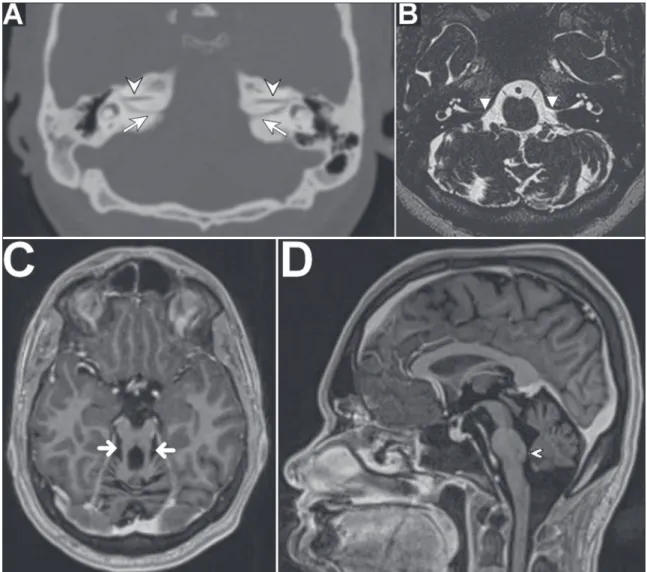
the vestibulocochlear nerve (Figure 1A), together with a discrete reduction in the volume of the pons and cerebellum. In addi -tion to the duplicated internal auditory canal, magnetic reso-nance imaging (MRI) of the brain and ears revealed the follow -ing: absence of the eighth cranial nerve (Figure 1B); elongated, discretely lateralized superior cerebellar peduncles, with an ap -pearance similar to the molar tooth sign (Figure 1C); pons with a dysplastic aspect and a reduction in its volume, especially in the ventral region, presenting a small prominence, on the poste-rior surface, projecting into the fourth ventricle; and cerebellar hypoplasia, mainly in the vermis (Figure 1D). On the basis of those indings, the patient was diagnosed with pontine tegmen -tal cap dysplasia (PTCD).
Cerebellar hypoplasia/hypogenesis can be seen in cases of metabolic disorder, exposure to teratogens, congenital infec -tion or genetic disorders(1). The molar tooth sign is observed in the axial plane of CT scans and, more clearly. of MRI scans at the junction between the rhombencephalon and mesencepha -lon, classically in the presence of cerebellar vermis hypoplasia/ agenesis, deep interpeduncular fossa; Superior, poorly oriented, thickened and elongated superior cerebellar peduncles(2).
PTCD is a brainstem malformation(1,3,4), initially described in 2007 by Barth et al.(5); to date, fewer than 50 cases have been reported(4). The main signs and symptoms are auditory de -iciency, in 92% of cases; cognitive deicit, in 76%; deglutition disorders, in 64%; facial paralysis, in 60%; abnormal eye move -ment, in 60%; trigeminal paresthesia, in 60%; ataxia, in 56%; hypotonia; cyclic vomiting syndrome; and various neurological disorders of the third to the eighth cranial nerves(4,6,7). Other po -tential characteristics of PTCD include hypoplasia of the pons (notably in its ventral aspect); a mass of ectopic dorsal pontine ibers protruding into the fourth ventricle; hypoplasia/agenesis of the middle and inferior cerebellar peduncles; elongation of the superior cerebellar peduncles; cerebellar vermis hypoplasia/ agenesis; absence or malformation of the inferior olivary nuclei; hypogenesis/absence of the third to eighth cranial nerves; costo -vertebral deformities; and cardiovascular anomalies(1,3–8). PTCD can exhibit a feature similar to the molar tooth sign, although with lateralized, tapered superior cerebellar peduncles(1,4,6). The differential diagnoses include pontocerebellar hypoplasia, as well as a number of syndromes(2,8,9): Joubert; Dekaban-Arima; Senior-Loken; COACH; Váradi-Papp; Malta; and Moebius.
276
Radiol Bras. 2017 Jul/Ago;50(4):266–276 Letters to the EditorRodolfo Mendes Queiroz1, Lara Zupelli Lauar1, Luiz Carlos Alves de Souza2, Rafael Gouvêa Gomes de Oliveira1, Lucas Giansante Abud1
1. MED – Medicina Diagnóstica / Hospital São Lucas, Ribeirão Preto, SP, Brazil. 2. Clínica Paparella de Otorrinolaringologia, Ribeirão Preto, SP, Brazil. Mailing address: Dr. Rodolfo Mendes Queiroz. MED – Medicina Diagnóstica. Rua Ber-nardino de Campos, 1426, Vila Seixas. Ribeirão Preto, SP, Brazil, 14015-130. E-mail: [email protected].
Although a duplicated internal auditory canal is extremely rare, it is found in at least 46% of all cases of PTCD. The two canals are often narrow (with a caliber of less than 2.0 mm) and accompanied by hypogenesis/agenesis of the eighth cra-nial nerve, which typically contraindicates cochlear implanta-tion(4,8,10,11). Some authors have reported differentiated cases in which the division is made by a bony septum, proposing that the term “partitioned” (rather than “duplicated”) be used in such cases(11).
REFERENCES
1. Poretti A, Boltshauser E, Doherty D. Cerebellar hypoplasia: differen -tial diagnosis and diagnostic approach. Am J Med Genet C Semin Med Genet. 2014;166C:211–26.
2. Gleeson JG, Keeler LC, Parisi MA, et al. Molar tooth sign of the mid -brain-hindbrain junction: occurrence in multiple distinct syndromes. Am J Med Genet A. 2004;125A:125–34.
3. Amaral LLF, Yared JH, Lopes BSC. Malformações congênitas infraten -toriais. In: Rocha AJ, Vedolin L, Mendonça RA, editores. Encéfalo. Série CBR. São Paulo: Elsevier; 2012. p. 79–80.
4. Nixon JN, Dempsey JC, Doherty D, et al. Temporal bone and cranial nerve indings in pontine tegmental cap dysplasia. Neuroradiology. 2016; 58:179–87.
5. Barth PG, Majoie CB, Caan MW, et al. Pontine tegmental cap dyspla -sia: a novel brain malformation with a defect in axonal guidance. Brain. 2007;130(Pt 9):2258–66.
6. Chong PF, Haraguchi K, Torio M, et al. A case of pontine tegmental cap dysplasia with comorbidity of oculoauriculovertebral spectrum. Brain Dev. 2015;37:171–4.
7. Singh D, Hsu CC, Kwan GN, et al. Pontine tegmental cap dyspla -sia: MR evaluation of vestibulocochlear neuropathy. J Neuroimaging. 2015;25:1038–43.
8. Desai NK, Young L, Miranda MA, et al. Pontine tegmental cap dyspla -sia: the neurotologic perspective. Otolaryngol Head Neck Surg. 2011; 145:992–8.
9. Barra FR, Gonçalves FG, Matos VL, et al. Signs in neuroradiology – Part 2. Radiol Bras. 2011;44:129–33.
10. Lee SY, Cha SH, Jeon MH, et al. Narrow duplicated or triplicated inter -nal auditory ca-nal (3 cases and review of literature): can we regard the separated narrow internal auditory canal as the presence of vestibuloco-chlear nerve ibers? J Comput Assist Tomogr. 2009;33:565–70. 11. Vincenti V, Ormitti F, Ventura E. Partitioned versus duplicated internal
auditory canal: when appropriate terminology matters. Otol Neurotol. 2014;35:1140–4.