Luis Eduardo Duarte Irala(a) Renata Grazziotin-Soares(b) Alexandre Azevedo Salles(b) Aline Zen Munari(c)
Joseani Santos Pereira(c)
(a)MSc; (b)PhD student; (c)Dental Student – School of Dentistry, Universidade Luterana do Brasil (ULBRA), Canoas, RS, Brazil.
Corresponding author: Renata Grazziotin-Soares
Rua Bento Gonçalves, 1624 - Centro Caxias do Sul - RS - Brazil
CEP: 95020-412
Email: [email protected]
Received for publication on Apr 21, 2010 Accepted for publication on Jun 13, 2010
Dissolution of bovine pulp tissue in
solutions consisting of varying NaOCl
concentrations and combined with EDTA
Abstract: This in vitro study evaluated (1) the dissolution of bovine pulp tissue in solutions consisting of varying NaOCl concentrations and com-bined with EDTA; and (2) the pH of these solutions before and after the experiment. The independent variables were the concentration and the volume of the solution. Thirty bovine pulps were divided in equal fragments, resulting in 90 fragments of pulp tissue. Each fragment was immersed in one of the following solutions: 1% NaOCl (4 ml), 2.5% NaOCl (4 ml), 1% NaOCl + 17% EDTA (2 ml : 2 ml), 1% NaOCl + 17% EDTA (1 ml : 3 ml), 2.5% NaOCl + 17% EDTA (2 ml : 2 ml), and 2.5% NaOCl + 17% EDTA (1 ml : 3 ml). The test solutions were dichotomized as either able or not able to dissolve the tissue, the latter being attributed when the dissolution of the pulp tissue was not complete within 48 hours. When the samples were able to dissolve the tissue, the time required for complete tissue dissolution was submitted to statistical analysis. The pH of the solutions was measured before and after the experiment. The pH variable was dichotomized as either changed or unchanged. The results demonstrated that the NaOCl solutions combined with 17% EDTA were not able to dissolve the tissue. The t-test revealed that the 2.5% NaOCl solution presented a lower mean dissolution time than the 1% NaOCl so-lution (p < 0.001). The pH of the soso-lutions with equal volumes of NaOCl and EDTA decreased in 48 hours.
Descriptors: Hydrogen-ion concentration; Sodium hypochlorite; Dissolution; Dental pulp.
Introduction
Successful root canal therapy depends on thorough chemomechani-cal debridement of pulp tissue, dentin debris, and infective microorgan-isms.1,2 Mechanical instrumentation alone is not able to reduce the mi-crobial population and leave dentinal surfaces free of a smear layer.3,4 Even in well-shaped canals, there are remnants of pulp tissue and inor-ganic debris, especially in areas where instruments cannot reach.4
ca-nals to be illed,13 or may also be used alternately with sodium hypochlorite during the whole prepa-ration of the root canal.5,14
Many authors have reported the signiicant inlu-ence of a variety of factors, like concentration, time, temperature, tissue-irrigating contact area, canal preparation size, volume, tissue type, and mechani-cal action, on the ability of NaOCl to dissolve both necrotic and vital tissues.10,15,16,17,18,19 Christensen et al.20 reported that concentration, time, and pH all play important roles in determining the amount of tissue dissolution. Higher concentrations and great-er time pgreat-eriods all lead to greatgreat-er amounts of tissue dissolution.Although antibacterial power may in-crease, tissue dissolution decreases as pH decreases. However, the related literature lacks more con-sistent information on the tissue dissolution results obtained when NaOCl is combined with EDTA.
Thus, the purpose of this in vitro study was to evaluate (1) the dissolution of bovine pulp tissue in solutions consisting of varying NaOCl concentra-tions and combined with EDTA; and (2) the pH of the solutions before and after the experiment. The independent variables of this study were the concen-tration and the volume of the solution.
Materials and Methods
This study was submitted to the local Ethical Committee for evaluation and was approved. Bovine pulp tissue was used to test the dissolving ability of different NaOCl concentrations, and of NaOCl combined with EDTA.
Pulp tissue collection
The pulp tissue of the animals used was obtained from a meat packing plant in the city of Montene-gro, RS, Brazil. The bone tissue containing the up-per incisors was collected immediately after their death. The tissue was kept refrigerated in 100% hu-midity until the teeth were removed with the help of a number 7 carver (Golgran, São Paulo, SP, Brazil) and a Lecron carver (Golgran, São Paulo, SP, Bra-zil). Subsequently, pilot grooves were prepared on the buccal and oral aspects of the crowns using a diamond-coated issure bur (Dentsply Maillefer, Ballaigues, Switzerland) without entering the pulp
chamber. The teeth were then carefully split into two fragments using a number 2 Ochsenbein mi-cro-chisel (Golgran, São Paulo, SP, Brazil), and the entire pulp tissue was removed from the pulp cham-ber and the root canals using a numcham-ber 17 spoon excavator (Golgran, São Paulo, SP, Brazil). Thirty pulp specimens were immersed in a 0.9% saline so-lution (Pharmacy-School of the Lutheran University of Brazil/ULBRA, Canoas, RS, Brazil) at room tem-perature for 30 min. Next, the 30 pulps were divid-ed into equal 5-mm-long fragments by a number 15 scalpel blade (Swann-Morton, Shefield, England), resulting in 90 fragments of pulp tissue.
Three fragments from each pulp were removed from the irst 15 mm of the coronal region. The tissue remainder (apical third) was discarded. The sizes of 90 fragments were very similar. Length, as well as buccolingual and mesiodistal diameters were measured with a digital caliper (799 Starret, Itu, SP, Brazil). Uniform volumes of all fragments were pre-pared to prevent any interference with the dissolu-tion time.
pH of the solutions
The solutions of NaOCl and EDTA were ob-tained from the Pharmacy-School of the University where the experiment was conducted, the Lutheran University of Brazil/ULBRA, Canoas, RS, Brazil. The pH of the solutions was measured before and after the experiment was performed, by pH measur-ing tapes (Merck KgaA, Darmstadt, Germany). The variable pH was dichotomized as either changedor
unchanged. In samples where there was change, the inal and initial pH values were recorded.
Tissue dissolution
Each fragment of bovine pulp tissue was im-mersed in 4 ml of solution. The solutions of NaOCl combined with EDTA were mixed with a plastic spatula (Golgran, São Paulo, SP, Brazil) before im-mersion of the fragment. Next, the solution was left to rest. The fragments were immersed in the test so-lutions through a randomized process conducted by someone who did not belong to the research group.
rub-ber stopper (Vidraria Sul Brasil S/A, Canoas, RS, Brazil). All glass containers were kept on a shelf at room temperature and protected from light. Fifteen samples were prepared for each type of solution (Ta-ble 1).
The room temperature measured by thermom-eter (Cotergavi, São Paulo, SP, Brazil) on the experi-ment days was 22°C. The variable dissolution abil-ity was dichotomized as either able or not able to dissolve the tissue. A chronometer (Casio Computer Co. Ltd., Shibuyaku, Japan) measured the time from pulp tissue immersion to its complete dissolution. Complete dissolution meant that the tissue was not visible through a 4X magniication loupe (Pró Vista Imp Com Materiais Óticos Ltda, São Paulo, SP, Bra-zil).
The maximum time set for the experiment was 48 hours. The dissolution process was followed dur-ing this time by two researches (A.Z.M. and J.S.P.) in 6-hour shifts. If the pulp tissue was not com-pletely dissolved after 48 hours, the solution sample was considered as not able to dissolve the tissue.
For samples that were able to dissolve the tissue, the time required for complete dissolution was submit-ted to statistical analysis.
Results
Table 2 shows the mean times (in minutes) of the
bovine pulp tissue dissolution for each solution. Since the solutions of NaOCl combined with 17% EDTA were unable to dissolve the pulp tissue, the statistical analysis was applied only to the pure solutions of NaOCl in order to assess if there was any statistically signiicant difference in dissolu-tion ability between the 1% NaOCl and the 2.5% NaOCl solutions. The t-test revealed that the 2.5% NaOCl (4 ml) solution presented a signiicantly low-er mean dissolution time (p < 0.001) than the 1% NaOCl (4 ml) solution (Table 3).
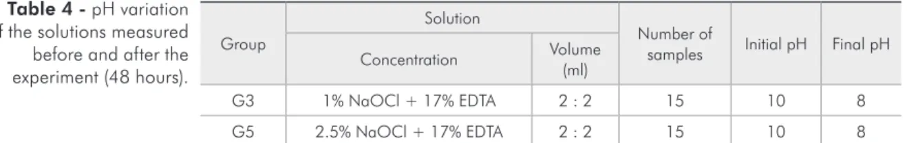
Only the pH values that changed after the exper-iment are reported in Table 4. The pH in all other solutions remained the same at the end of the ex-periment as in the beginning.
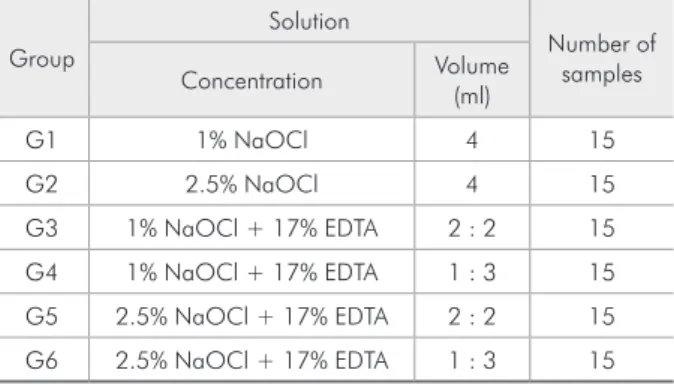
Table 1 - Concentration and volume of the solutions ana-lyzed.
Group
Solution
Number of samples Concentration Volume
(ml)
G1 1% NaOCl 4 15
G2 2.5% NaOCl 4 15
G3 1% NaOCl + 17% EDTA 2 : 2 15
G4 1% NaOCl + 17% EDTA 1 : 3 15
G5 2.5% NaOCl + 17% EDTA 2 : 2 15
G6 2.5% NaOCl + 17% EDTA 1 : 3 15
Group
Solution
Number of
samples Mean dissolution time (min) Concentration Volume
(ml)
G1 1% NaOCl 4 15 76.44
G2 2.5% NaOCl 4 15 34.95
G3 1% NaOCl + 17% EDTA 2 : 2 15 Not able to dissolve the tissue
G4 1% NaOCl + 17% EDTA 1 : 3 15 Not able to dissolve the tissue
G5 2.5% NaOCl + 17% EDTA 2 : 2 15 Not able to dissolve the tissue
G6 2.5% NaOCl + 17% EDTA 1 : 3 15 Not able to dissolve the tissue Table 2 - Mean times (in
minutes) of bovine pulp tissue dissolution promoted by the solutions analyzed.
Group n Dissolution time (min) p
Mean Standard deviation
G1 1% NaOCl 15 76.44 10.18
< 0.001
G2 2.5% NaOCl 15 34.95 4.89
Table 3 - Application of the t-test
Discussion
Many authors have reported the signiicant in-luence of a variety of factors, like concentration, time, temperature, tissue-irrigating contact area, canal preparation size, volume, tissue type, and me-chanical action, on the ability of NaOCl to dissolve both necrotic and vital tissues.10,15,16,17,18,19 Our study focused on the effect of varying NaOCl concentra-tions, on the effect of varying NaOCl concentrations combined with EDTA, and on measuring possible pH variations during the experiment.
The validity of the in vitro experimental outline is conirmed by the possibility of standardizing not only the solutions but also the fragments of pulp tis-sue in terms of their origin, surface size and storage conditions. In so doing, an evaluation can be made of the dissolution ability of NaOCl solutions and possible variations in pH for each solution without the interference of other factors. In vitro studies are also extremely valid because of their ability to gen-erate hypotheses that may be tested later in other studies.
The results of the present study demonstrated that when NaOCl, in any of the concentrations and volumes tested, was combined with 17% EDTA, the solution was unable to dissolve the bovine pulp tis-sue in 48 hours. EDTA strongly reduces the available chlorine in the NaOCl solutions, possibly rendering them ineffective.21,22 Zehnder et al.23 conirmed that EDTA solutions caused an almost complete loss of free available chlorine immediately upon mixing with NaOCl.
Because of its elevated alkalinity, the sodium hy-droxide resulting from the dissociation of NaOCl has the ability of dissolving organic matter through the saponiication of fats.8,9 Another explanation for the non-dissolution of the organic matter in sam-ples where hypochlorite was combined with EDTA is that the sodium hydroxide provided ions to the
EDTA, which is originally dissodium, turning it into trissodium, tetrassodium, and pentassodium. Some authors have already demonstrated that, in the pres-ence of NaOCl, the chemical structure of EDTA has increased sodium.24
Although it is dificult to make clinical practice inferences from the results of in vitro studies be-cause of the different variables that may be present in studies with patients, the authors suggest that the ability to dissolve organic tissue in an alternating ir-rigating regimen is damaged because of the reactivi-ty of NaOCl with EDTA. This fact has prompted us to indicate the use of EDTA as a inal lush, instead of using it alternately with sodium hypochlorite dur-ing canal preparation.
Although possible changes in the antimicrobial ability of solutions combining NaOCl and EDTA were not evaluated in this study, other authors have already demonstrated that the reduction in the available chlorine content possibly damages the formation of chloramines, impairing the power of the solution to inhibit bacterial enzymes, ultimately leading to reduced antiseptic action.18,23
Studies have shown that both the antibacte-rial properties and the tissue-dissolving properties of 5.25% NaOCl decrease when diluted. Based on the results of the present study, the ability to dis-solve organic matter is directly proportional to the NaOCl concentration in the solution.9,10,17,19,25,26,27,28 However, it is important to remember that serious incidents have been reported when concentrated hy-pochlorite solutions were inadvertently forced into periodontal tissues,29 or when such a solution leaked through the rubber dam onto the patient’s skin.30
As regards the volume of the solutions, since our study used the same volume (4 ml) for all pure solu-tions of NaOCl, no dissolution ability variation was observed owing to this factor. Further studies are recommended to analyze the effect of different vol-Group
Solution
Number of
samples Initial pH Final pH Concentration Volume
(ml)
G3 1% NaOCl + 17% EDTA 2 : 2 15 10 8
G5 2.5% NaOCl + 17% EDTA 2 : 2 15 10 8
umes of pure solutions of NaOCl, while maintain-ing the same concentration.
Regarding pH change, the pH of the solution de-creased in all the samples where NaOCl was com-bined with EDTA in an equal volume proportion. An interesting fact was that the pH did not change in the solutions where the volume of EDTA was greater than that of NaOCl. We believe that EDTA induces an increased availability of negative ions by removing Na+ from NaOCl, leading to a reduction in pH. When a greater volume of EDTA was used, the EDTA also removed sodium ions, but the lower concentration of NaOCl led to a reduced availabil-ity of negative ions, thus leaving the pH unchanged. Further studies to elucidate this inding should be conducted. The fact that the samples presented a de-crease in pH also may have contributed to the
non-dissolution of the bovine pulp tissue. Cristhensen
et al.20 and Zehnder23 demonstrated that higher pH levels resulted in greater tissue dissolution.
Conclusion
In conclusion, the concentration of NaOCl and its association with EDTA had an important effect on the ability to dissolve bovine pulp tissue. Only the samples with 1% and 2.5% NaOCl not combined with EDTA and at equal volumes (4 ml) were able to dissolve the tissue in the time set for the experiment. A greater dissolution ability (measured by the mean time necessary for dissolution) was observed in the samples with greater concentration (2.5%). The pH of the solutions with equal volumes of NaOCl and EDTA decreased after the test time of 48 hours.
References
1. Abbott PV. The periapical space: a dynamic interface. Aust Endod J. 2002 Dec;28(3):96-107.
2. Kakehashi S, Stanley HR, Fitzgerald RJ. The effects of surgi-cal exposures of dental pulps in germ-free and convention-al laboratory rats. Orconvention-al Surg Orconvention-al Med Orconvention-al Pathol. 1965 Sep;20(3):340-9.
3. McComb D, Smith DC. A preliminary scanning electron mi-croscopic study of root canals after endodontic procedures. J Endod. 1975 Jul;1(7):238–42.
4. Davis SR, Brayton SM, Goldman M. The morphology of the prepared root canal: a study utilizing injectable silicone. Oral Surg Oral Med Oral Pathol. 1972 Oct;34(4):642–8. 5. Baumgartner JC, Mader CL. A scanning electron microscopic
evaluation of four root canal irrigation regimens. J Endod. 1987 Apr;13(4):147–57.
6. Cengiz T, Aktener BO, Piskin B. The effect of dentinal tubule orientation on the removal of smear layer by root canal ir-rigants. A scanning electron microscopic study. Int Endod J. 1990 May;23(3),163-71.
7. Medici MC, Fröner IC. A scanning electron microscopic evalu-ation of different root canal irrigevalu-ation regimens. Braz Oral Res. 2006 Jul-Sep.;20(3):235-40.
8. Liolios E, Economides N, Parissis-Messimeris S, Boutsioukis A. The efectiveness of three irrigating solutions on root canal cleaning after hand and mechanical preparation. Int Endod J. 1997 Jan;30(1):51-7.
9. Hand RE, Smith ML, Harrison JW. Analysis of the effect of dilution on the necrotic tissue dissolution property of sodium hypochlorite. J Endod. 1978 Feb;4(2):60–4.
10. Clarkson RM, Moule AJ, Podlich H, Kellaway R, Macfar-lane R, Lewis D, et al. Dissolution of porcine incisor pulps in sodium hypochlorite solution of varying compositions and concentrations. Aust Dent J. 2006 Sep; 51(3):245-51. 11. Torabinejad M, Handysides R, Ali Khademi A, Bakland LK.
Clinical implications of the smear layer in endodontics: a review. Oral Surg Oral Med Oral Pathol Oral Radiol Endod. 2002 Dec;94(6):658–66.
12. Hülsmann M, Heckendorff M, Lennon A. Chelating agents in root canal treatment: mode of action and indication for their use. Int Endod J. 2003 Dec;36(12):810-30.
13. Villegas JC, Yoshioka T, Kobayashi C, Suda H. Obturation of accessory canals after four different final irrigation regimes. J Endod. 2002 Jul;28(7):534–6.
14. Peters LB, Van Winkelhoff AJ, Buijs JF, Wesselink PR. Ef-fects of instrumentation, irrigation and dressing with calcium hydroxide on infection in pulpless teeth with periapical bone lesions. Int Endod J. 2002 Jan;35(1):13–21.
15. Baker NA, Eleazer PD, Averbach RE, Seltzer S. Scanning electron microscopic study of the efficacy of various irrigation solutions. J Endod. 1975 Apr;1(4):127–35.
16. Louis I. Grossman, Benjamin W. Meiman. Solution of pulp tissue by chemical agents. J Endod.1982 Jan; 8:10-12. 17. Abou-Rass M, Oglesby SW. The effects of temperature,
con-centration, and tissue type on the solvent ability of sodium hypochlorite. J Endod. 1981 Aug;7(8):376 –7.
19. Rossi-Fedele G, De Figueiredo JA. Use of a bottle warmer to increase 4% sodium hypochlorite tissue dissolution ability on bovine pulp. Aust Endod J 2008 Apr;34(1):39-42.
20. Christensen CE, McNeal SF, Eleazer P. Effect of lowering the pH of sodium hypochlorite on dissolving tissue in vitro. J Endod. 2008 Apr;34(4):449-52. Epub 2008 Feb 7. 21. Sjögren U, Figdor D, Persson S, Sundqvist G. Influence of
in-fection at the time of root filling on the outcome of endodontic treatment of teeth with apical periodontitis. Int Endod J. 1997 Sep;30(5):297–306.
22. Molander A, Reit C, Dahlén G, Kvist T. Microbiological sta-tus of root-filled teeth with apical periodontitis. Int Endod J. 1998 Jan;31(1):1–7.
23. Zehnder M. Root Canal Irrigants. J Endod. 2006 May;32(5): 389–98.
24. Basset J, Mendham J. Vogel - Análise química quantitativa. 6rd ed. Rio de Janeiro: LTC; 2002. 462 p.
25. Radcliffe CE, Potouridou L, Qureshi R, Habahbeh N, Qual-trough A, Worthington H et al. Antimicrobial activity of vary-ing concentrations of sodium hypochlorite on the endodontic microorganisms Actinomyces israelii, A. naeslundii, Candida
albicans and Enterococcus faecalis. Int Endod J. 2004 Jul;
37(7):438–46.
26. Zehnder M, Grawehr M, Hasselgren G, Waltimo T. Tissue-dissolution capacity and dentin disinfecting potential of calci-um hydroxide mixed with irrigating solutions. Oral Surg Oral Med Oral Pathol Oral Radiol Endod. 2003 Nov;96(5):608-13.
27. Spångberg L, Engstrom B, Langeland K. Biologic effects of dental materials. 3. Toxicity and antimicrobial effect of end-odontic antiseptics in vitro. Oral Surg Oral Med Oral Pathol. 1973 Dec;36(6):856 –71.
28. Okino L , Siqueira EL , Santos M, Bombana AC , Figueiredo JAP. Dissolution of pulp tissue by aqueos solution of chlorhexidine digluconate gel. Int Endod J. 2004 Jan;37(1):38-41.
29. Hulsmann M, Hahn W. Complications during root canal irrigation—literature review and case reports. Int Endod J. 2000 May;33(3):186 –93.