Research Article
Three-Dimensional Aspects of the Lingual Papillae
and Their Connective Tissue Cores in the Tongue of
Rats: A Scanning Electron Microscope Study
Gabriela de Souza Reginato,
1Cristina de Sousa Bolina,
1Ii-sei Watanabe,
1,2and Adriano Polican Ciena
2,31Departamento de Cirurgia, Faculdade de Medicina Veterin´aria e Zootecnia da Universidade de S˜ao Paulo,
Avenida Prof. Dr. Orlando Marques de Paiva n. 87, Cidade Universit´aria, 05508-270 S˜ao Paulo, SP, Brazil
2Departamento de Anatomia, Instituto de Ciˆencias Biom´edicas (ICB-III), Avenida Prof. Lineu Prestes n. 2415, Cidade Universit´aria,
Butant˜a, 05508-900 S˜ao Paulo, SP, Brazil
3Laborat´orio de Morfologia e Atividade F´ısica, Instituto de Biociˆencias, Universidade Estadual Paulista (UNESP),
Avenida 24 A n. 1515, Bela Vista, 13506-900 Rio Claro, SP, Brazil
Correspondence should be addressed to Adriano Polican Ciena; [email protected]
Received 30 July 2014; Accepted 15 October 2014; Published 10 November 2014
Academic Editor: Romeu Rodrigues de Souza
Copyright © 2014 Gabriela de Souza Reginato et al. his is an open access article distributed under the Creative Commons Attribution License, which permits unrestricted use, distribution, and reproduction in any medium, provided the original work is properly cited.
he aim of the present study was to describe the tridimensional morphological characteristics of the lingual papillae and their connective tissue cores (CTCs) in Sprague Dawley rats. Four types of papillae were reported on the dorsal surface. Filiform papillae were distributed on the tongue surface and ater epithelial maceration a conic and multiilamentary shape of the CTCs was revealed. Fungiform papillae were reported on the rostral and middle regions covered by a squamous epithelium. Ater the removal of the epithelium, the shape of a volcano with the taste oriice at its top was noted. Foliate papillae were composed of ive pairs of epithelial folds situated on the lateral-caudal margin of the tongue. Ater the removal of the epithelium, they were shown to be limited by thin laminar projections. he vallate papilla with an oval shape was present in the caudal region and delimited by an incomplete groove. he morphological characteristics of the lingual papillae of Sprague Dowley rats, three-dimensional SEM images, and the types of papillae on the dorsal surface were similar to those reported previously in other rodent mammals. he maceration technique revealed the details of extracellular matrix with varied shapes form of connective tissue cores.
1. Introduction
he tongue ills most of the oral cavity and extends itself to the mouth pharynx. Its root is linked to a body and a free apex. It is in fact a muscular organ capable of vigorous and precise movements, such as holding, lapping, grooming and manip-ulation of food within the oral cavity, and vocalization [1].
he body of the tongue consists of a mass of bundles inter-woven with varied disposition of skeletal striated muscular ibers which permit a great variation of tongue movements. he ibers may be classiied into two groups: ibers that originate outside of the tongue (extrinsic muscles) and those that originate within the tongue and which are inserted into it.
he latter form the intrinsic muscles that change the tongue’s shape [2].
he tongue exhibits a lining or rather a continuous mucous of diferent thicknesses, throughout its surface. he epithelium is thick and rigid on the dorsal surface where tongue wear is greater due to friction with food [3]. he ultra-structural elements of the epithelium vary in the morphology of diferent mammals and show several types of papillae. Besides being associated with animal species, variations may be also related to type of food and to the animal’s adaptation to environmental conditions [4].
he lingual epithelium is made up of four types of tongue papillae, namely, iliform, fungiform, foliate, and vallate
he iliform papillae are distributed thickly on all the dorsal and lateral surfaces of the tongue. hey assist in the manipulation of food and increase friction during chewing. hey are predominant on the apex and generally are inclined at the caudal region, whereas they are modiied and presented various shapes at the root [7]. Diferences in the morphology of iliform papillae may be observed on the apex and in the middle region of tongues of several species. he conical papillae are elongated shaped with a wide base and lat apex. heir lining consists of well-developed keratinized stratiied epithelial tissues with abrasion and protection as their main role [8]. he fungiform papillae may be identiied on the apex, featuring a great quantity of connective tissues and adjacent epithelial layer. hey are well vascularized but in a lesser number than the iliform papillae, taking the shape of a “mushroom.” hey may present taste buds generally situated on the top of the papillae and are involved in the sensorial system related to taste [9].
he foliate papillae when present are situated at the bilat-eral margins of the caudal region, are leaf-shaped structures, separated from one another by an invagination of the mucous membrane, and have taste buds. he amount of foliate papil-lae may vary according to the evolution of each species [10].
he vallate papilla is the biggest of the papillae and is situated on the caudal region of the dorsal surface, involved in a deep continuous groove. he quantity and shape of the vallate papillae vary widely and depend on the species analyzed [11]. hey vary in size and shape, from round shaped to lat shaped, either lying parallel or in rows at each side of the caudal region. Groups of glands, also known as von Ebner glands, are detected in the lower part of the vallate papillae. hey are salivary serous glands where ducts open at the base of the papillae grooves and secrete a watery liquid that dissolves food contents and makes easy taste perception [12]. In the order Rodentia, several studies have been undertaken on mice [13, 14], cavy [15], agouti [12], lying squirrel [16], American beaver [17], and lowland paca [7]. However, studying the lingual papillae of rats may note diferent shapes on the surface and their connective tissue cores (CTCs) in the treated samples with NaOH solution. he types and subtypes of tongue’s papillae of rodent mammals may exhibit several diferences in the morphological characteristics to classiication, shape, and taste buds. he aim of the present study was to describe the three-dimensional morphological characteristics of the lingual papillae and their connective tissue cores (CTCs) of Sprague Dawley rats employing scan-ning electron microscope methods.
2. Material and Methods
Four tongues of four-month-old male rats, species, order: Rodentia, were investigated. he animals were kept in poly-propylene cages with water and ration “ad libitum,” main-tained in 12-hour-light/dark periods, at a mean temperature of 25 ± 2∘C. his study was approved by the Committee
2.1. Scanning Electron Microscopy. he tongues (� = 4)
were immersed in modiied Karnovsky ixative solution according to Watanabe and Yamada method [18]. he samples were then washed in a bufer solution and divided for the diferent techniques. Conventional technique comprised the washing of samples in distilled water for 2 hours at room temperature to analyze the epithelium surface. For maceration, the other samples were washed in distilled water and immersed in a 10% sodium hydroxide (NaOH) aqueous solution for 4 days at room temperature [19–21] for the removal of the epithelial surface and the analysis of the connective tissue core (CTC). hey were then washed in distilled water, with frequent changes, for two days, at 4∘C. Ater this stage, all samples were dehydrated in an increasing series of alcohols and dried in a critical point dryer (Balzers CPD-030) with liquid CO2. Samples were mounted on a metallic base, coated with gold ions (Balzers-040 SDC) [22], and examined using a scanning electron microscope LEO 435 VP.
3. Results
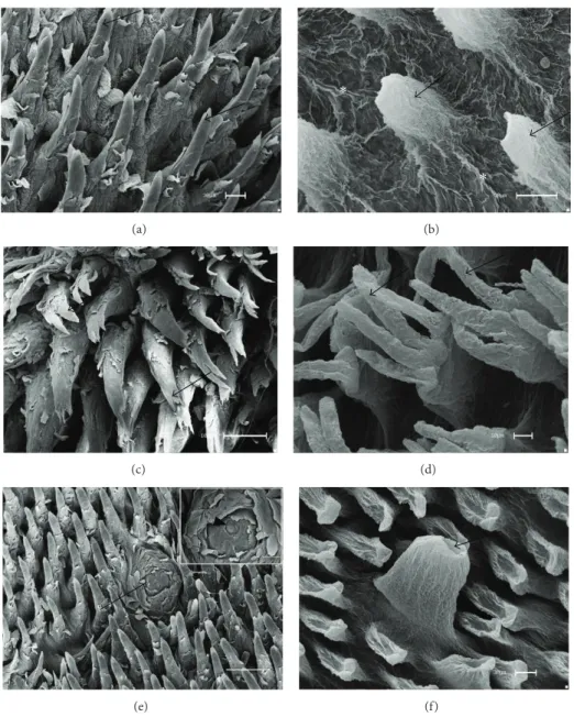
he analyses by scanning electron microscope showed that iliform papillae were the most numerous on the dorsal surface of the tongue with a decreased amount on the caudal region. hey are shaped and have diferent diameters according to the region. In the rostral region the papillae are low cone shaped, with a rounded apex (Figure1(a)). Elon-gated and multiilamentary papillae may be found in the middle and caudal regions, with their respective long and thin ilaments (Figure1(c)). Ater the removal of the epithelium, the CTCs of the iliform papillae (rostral region) revealed low, with a cone-shaped apex, a base wider than the apex, and thin bundles in the interpapillae zone (Figure1(b)), diferent from other iliform papillae. A greater magniication revealed three to four ilaments (multiilamentary) starting from the third mid-upper part of the CTCs (Figure1(d)). Fungiform papillae were distributed in the rostral and middle regions of the dorsal surface. hey are very numerous in the rostral region, with a rounded shape and dome-like top. In the details one may note that the papillae surface comprises a squamous epithelium and a taste oriice on the top of the surface (Figure1(e)). Ater the epithelium removal, the volcano shape was revealed and a cavity for the taste bud on the papillae top was observed (Figure1(f)).
(a)
∗
∗
(b)
(c) (d)
(e) (f)
Figure 1: Scanning electron micrographs—iliform and fungiform papillae of rats. (a) Conical iliform (arrows) and (b) ater maceration with NaOH the CTCs of the iliform papillae presented conical (arrows) and bundles (∗) in the interpapillary zone. (c) Multiilamentary papillae (arrows) and (d) their CTCs revealed three to four ilaments (arrows) in the third upper part. (e) he fungiform papillae (arrow) exhibited dome-like shape between conical iliform papillae and the highlighted taste oriice (arrowhead) may be seen in the surface. (f) Ater the removal of the epithelium, the CTCs of the fungiform papillae revealed volcano-like shape, with a cavity on the top for the taste buds (arrow). Bars: 30�m (a, b, f), 100�m (c, e), and 10�m (d).
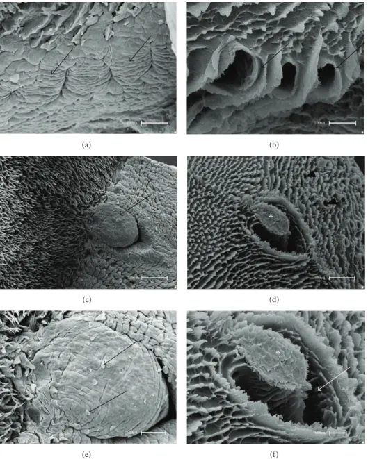
of the epithelium the CTC constitution by a thick bundle margined by a wide groove was observed. he aperture of salivary ducts was seen on the side walls (Figures 2(d)
and2(f)).
4. Discussion
he results obtained in this study revealed mainly the three-dimensional characteristics of the tongue’s papillae and the original disposition of connective tissue cores (CTCs) in the Sprague Dawley rats. In these data four types of papillae, iliform, fungiform, foliate, and vallate, were observed on the
dorsal surface of the lingual epithelial layer similar to those reported by Ciena et al. [12].
(a) (b)
(c)
∗
(d)
(e)
∗
(f)
Figure 2: Scanning electron micrographs—foliate and vallate papillae of rats. (a) he foliate papillae are constituted by epithelial folds (arrows) and separated by parallel grooves. (b) Ater the removal of the epithelial tissue, the CTCs of the foliate papillae showed wide grooves limited by laminar projections (arrows). (c) he vallate papilla (arrow) is situated in the caudal region and (d) ater the removal of the epithelium the general aspects of the CTCs (∗), delimited by CTCs of the iliform papillae and salivary glands ducts (arrowheads), were observed. (e) At higher magniication, squamous epithelium (arrows) in the surface was noted. (f) At higher magniication the constitution of the CTC by a thick bundle (∗) margined by a wide groove (arrow) was revealed. Bars: 100�m (a, b, e, f) and 300�m (c, d).
the tongue of rodent mammals, distributed throughout the dorsal epithelium surface and decreased in the caudal region [12]. he morphological characteristics of the iliform papillae of the rats difered from those of other rodents as has been reported in the lying squirrel. Emura et al. [16] reported that the latter species presented conical papillae in the caudal region and elongated papillae in the rostral region. hese morphological characteristics were similar to those of the il-iform papillae reported in the cavy’s tongue, as described by Watanabe et al. [15], and in the Patagonian cavy’s tongue, as described by Emura et al. [23].
he foliate papillae revealed ive epithelium folds sep-arated by parallel grooves on the lateral-caudal margin of the tongue. Ater the removal of the epithelium, wide oval grooves limited by laminar projections were observed. he localization of such papillae was similar to that in the paca [7], lying squirrel [16], and cavy [15]. According to Emura et al. [23], foliate papillae in Rodentia are well developed. Further, the number of epithelial folds may vary according to each species. here were 12 pairs of well-designed foliate papillae in the tongue of the agouti [12] and between 22 and 25 pairs in the tongue of the American castor [17].
A vallate papilla was observed in the central caudal region, limited by an incomplete groove at the upper part. Ater the removal of the epithelium, the constitution of a thick bundle margined by a wide groove of the CTC was noted. he quantity and the shape of these papillae difered according to the species. he number of papillae in the bank vole [25] is similar to that in the rat, with only one papilla. Further, the paca [7], Patagonian cavy [23], and blind mole rat [9] present a pair of parallel papillae, whereas the lying squirrel has three papillae [16]. However, four papillae were reported in the agouti, the largest amount in rodents as described by Ciena et al. [12].
5. Conclusion
he morphological characteristics of the lingual papillae of Sprague Dawley rats, three-dimensional SEM images, and the types of papillae on the dorsal surface were similar to those reported previously in other rodent mammals. he maceration technique revealed the details of extracellular matrix with varied shapes form of connective tissue cores.
Conflict of Interests
he authors declare that there is no conlict of interests regarding the publication of this paper.
References
[1] K. M. Dyce, W. O. Sack, and C. J. G. Wensing,Tratado de anat-omia veterin´aria, Elsevier, Rio de Janeiro, Brazil, 4th edition, 2010.
[2] P. L. Gartner and L. J. Hiatt,Tratado de Histologia em Cores, Elsevier, Rio de Janeiro, Brazil, 3rd edition, 2007.
[3] R. J. Ferreira, A. E. Carvalho, W. Souza, F. B. Alvarenga, and F. B. Rodrigues, “Anatomia da Art´eria Lingual Profunda em Sus scrofa domestica, LINNAEUS, 1758,”Ciˆencia Animal Brasileira, vol. 12, no. 2, pp. 298–305, 2011.
[4] Y. Miyawaki, K. Yoshimura, J. Shindo, and I. Kageyama, “Light and scanning electron microscopic study on the tongue and lin-gual papillae of the common raccoon,Procyon lotor,”Okajimas Folia Anatomica Japonica, vol. 87, no. 2, pp. 65–73, 2010. [5] E. T. Fonseca, C. M. Oliveira, A. L. R. Franciolli, and M. A.
Miglino, “Caracteristicas das papilas do dorso da lingua de cabras (Capra hircus): estudo por de microscopia eletrˆonica de varredura e luz,”Pesquisa Veterin´aria Brasileira, vol. 31, no. 1, pp. 67–73, 2011.
[6] J. Zheng and K. Kobayashi, “Comparative morphological study on the lingual papillae and their connective tissue cores (CTC) in reeves’ muntjac deer (Muntiacus reevesi),”Annals of Anatomy, vol. 188, no. 6, pp. 555–564, 2006.
[7] B. C. M. Massoli, Q. R. P. Ribeiro, G. L. Vieira et al., “Morfologia da l´ıngua e caracter´ısticas das papilas linguais deCuniculus paca
(Rodentia: Cuniculidae),”Revista Biotemas, vol. 26, no. 4, pp. 167–177, 2013.
[8] K. Kobayashi, M. Kumakura, K. Yoshimura, K. Nonaka, T. Murayama, and M. Henneberg, “Comparative morphological study of the lingual papillae and their connective tissue cores of the koala,”Anatomy and Embryology, vol. 206, no. 4, pp. 247– 254, 2003.
[9] M. Kilinc, S. Erdogan, S. Ketani, and M. A. Ketani, “Morpho-logical study by scanning electron microscopy of the lingual papillae in the middle east blind mole rat (Spalax ehrenbergi, Nehring, 1898),”Journal of Veterinary Medicine Series C: Anato-mia Histologia Embryologia, vol. 39, no. 6, pp. 509–515, 2010. [10] S. Emura, D. Hayakawa, H. Chen, and S. Shoumura,
“Morphol-ogy of the dorsal lingual papillae in the Japanese macaque and savanna monkey,”Anatomia, Histologia, Embryologia, vol. 31, no. 5, pp. 313–316, 2002.
[11] A. A. El Sharaby, S. A. El-Gendy, M. A. Alsafy, A. G. Nomir, and S. Wakisaka, “Morphological variations of the vallate papillae in some mammalian species,”Anatomical Science International, vol. 89, pp. 161–170, 2014.
[12] A. P. Ciena, C. de Sousa Bolina, S. R. Y. de Almeida et al., “Structural and ultrastructural features of the agouti tongue (Dasyprocta agutiLinnaeus, 1766),”Journal of Anatomy, vol. 223, no. 2, pp. 152–158, 2013.
[13] J.-Y. Kim, T. Mochizuki, K. Akita, and H.-S. Jung, “Morphologi-cal evidence of the importance of epithelial tissue during mouse tongue development,”Experimental Cell Research, vol. 290, no. 2, pp. 217–226, 2003.
[14] M. Abe and T. Osawa, “he structure of the interstitial surfaces of the epithelial basement membranes of mouse oral mucosa, gingiva and tongue,”Archives of Oral Biology, vol. 44, no. 7, pp. 587–594, 1999.
[15] I.-S. Watanabe, C. A. dos Santos Haemmerle, F. J. Dias et al., “Structural characterization of the capybara (Hydrochaeris hydrochaeris) tongue by light, scanning, and transmission electron microscopy,”Microscopy Research and Technique, vol. 76, no. 2, pp. 141–155, 2013.
[16] S. Emura, A. Tamada, D. Hayakawa et al., “SEM study on the dorsal lingual surface of the lying squirrel,Petaurista leu-cogenys,”Annals of Anatomy, vol. 181, no. 5, pp. 495–498, 1999. [17] J. Shindo, K. Yoshimura, and K. Kobayashi, “Comparative
mor-phological study on the stereo—structure of the lingual papillae and their connective tissue cores of the American beaver (Castor canadensis),”Okajimas Folia Anatomica Japonica, vol. 82, no. 4, pp. 128–137, 2006.
[18] I. Watanabe and E. Yamada, “he ine structure of lamellated nerve endings found in the rat gingiva,”Archivum Histologicum Japonicum, vol. 46, no. 2, pp. 173–182, 1983.
[19] O. Ohtani, “hree-dimensional organization of the connective tissue ibers of the human pancreas: a scanning electron micro-scopic study of NaOH treated-tissues,”Archivum Histologicum Japonicum, vol. 50, no. 5, pp. 557–566, 1987.
[21] E. J. Benetti, L. C. P´ıcoli, J. P. Guimar˜aes, A. A. Motoyama, M. A. Miglino, and L.-S. Watanabe, “Characteristics of ili-form, fungiform and vallate papillae and surface of interface epithelium-connective tissue of the maned sloth tongue mucosa (Bradypus torquatus, Iliger, 1811): light and scanning electron microscopy study,” Journal of Veterinary Medicine Series C: Anatomia Histologia Embryologia, vol. 38, no. 1, pp. 42–48, 2009. [22] C. C. Duro, A. P. Ciena, S. R. Y. de Almeida et al., “Qualitative study of young, adult, and aged wistar rats temporomandibular synovial membrane employing light, scanning, and transmis-sion electron microscopy,”Microscopy Research and Technique, vol. 75, no. 11, pp. 1522–1527, 2012.
[23] S. Emura, T. Okumura, and H. Chen, “Morphology of the lin-gual papillae in the Patagonian cavy,”Okajimas Folia Anatomica Japonica, vol. 88, no. 3, pp. 121–125, 2011.
[24] K. Kobayashi, “hree-dimensional architecture of the con-nective tissue core of the lingual papillae in the guinea pig,”
Anatomy and Embryology, vol. 182, no. 3, pp. 205–213, 1990. [25] H. Jackowiak and S. Godynicki, “he distribution and structure
Submit your manuscripts at
http://www.hindawi.com
Hindawi Publishing Corporation
http://www.hindawi.com Volume 2014
Anatomy
Research International
Peptides
Hindawi Publishing Corporation
http://www.hindawi.com Volume 2014
Hindawi Publishing Corporation http://www.hindawi.com
International Journal of
Volume 2014
Zoology
Hindawi Publishing Corporation
http://www.hindawi.com Volume 2014 Molecular Biology International
Genomics
International Journal of
Hindawi Publishing Corporation
http://www.hindawi.com Volume 2014
The Scientiic
World Journal
Hindawi Publishing Corporationhttp://www.hindawi.com Volume 2014
Hindawi Publishing Corporation
http://www.hindawi.com Volume 2014
Bioinformatics
Advances inMarine Biology
Journal of Hindawi Publishing Corporationhttp://www.hindawi.com Volume 2014
Hindawi Publishing Corporation
http://www.hindawi.com Volume 2014
Signal Transduction
Journal ofHindawi Publishing Corporation
http://www.hindawi.com Volume 2014
BioMed
Research International
Evolutionary Biology International Journal of
Hindawi Publishing Corporation
http://www.hindawi.com Volume 2014
Hindawi Publishing Corporation
http://www.hindawi.com Volume 2014 Biochemistry Research International
Archaea
Hindawi Publishing Corporation
http://www.hindawi.com Volume 2014
Hindawi Publishing Corporation
http://www.hindawi.com Volume 2014
Genetics
Research International
Hindawi Publishing Corporation
http://www.hindawi.com Volume 2014
Advances in
Virology
Hindawi Publishing Corporation http://www.hindawi.com
Nucleic Acids
Journal ofVolume 2014
Stem Cells
International
Hindawi Publishing Corporation
http://www.hindawi.com Volume 2014
Hindawi Publishing Corporation
http://www.hindawi.com Volume 2014
Enzyme
Research
Hindawi Publishing Corporation
http://www.hindawi.com Volume 2014