MicroRNA Expression Profiling in Clear Cell
Renal Cell Carcinoma: Identification and
Functional Validation of Key miRNAs
Haowei He1☯, Linhui Wang2☯, Wenquan Zhou1, Zhengyu Zhang1, Longxin Wang1, Song Xu1, Dong Wang1, Jie Dong1, Chaopeng Tang1, Hao Tang1, Xiaoming Yi1, Jingping Ge1*
1Department of Urology, Jinling Hospital, Nanjing University Medical School, Nanjing, Jiangsu, China, 2Department of Urology, Changzheng Hospital, Second Military Medical University, Shanghai, Shanghai, China
☯These authors contributed equally to this work. *[email protected]
Abstract
Objective
This study aims to profile dysregulated microRNA (miRNA) expression in clear cell renal cell carcinoma (ccRCC) and to identify key regulatory miRNAs in ccRCC.
Methods and Results
miRNA expression profiles in nine pairs of ccRCC tumor samples at three different stages and the adjacent, non-tumorous tissues were investigated using miRNA arrays. Eleven miR-NAs were identified to be commonly dysregulated, including three up-regulated (miR-487a, miR-491-3p and miR-452) and eight down-regulated (miR-125b, miR-142-3p, miR-199a-5p, miR-22, miR-299-3p, miR-29a, miR-429, and miR-532-5p) in tumor tissues as compared with adjacent normal tissues. The 11 miRNAs and their predicted target genes were analyzed by Gene Ontology and Kyoto Encyclopedia of Genes and Genomes (KEGG) pathway enrich-ment analysis, and three key miRNAs (miR-199a-5p, miR-22 and miR-429) were identified by microRNA-gene network analysis. Dysregulation of the three key miRNAs were further validated in another cohort of 15 ccRCC samples, and the human kidney carcinoma cell line 786-O, as compared with five normal kidney samples. Further investigation showed that over-expression of miR-199a-5p significantly inhibited the invasion ability of 786-O cells. Lu-ciferase reporter assays indicated that miR-199a-5p regulated expression of TGFBR1 and JunB by directly interacting with their 3’untranslated regions. Transfection of miR-199a-5p successfully suppressed expression of TGFBR1 and JunB in the human embryonic kidney 293T cells, further confirming the direct regulation of miR-199a-5p on these two genes.
Conclusions
This study identified 11 commonly dysregulated miRNAs in ccRCC, three of which (miR-199a-5p, miR-22 and miR-429) may represent key miRNAs involved in the pathogenesis of
OPEN ACCESS
Citation:He H, Wang L, Zhou W, Zhang Z, Wang L, Xu S, et al. (2015) MicroRNA Expression Profiling in Clear Cell Renal Cell Carcinoma: Identification and Functional Validation of Key miRNAs. PLoS ONE 10 (5): e0125672. doi:10.1371/journal.pone.0125672
Academic Editor:Kandiah Jeyaseelan, National University of Singapore, SINGAPORE
Received:October 1, 2014
Accepted:March 17, 2015
Published:May 4, 2015
Copyright:© 2015 He et al. This is an open access article distributed under the terms of theCreative Commons Attribution License, which permits unrestricted use, distribution, and reproduction in any medium, provided the original author and source are credited.
Data Availability Statement:All relevant data are within the paper and its Supporting Information files.
Funding:The authors have no support or funding to report.
ccRCC. Further studies suggested that miR-199a-5p plays an important role in inhibition of cell invasion of ccRCC cells by suppressing expression of TGFBR1 and JunB.
Introduction
Renal cell carcinoma (RCC) constitutes about 3% of all human cancers [1]. Of the various his-tological subsets, clear cell renal cell carcinoma (ccRCC) is the most common subtype at diag-nosis and accounts for 75–88% of RCCs in contemporary surgical series [2,3]. Although the average age at diagnosis of ccRCC is 60–64 years [2,4,5], there are 7% of sporadic ccRCC cases diagnosed at ages younger than 40 years [6]. One third of ccRCC patients present with metasta-ses, and another third are expected to develop metastases eventually. Current approaches of chemotherapy and radiotherapy have only limited efficacy on ccRCC [7], and novel effective targeted agents for metastatic disease fail to work on ccRCC patients with distant metastases [8,9]. There is an urgent need to elucidate the molecular basis of ccRCC so as to identify effec-tive therapeutic targets in the future.
MicroRNAs (miRNAs) are small noncoding RNAs that play important roles in the control of gene expression by binding, with imperfect base pairing, to complementary sequences in the 3’untranslated regions (3’UTRs) of their target mRNAs, resulting in down-regulation of target gene expression at either transcriptional or translational levels [10]. Through their specific gene regulatory networks, miRNAs have important functions in controlling eukaryotic cell proliferation, differentiation and metabolism. Over the last decade, miRNAs have emerged as important and evolutionarily conserved regulators of various physiopathological processes, in-cluding carcinogenesis [11,12].
Recently, efforts have been made to identify miRNAs that are dysregulated and play poten-tial roles in the pathogenesis of ccRCC [13–22]. Some of the miRNAs have been frequently demonstrated to be functionally involved in ccRCC, such as members of the miR-200 family [13,21,22], miR-210 [21–23], and miR-17-92 cluster [24,25], indicating that these dysregulated miRNAs may play pivotal roles in the tumorigenesis of ccRCC. However, due to variations in sample size, sample selection (groupedvsungrouped samples according to the stage of disease; use of autologousvsallogeneic controls), sample preparation (frozenvsformalin fixed tissues), ethnic origin (identicalvsmixed), and detection sensitivity, inconsistency between different studies often occurs, thus the function of majority of the miRNA candidates remains to be determined.
In this study, we performed a comprehensive profiling of miRNA expression and investigat-ed the differential expression of miRNAs in tumor samples and adjacent normal tissues from patients with ccRCC at different stages. Commonly dysregulated miRNAs were subjected to miRNA-gene network analysis to identify key miRNAs which have potential pivotal roles in cancer development. Candidate key miRNAs were then validated in clinical samples and human kidney carcinoma cell lines. The function and molecular mechanism of a selected miRNA (miR-199-5p) were further investigated.
Materials and Methods
ccRCC tissue sample selection and RNA preparation
system [26]. Adjacent non-tumorous tissues were obtained at the same time. For the subse-quent miRNA candidate validation study, normal kidney samples were collected from 8 indi-viduals under nephrectomy due to injury. Written informed consents were obtained from all patients involved before the collection of tissue samples. This study was approved by the ethics committee of the Second Military Medical University. All samples were stored in liquid nitro-gen until use. Total RNA was isolated using the TRIzol reanitro-gent (Invitronitro-gen, Carlsbad, CA, USA) according to the manufacturer’s instruction.
miRNA expression profiling using microarrays
In order to identify ccRCC-associated miRNAs, RNA from 9 randomized ccRCC tumor tissues (3 GI, 3 GII, and 3 GIII) and the adjacent nontumorous tissues were subjected to global miRNA expression profiling using miRCURY LNA microRNA Arrays (Exiqon, Woburn, MA, USA). Briefly, miRNA probes labeled with Hy3 were synthesized from RNA samples using miRCURY LNA microRNA Power Labeling Kit (Exiqon) and then hybridized to miRCURY arrays. The miRNA array scanning was performed with the Axon GenePix 4000B microarray scanner. miRNA expression levels were then analyzed using the tool GenePix pro V6.0. Differ-ential miRNA expression profiles were analyzed by comparing the tumors of each stage with the adjacent normal tissues. The miRNAs with more than two-fold change in tumor tissues compared with the adjacent normal tissues were considered as differentially expressed. The miRNAs which were differentially expressed in all nine ccRCC tumors were considered to be commonly dysregulated miRNAs in ccRCC.
Cell lines and antibodies
The human kidney carcinoma cell line, 786-O, and the human embryonic kidney cell line, 293T, were purchased from American Type Culture Collection (ATCC). Cells were cultured in RPMI-1640 medium (Genepharma, Shanghai, China) supplemented with 10% fetal bovine serum (Invitrogen) in 5% CO2at 37°C. The primary antibodies anti-TGFBR1 and anti-JunB were purchased from Santa Cruz Biotechnology (Dallas, TX, USA), and primary antibody anti-Actin was purchased from Beyotime Institute of Biotechnology (Jiangsu, China). Secondary antibodies anti-rabbit IgG and anti-mouse IgG were purchased from Beyotime and Santa Cruz, respectively.
Gene Ontology and KEGG pathway enrichment analysis and
microRNA-gene network analysis
To identify the key miRNAs in ccRCC, microRNA-gene network analysis was conducted for the commonly dysregulated miRNAs identified from the microRNA arrays and their predicted downstream target genes. Downstream target genes of the miRNA candidates were predicted using the tool TargetScan (www.targetscan.org). Selection criteria for target prediction includ-ed: predicted targeting efficacy (context + scores method) [27], positioning of the potential reg-ulatory elements, evolutionary conservation, and probability of off-target effects. Predicted target genes were then sorted by the number of their upstream miRNAs out of the miRNA can-didates. The top 25% of the target genes were then subjected to Gene Ontology (GO) analysis and KEGG Pathway enrichment analysis using DAVID Bioinformatics Resources (http://
david.abcc.ncifcrf.gov/). Genes included in both the significant GO terms and KEGG pathways
Dual-luciferase reporter assay and transfection of miRNA mimics
To further examine whether the predicted target genes are directly regulated by miR-199a-5p, 3’UTR segments of the corresponding genes containing the miR-199a-5p recognition region mutated region not recognizable by miR-199a-5p were sub-cloned downstream of the lucifer-ase reporter in psiCHECK-2 vectors (Applied Biosystems, Grand Island, NY, USA). The mu-tated sequences in the 3’UTR segments of JunB and TGFBR1 were CATTACC and ACTAAC, respectively. The hsa-mir-199a-5p mimics and negative control (NC) mimics were purchased from Genepharma (Shanghai, China). 293T cells that were transiently transfected with hsa-mir-199a-5p or NC mimics at 15 or 50 nM of final concentration were co-transfected with
Renillaluciferase reporter vector (195 ng/well) andFireflyluciferase reporter vector (5 ng/well) using Lipofectamine 2000 (Invitrogen) in 24-well plates. Cells were harvested at 48 hours post transfection and luciferase activities were analyzed by psiCHECK Dual Luciferase Reporter Assay (Promega, Madison, WI, USA) according to the manufacturer’s instruction. Transfec-tion of miRNA or NC mimics into 786-O cells was carried out using Lipofectamine 2000 (Invi-trogen) according to manufacturer’s guide. Cells were collected for RNA and protein
extraction at 48 hours post transfection.
Transwell assay
Cells in RPMI-1640 media supplemented with 1% FBS were seeded into the upper compart-ments of the 24-transwell Boyden chamber (Costar, Bedford, MA, USA). Culture media supple-mented with 10% FBS was loaded into the lower compartments to be used as a chemoattractant. After incubation for 12 hours, the non-invaded cells were removed from the upper compart-ments, and the invaded cells on the lower side were fixed, stained with 0.1% crystal violet, and photographed. The number of invaded cells per field was counted.
miRNA quantitation by quantitative RT-PCR (qRT-PCR)
Reverse transcription (RT) of target miRNAs was performed using gene-specific RT primers and MMLV Reverse Transcriptase 1st-Strand cDNA Synthesis Kit (Epicentre Biotechnologies, Madison, WI, USA) according to manufacturer’s instructions. Relative qRT-PCR of individual miRNA was then performed using Platinum SYBR Green qPCR SuperMix-UDG w/ROX (Invi-trogen). Expression levels of target miRNAs were normalized to the small RNA U6 internal control. Primers used were listed inS1 Table. All experiments were performed in triplicate.
Western blotting
Statistical analysis
All values were expressed as means ± standard deviation (SD). Student's t-test was performed using the SPSS 13.0 Statistical Package (IBM, New York, NY, USA).
Results
MiRNA expression profiling in ccRCC using microarray analysis
Nine pairs of ccRCC tumor tissues (3 GI, 3 GII and 3 GIII) and the adjacent nontumorous tis-sues were analyzed for global miRNA expression profiling, and the results are presented in
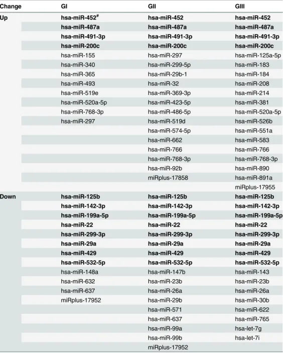
Table 1andFig 1. Twenty-four miRNAs were found to be differentially expressed in GI tumor
samples as compared with adjacent normal tissues, with 12 up-regulated and 12 down-regulat-ed. In GII group, 35 miRNAs were found to be dysregulated in tumor samples as compared with adjacent normal tissues, including 18 up-regulated and 17 down-regulated. Thirty-four miRNAs were differentially expressed in GIII tumor samples, with 19 up-regulated and 16 down-regulated compared with adjacent normal samples. While expression of some of these miRNAs seemed to be specifically dysregulated in certain tumor stages, 12 miRNAs, including 4 up-regulated (200c, 487a, 491-3p and 452) and 8 down-regulated miR-NAs (miR-125b, miR-142-3p, miR-199a-5p, miR-22, miR-299-3p, miR-29a, miR-429, and miR-532-5p), were identified to be commonly dysregulated in all ccRCC tumors at different stages (Table 1). Up-regulated expression of miR-200c appeared to contradict previous studies, which often linked down-regulation of miR-200c with ccRCC malignancy [13,21,22]. There-fore, these miRNA candidates were further validated by qRT-PCR analysis. With the only ex-ception of miR-200c, the results for all the other 11 miRNA candidates were consistent with the results from microarray profiling, which demonstrated a low false discovery rate and sup-ported the validity of the profiling (data not shown). The expression of miR-200c was indeed down-regulated in ccRCC, as illustrated using GII tumor samples, and the representative result is shown inS1 Fig.
MicroRNA-gene network analysis identified key miRNAs in ccRCC
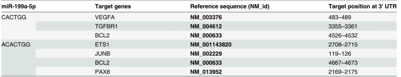
The top 10 significant GO terms and KEGG pathways for the 11 validated miRNA candidates and putative target genes are shown inFig 2A & 2B, respectively. The most significantly en-riched GO terms included cell communication, signaling, signal transduction, enzyme linked receptor protein signaling pathway, transmembrane receptor protein tyrosine kinase signaling pathway, regulation of cellular process, and cell development. The most significantly enriched KEGG pathways included pathways in cancers (pathway Id, hsa05200), Wnt signaling (hsa04310), actin cytoskeleton (hsa04810), adherens junction (hsa04520), focal adhesion (hsa04510), and ErbB signaling (hsa04012).involved in the regulation of disease progression. As miR-199a-5p and miR-22 were more greatly down-regulated in 786-O cells compared to miR-429, and the function of miR-22 has been widely studied in other cancers, we selected the less characterized miR-199a-5p for func-tional validation. Using the tool TargetScan, we found that six genes involved in cancer-related pathways were predicted to be potential targets of miR-199a-5p (Table 3).
Table 1. Differentially expressed miRNAs in ccRCC as compared with adjacent normal tissues.
Change GI GII GIII
Up hsa-miR-452# hsa-miR-452 hsa-miR-452
hsa-miR-487a hsa-miR-487a hsa-miR-487a hsa-miR-491-3p hsa-miR-491-3p hsa-miR-491-3p hsa-miR-200c hsa-miR-200c hsa-miR-200c
hsa-miR-155 hsa-miR-297 hsa-miR-125a-5p
hsa-miR-340 hsa-miR-299-5p hsa-miR-183
hsa-miR-365 hsa-miR-29b-1 hsa-miR-184
hsa-miR-493 hsa-miR-32 hsa-miR-208
hsa-miR-519e hsa-miR-369-3p hsa-miR-214
hsa-miR-520a-5p hsa-miR-423-5p hsa-miR-381
hsa-miR-768-3p hsa-miR-486-5p hsa-miR-520a-5p
hsa-miR-297 hsa-miR-519d hsa-miR-526b
hsa-miR-574-5p hsa-miR-551a
hsa-miR-662 hsa-miR-583
hsa-miR-766 hsa-miR-766
hsa-miR-768-3p hsa-miR-768-3p
hsa-miR-92b hsa-miR-890
miRplus-17858 hsa-miR-891a miRplus-17955 Down hsa-miR-125b hsa-miR-125b hsa-miR-125b hsa-miR-142-3p hsa-miR-142-3p hsa-miR-142-3p hsa-miR-199a-5p hsa-miR-199a-5p hsa-miR-199a-5p hsa-miR-22 hsa-miR-22 hsa-miR-22 hsa-miR-299-3p hsa-miR-299-3p hsa-miR-299-3p hsa-miR-29a hsa-miR-29a hsa-miR-29a hsa-miR-429 hsa-miR-429 hsa-miR-429 hsa-miR-532-5p hsa-miR-532-5p hsa-miR-532-5p
hsa-miR-148a hsa-miR-147b hsa-miR-143
hsa-miR-632 hsa-miR-23b hsa-miR-23b
hsa-miR-637 hsa-miR-26a hsa-miR-26a
miRplus-17952 hsa-miR-29b hsa-miR-30b
hsa-miR-571 hsa-miR-622
hsa-miR-637 hsa-miR-765
hsa-miR-99a hsa-let-7g
hsa-miR-99b hsa-let-7i
miRplus-17952
#
miRNAs in bold are commonly up- or down-regulated in all samples at three different stages.
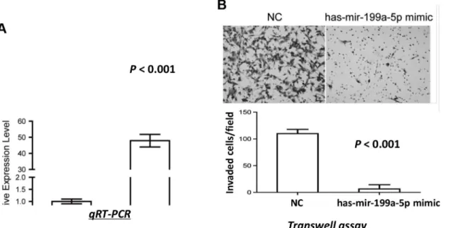
miR-199a-5p decreased invasive ability of ccRCC cells
786-O cells were transfected with either miR-199a-5p or negative control (NC) mimics, and significantly higher expression of miR-199a-5p was confirmed in the miR-199a-5p mimics-transfected cells (Fig 4A). No significant difference in cell viability was observed between the miR-199a-5p mimics-transfected cells and the control cells (P>0.05, data not shown). In
addi-tion, no significant changes were found in either cell cycle phase distribution or apoptosis by overexpression of miR-199a-5p (P>0.05, data not shown). However, 786-O cells with
overex-pressed miR-199a-5p showed significantly reduced invasive ability as compared with the con-trol cells (P<0.001;Fig 4B). These results demonstrated that miR-199a-5p played an
important role in suppressing cell invasion of ccRCC.
miR-199a-5p directly suppressed expression of TGFBR1 and JunB in
ccRCC
Luciferase assays were performed to assess the direct regulation of miR-199a-5p on the expres-sion of the target genes identified using the TargetScan algorithm. The suppressive effects of
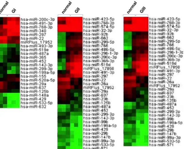
Fig 1. MiRNA expression profiling in ccRCC using microarray analysis.Unsupervised clustering of differentially expressed miRNAs in the three stages of ccRCC tumors (GI, GII and GIII) as compared with adjacent normal tissues. Three ccRCC tumor tissues from each stage, as well as the adjacent nontumorous tissues were randomly selected and subjected to global miRNA expression profiling. Differential miRNA expression was analyzed by
comparison of the tumor tissues with adjacent normal tissues. The miRNAs with more than two-fold change in expression were considered to be differentially expressed. The expression of these miRNA candidates is illustrated in the heat map. The brightest green, black, and brightest red colors represent low, medium, and high expression of miRNAs, respectively.
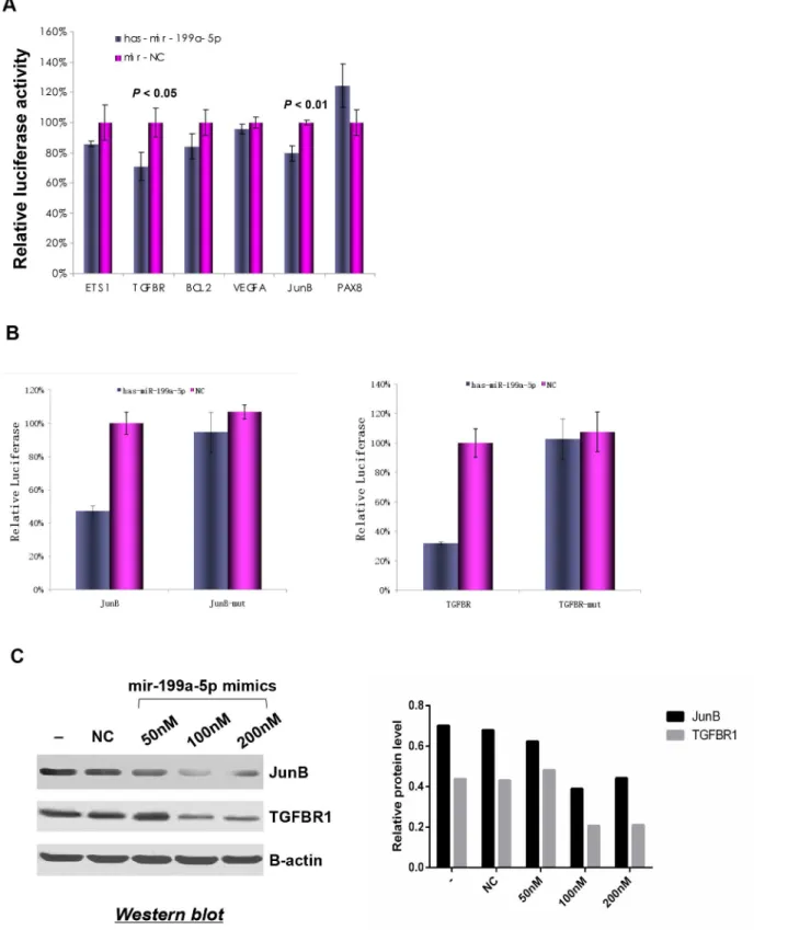
miR-199a-5p on the 3’UTRs of TGFBR1 and JunB were observed, as indicated by significant decrease in the luciferase activities of these genes in 293T cells transfected with miR-199a-5p. However, no significant suppressive effects were observed on 3’UTRs of the other genes
(Fig 5A). To demonstrate that the suppressive effect of miR-199a-5p was mediated through
di-rect targeting of the recognition regions in 3’UTR of target genes, the recognition sequences for JunB and TGFBR1 were mutated and the luciferase activities were measured. As shown in
Fig 5B, the hsa-mir-199a-5p mimics showed significantly inhibitory effects on 3’UTRs of JunB
and TGFBR1 containing the miR-199a-5p recognition sequences, whereas no inhibitory effects were observed on those containing the mutated recognition sequences. To further validate the regulatory role of miR-199a-5p on the expression of TGFBR1 and JunB, we transfected hsa-mir-199a-5p mimics into the human kidney carcinoma 786-O cells, which express high levels of TGFBR1 and JunB, and low level of miR-199a-5p as shown inFig 3. The protein expression of TGFBR1 and JunB decreased significantly in a dose-dependent manner after transfection of hsa-mir-199a-5p in 786-O cells (Fig 5C). These results confirmed that TGFBR1 and JunB are the direct targets of miR-199a-5p.
Discussion
In this study, we systematically investigated the miRNA expression profiles in a panel of differ-ently staged ccRCC samples by miRNA arrays. Eleven commonly dysregulated miRNAs, in-cluding 3 up-regulated 487a, miR-491-3p and miR-452) and 8 down-regulated (miR-125b, miR-142-3p, miR-199a-5p, miR-22, miR-299-3p, miR-29a, miR-429, and miR-532-5p), were identified in ccRCC tumor samples as compared with adjacent nontumorous samples. The discrepancy of miR-200c expression measured between microarray and qRT-PCR may re-flect the detection or analysis errors that frequently occur in microarray studies. qRT-PCR analysis is more sensitive and remains the golden standard for gene expression quantification
Fig 2. MicroRNA-gene network in ccRCC. (A)The top 10 Gene Ontology terms significantly enriched by predicted target genes of the up-regulated (A1) and down-regulated (A2) miRNAs in ccRCC.(B)The top 10 KEGG pathways significantly enriched by predicted target genes of the up-regulated (B1) and down-reguated (B2) miRNAs in ccRCC.(C)Interactive miRNA-gene networks between up-regulated (C1) or down-regulated (C2) miRNAs and their target genes. The red nodes represent the regulators (miRNAs), the grey nodes represent the targets (genes), and the green edges indicate direct interaction.
doi:10.1371/journal.pone.0125672.g002
Table 2. Three key miRNAs and their predicted target genes in cancer-related pathways.
Key miRNAs
Target genes
miR-199-5p ABL1,AKT2,APC,APPL1,BCL2,BID,CBL,CDKN1A,CDKN2B,CEBPA,COL4A4,CRKL, CYCS,E2F2,E2F3,ERBB2,ETS1,FGF23,FGFR1,FZD4,FZD5,FZD6,GLI3,GRB2,IGF1R, ITGA2,ITGA6,KITLG,KRAS,LAMB3,LAMC1,MAPK1,MMP2,PAX8,PIK3R1,PIK3R5,PML, PRKCA,PTCH1,RARA,RUNX1,RXRA,SMAD2,SMAD3,STAT1,STK4,TGFA,TGFBR1, TPM3,TRAF1,VEGFA,WNT4,WNT5A
miR-22 ABL1,AKT2,AKT3,APPL1,BCL2L1,CBL,CDK6,CDKN1A,CEBPA,COL4A4,CRK,CRKL, CYCS,E2F2,ETS1,FAS,FGF23,FGF5,FGFR1,FGFR2,FGFR3,FOXO1,FZD5,FZD6,GLI3, GRB2,IGF1R,LAMC1,MAPK1,MAPK10,MAX,PAX8,PIK3R5,PLD1,PML,PRKCA,PTCH1, RET,RUNX1,RUNX1T1,RXRA,SMAD3,STAT1,STK4,TCF7,TGFBR1,TP53,TPM3,TRAF1, TRAF6,WNT4,WNT5A,XIAP
miR-429 AKT2,AKT3,APPL1,BCL2,CBL,CDC42,CDK6,CDKN1A,CDKN2B,CEBPA,COL4A4, COL4A6,CRK,CRKL,CYCS,E2F3,ETS1,FAS,FGF23,FN1,FOXO1,FZD4,FZD5,FZD6,GLI3, IGF1,ITGA2,KITLG,KRAS,LAMC1,MAPK1,MITF,PIK3R3,PLCG1,PRKCA,PTCH1,RAC1, RET,RUNX1,RXRA,SMAD2,SMAD3,SOS1,STAT1,STK4,TGFBR1,TRAF6,VEGFA,WNT4, WNT5A,XIAP
[29]. Since the results for the other 11 miRNA candidates were consistent between the microar-ray and qRT-PCR analysis, the validity of the microarmicroar-ray screen was proved. Our study not only confirmed some of the results from previous studies, but further revealed many novel miRNAs that have not been previously reported in ccRCC tumorigenesis. For example, miR-142-3p has been shown to function as a tumor suppressor in colon cancer [30]. Due to its down-regulation in ccRCC in our study, we hypothesized that miR-142-3p may also exert tumor suppressive functions in ccRCC. miR-29a has been reported to be one of the predictors distinguishing metastatic and non-metastatic ccRCC [31], and to suppress cell proliferation in hepatocellular carcinoma [32]. Low miR-29a expression has been significantly associated with poorer survival in pediatric acute myeloid leukemia patients [33]. These findings suggest an important function of miR-29a in pathogenesis of multiple cancers.
We conducted GO and KEGG pathway analyses and miRNA-gene network analysis to identify the potential function of the dysregulated expression of miRNAs and their predicted target genes. The most significantly enriched GO terms included cell communication, signal-ing, signal transduction, enzyme linked receptor protein signaling pathway, transmembrane re-ceptor protein tyrosine kinase signaling pathway, regulation of cellular process, and cell development. The most significantly enriched KEGG pathways included pathways in cancers, Wnt signaling, actin cytoskeleton, adherens junction, focal adhesion, and ErbB signaling.
Fig 3. Down-regulation of key miRNAs in ccRCC.Down-regulation of miR-199a-5p, miR-22 and miR-429 in ccRCC of all three stages and in 786-O cells as compared with normal kidney samples.**P<0.001.
doi:10.1371/journal.pone.0125672.g003
Table 3. Candidate target genes of miR-199a-5p and the inserted sequences in psiCHECK vectors.
miR-199a-5p Target genes Reference sequence (NM_id) Target position at 3' UTR
CACTGG VEGFA NM_003376 483–489
TGFBR1 NM_004612 3355–3361
BCL2 NM_000633 4526–4532
ACACTGG ETS1 NM_001143820 2708–2715
JUNB NM_002229 119–126
BCL2 NM_000633 4667–4673
PAX8 NM_013952 2169–2175
These findings suggested critical roles of the miRNAs and their predicted target genes in the pathogenesis of ccRCC.
Three key miRNAs were refined according to miRNA-gene network analysis and the en-richment of predicted target genes in the“Pathway in cancers”(pathway Id, hsa05200), includ-ing miR-199a-5p, miR-22 and miR-429. Down-regulation of the three miRNAs were further confirmed in a panel of 15 ccRCC samples (consisting of 5 GI, 5 GII and 5 GIII samples) and in a human kidney carcinoma cell line 786-O in comparison with normal kidney samples. It has been reported that miR-199a-5p is down-regulated in prostate, colon and bladder tumors, and a variety of human cancer cell lines, and delivery of miR-199a-5p with other miRNAs can efficiently suppress GRP78-mediated chemoresistance [34]. Tumor suppressive function of miR-22 has been reported in gastric cancer and colon cancer. miR-22 expression inhibits cell migration and invasionviatargeting transcription factor Sp1 [35], while it suppresses colon cancer cell migration and invasion by inhibiting the expression of T-cell lymphoma invasion and metastasis 1 (TIAM1) [36]. Down-regulation and tumor suppressive function of miR-429 have been reported in gastric cancer [37,38]. These findings of tumor suppressive functions/ potentials of the three key miRNAs in other cancers imply that they may also possess tumor suppressive properties in ccRCC.
By over-expressing miR-199a-5p, we found that the invasive ability was significantly inhib-ited in 786-O cells, while cell proliferation, cell cycle distribution or apoptosis were not signifi-cantly changed by miR-199a-5p, demonstrating that miR-199a-5p functions in ccRCC cells mainlyviamodulating cell invasion. We further confirmed that miR-199a-5p directly regulat-ed the expression of TGFBR1 and JunB. Notably, a variant of TGFBR1 (TGFBR16A) has been
found to enhance the migration and invasion of breast cancer cells [39]. Reduced expression of TGFBR1 by siRNA suppressed the invasive ability of a human lung carcinoma cell line, A549 cells [40]. Additionally, the ability of JunB to promote renal cell carcinoma cell invasion has been reported, and knockdown of JunB expression by shRNA greatly inhibited the invasiveness
Fig 4. miR-199a-5p decreased invasive ability of ccRCC cells. (A)Overexpression of miR-199a-5p in 786-O cells after miR-199a-5p mimics transfection compared with that in NC cells.(B)miR-199a-5p suppressed the invasive ability of 786-O cells as indicated by the transwell assay. Representative images from transwell assay (upper panel) and quantitative comparison of the invasive ability of cells transfected with miR-199a-5p mimics and NC mimics (lower panel) are shown.
of the cells [41]. These findings collectively demonstrate that miR-199a-5p inhibits ccRCC cell invasion by directly suppressing expression of TGFBR1 and JunB.
In recent years, mir-199a-5p has been found to be down-regulated in many cancers includ-ing colorectal cancer, and can regulate CAC1, a cell cycle-related protein which contributes to tumorigenesis in patients with colorectal cancer [42]. Huanget al. [43] found that down-regu-lation of mir-199a-5p was associated with advanced stage, lymph node metastasis and reduced survival in small cell carcinoma of the cervix. Shenet al. [44] found that decreased expression of miR-199a-5p contributes to increased cell invasion by functional deregulation of DDR1 ac-tivity in hepatocellular carcinoma. Wanget al. [45] identified 10 up-regulated and 11 down-regulated miRNAs by Taqman miRNAs array and confirmed quantitatively by RT-PCR in HCC and adjacent non-tumor tissues. GO and KEGG pathway analysis revealed that "regula-tion of actin cytoskeleton" and "pathway in cancer" are most likely to play critical roles in HCC tumorigenesis, which are also present in our study. Furthermore, Wanget al. found that mir-199a-5p targets clathrin heavy chain in HCC tumorigenesis [45]. These findings suggest that miR-199a-5p can function as a tumor suppressor in many cancers and contributes to anti-carcinogenesis.
In conclusion, this study identified 11 miRNAs that were commonly dysregulated in ccRCC samples by miRNA expression profiling. Three miRNAs (miR-199a-5p, miR-22 and miR-429) were further refined and may represent key miRNAs in the pathogenesis of ccRCC. Functional validation demonstrated that miR-199a-5p inhibited ccRCC cell invasionviasuppressing the expression of TGFBR1 and JunB. Further investigation could also emphasize on the miRNAs that are differentially expressed at certain stages of ccRCC and elucidate their roles in disease progression.
Supporting Information
S1 Fig. Relative expression of miR-200c in ccRCC GII tumor samples compared with adja-cent nontumorous tissues.RNAs from 4 different ccRCC GII tumor samples and adjacent nontumorous tissues were analyzed for relative expression of miR-200c by qRT-PCR. All ex-periments were performed in triplicates.P= 0.01 between the two groups.
(TIF)
S1 Table. Primers used in qRT-PCR assays.Forward primers were also used as the
gene-spe-cific primer in reverse transcription. (DOC)
Author Contributions
Conceived and designed the experiments: HH LW WZ ZZ JG. Performed the experiments: HH SX CT XY. Analyzed the data: LW HT. Contributed reagents/materials/analysis tools: DW JD HT. Wrote the paper: HH LW.
References
1. Gupta K, Miller JD, Li JZ, Russell MW, Charbonneau C. Epidemiologic and socioeconomic burden of metastatic renal cell carcinoma (mRCC): a literature review. Cancer Treat Rev. 2008; 34: 193–205. doi:10.1016/j.ctrv.2007.12.001PMID:18313224
2. Patard JJ, Leray E, Rioux-Leclercq N, Cindolo L, Ficarra V, Zisman A, et al. Prognostic value of histo-logic subtypes in renal cell carcinoma: a multicenter experience. J Clin Oncol. 2005; 23: 2763–2771. PMID:15837991
4. Cheville JC, Lohse CM, Zincke H, Weaver AL, Blute ML. Comparisons of outcome and prognostic fea-tures among histologic subtypes of renal cell carcinoma. Am J Surg Pathol. 2003; 27: 612–624. PMID: 12717246
5. Gudbjartsson T, Hardarson S, Petursdottir V, Thoroddsen A, Magnusson J, Einarsson GV. Histological subtyping and nuclear grading of renal cell carcinoma and their implications for survival: a retrospective nation-wide study of 629 patients. Eur Urol. 2005; 48: 593–600. PMID:15964127
6. Taccoen X, Valeri A, Descotes JL, Morin V, Stindel E, Doucet L, et al. Renal cell carcinoma in adults 40 years old or less: young age is an independent prognostic factor for cancer-specific survival. Eur Urol. 2007 51: 980–987. PMID:17092632
7. Vaziri SA, Grabowski DR, Hill J, Rybicki LR, Burk R, Bukowski RM, et al. Inhibition of proteasome activ-ity by bortezomib in renal cancer cells is p53 dependent and VHL independent. Anticancer Res. 200929: 2961–2969.
8. Rini BI, Rathmell WK, Godley P. Renal cell carcinoma. Curr Opin Oncol. 2008; 20: 300–306. doi:10. 1097/CCO.0b013e3282f9782bPMID:18391630
9. Coppin C, Kollmannsberger C, Le L, Porzsolt F, Wilt TJ. Targeted therapy for advanced renal cell can-cer (RCC): a Cochrane systematic review of published randomised trials. BJU Int. 2011; 108: 1556–
1563. doi:10.1111/j.1464-410X.2011.10629.xPMID:21952069
10. Bushati N, Cohen SM. microRNA functions. Annu Rev Cell Dev Biol. 2007; 23: 175–205. PMID: 17506695
11. Meola N, Gennarino VA, Banfi S. microRNAs and genetic diseases. Pathogenetics. 2009; 2: 7. doi:10. 1186/1755-8417-2-7PMID:19889204
12. Visone R, Croce CM. MiRNAs and cancer. Am J Pathol. 2009; 174: 1131–1138. doi:10.2353/ajpath. 2009.080794PMID:19264914
13. Nakada C, Matsuura K, Tsukamoto Y, Tanigawa M, Yoshimoto T, Narimatsu T, et al. Genome-wide microRNA expression profiling in renal cell carcinoma: significant down-regulation of 141 and miR-200c. J Pathol. 2008; 216: 418–427. doi:10.1002/path.2437PMID:18925646
14. Huang Y, Dai Y, Yang J, Chen T, Yin Y, Tang M, et al. Microarray analysis of microRNA expression in renal clear cell carcinoma. Eur J Surg Oncol. 2009; 35: 1119–1123. doi:10.1016/j.ejso.2009.04.010 PMID:19443172
15. Petillo D, Kort EJ, Anema J, Furge KA, Yang XJ, Teh BT. MicroRNA profiling of human kidney cancer subtypes. Int J Oncol. 2009; 35: 109–114. PMID:19513557
16. Chow TF, Youssef YM, Lianidou E, Romaschin AD, Honey RJ, Stewart R, et al. Differential expression profiling of microRNAs and their potential involvement in renal cell carcinoma pathogenesis. Clin Bio-chem. 2010; 43: 150–158. doi:10.1016/j.clinbiochem.2009.07.020PMID:19646430
17. Fridman E, Dotan Z, Barshack I, David MB, Dov A, Tabak S, et al. Accurate molecular classification of renal tumors using microRNA expression. J Mol Diagn; 2010; 12: 687–696. doi:10.2353/jmoldx.2010. 090187PMID:20595629
18. Youssef YM, White NM, Grigull J, Krizova A, Samy C, Mejia-Guerrero S, et al. Accurate molecular clas-sification of kidney cancer subtypes using microRNA signature. Eur Urol. 2011; 59: 721–730. doi:10. 1016/j.eururo.2011.01.004PMID:21272993
19. Wu X, Weng L, Li X, Guo C, Pal SK, Jin JM, et al. Identification of a 4-microRNA signature for clear cell renal cell carcinoma metastasis and prognosis. PLoS One. 2012; 7: e35661. doi:10.1371/journal.pone. 0035661PMID:22623952
20. Yi Z, Fu Y, Zhao S, Zhang X, Ma C. Differential expression of miRNA patterns in renal cell carcinoma and nontumorous tissues. J Cancer Res Clin Oncol. 2010; 136: 855–862. doi: 10.1007/s00432-009-0726-xPMID:19921256
21. White NM, Bao TT, Grigull J, Youssef YM, Girgis A, Diamandis M, et al. miRNA profiling for clear cell renal cell carcinoma: biomarker discovery and identification of potential controls and consequences of miRNA dysregulation. J Urol. 2011; 186: 1077–1083. doi:10.1016/j.juro.2011.04.110PMID:21784468
22. Jung M, Mollenkopf HJ, Grimm C, Wagner I, Albrecht M, Waller T, et al. MicroRNA profiling of clear cell renal cell cancer identifies a robust signature to define renal malignancy. J Cell Mol Med. 2009; 13: 3918–3928. doi:10.1111/j.1582-4934.2009.00705.xPMID:19228262
23. Huang X, Ding L, Bennewith KL, Tong RT, Welford SM, Ang KK, et al. Hypoxia-inducible mir-210 regu-lates normoxic gene expression involved in tumor initiation. Mol Cell. 2009; 35: 856–867. doi:10.1016/ j.molcel.2009.09.006PMID:19782034
25. Valera VA, Walter BA, Linehan WM, Merino MJ. Regulatory Effects of microRNA-92 (miR-92) on VHL Gene Expression and the Hypoxic Activation of miR-210 in Clear Cell Renal Cell Carcinoma. J Cancer. 2011; 2: 515–526. PMID:22043236
26. Fuhrman SA, Lasky LC, Limas C. Prognostic significance of morphologic parameters in renal cell carci-noma. Am J Surg Pathol. 1982; 6: 655–663. PMID:7180965
27. Grimson A, Farh KK, Johnston WK, Garrett-Engele P, Lim LP, Bartel DP. MicroRNA targeting specifici-ty in mammals: determinants beyond seed pairing. Mol Cell. 2007; 27: 91–105. PMID:17612493
28. Goncalves JP, Francisco AP, Mira NP, Teixeira MC, Sa-Correia I, Oliveira AL, et al. TFRank: network-based prioritization of regulatory associations underlying transcriptional responses. Bioinformatics. 2011; 27: 3149–3157. doi:10.1093/bioinformatics/btr546PMID:21965816
29. Peirson SN, Butler JN, Foster RG. Experimental validation of novel and conventional approaches to quantitative real-time PCR data analysis. Nucleic Acids Res. 2003; 31: e73. PMID:12853650
30. Shen WW, Zeng Z, Zhu WX, Fu GH. MiR-142-3p functions as a tumor suppressor by targeting CD133, ABCG2, and Lgr5 in colon cancer cells. J Mol Med. 2013; 91: 989–1000. doi: 10.1007/s00109-013-1037-xPMID:23619912
31. Heinzelmann J, Henning B, Sanjmyatav J, Posorski N, Steiner T, Wunderlich H, et al. Specific miRNA signatures are associated with metastasis and poor prognosis in clear cell renal cell carcinoma. World J Urol. 2011; 29: 367–373. doi:10.1007/s00345-010-0633-4PMID:21229250
32. Zhu XC, Dong QZ, Zhang XF, Deng B, Jia HL, Ye QH, et al. microRNA-29a suppresses cell prolifera-tion by targeting SPARC in hepatocellular carcinoma. Int J Mol Med. 2012; 30: 1321–1326. doi:10. 3892/ijmm.2012.1140PMID:23023935
33. Zhu C, Wang Y, Kuai W, Sun X, Chen H, Hong Z. Prognostic value of miR-29a expression in pediatric acute myeloid leukemia. Clin Biochem. 2013; 46: 49–53. doi:10.1016/j.clinbiochem.2012.09.002 PMID:22981932
34. Su SF, Chang YW, Andreu-Vieyra C, Fang JY, Yang Z, Han B, et al. 30d, 181a and miR-199a-5p cooperatively suppress the endoplasmic reticulum chaperone and signaling regulator GRP78 in cancer. Oncogene. 2013; 32:4694–701. doi:10.1038/onc.2012.483PMID:23085757
35. Guo MM, Hu LH, Wang YQ, Chen P, Huang JG, Lu N, et al. miR-22 is down-regulated in gastric cancer, and its overexpression inhibits cell migration and invasion via targeting transcription factor Sp1. Med Oncol. 2013; 30: 542. doi:10.1007/s12032-013-0542-7PMID:23529765
36. Li B, Song Y, Liu TJ, Cui YB, Jiang Y, Xie ZS, et al. miRNA-22 suppresses colon cancer cell migration and invasion by inhibiting the expression of T-cell lymphoma invasion and metastasis 1 and matrix metalloproteinases 2 and 9. Oncol Rep. 2013; 29: 1932–1938. doi:10.3892/or.2013.2300PMID: 23440286
37. Liu D, Xia P, Diao D, Cheng Y, Zhang H, Yuan D, et al. MiRNA-429 suppresses the growth of gastric cancer cells in vitro. J Biomed Res. 2012; 26: 389–393. doi:10.7555/JBR.26.20120029PMID: 23554776
38. Sun T, Wang C, Xing J, Wu D. miR-429 modulates the expression of c-myc in human gastric carcinoma cells. Eur J Cancer. 2011; 47: 2552–2559. doi:10.1016/j.ejca.2011.05.021PMID:21684154
39. Rosman DS, Phukan S, Huang CC, Pasche B. TGFBR1*6A enhances the migration and invasion of MCF-7 breast cancer cells through RhoA activation. Cancer Res. 2008; 68: 1319–1328. doi:10.1158/ 0008-5472.CAN-07-5424PMID:18316594
40. Wang LK, Hsiao TH, Hong TM, Chen HY, Kao SH, Wang WL, et al. MicroRNA-133a suppresses multi-ple oncogenic membrane receptors and cell invasion in non-small cell lung carcinoma. PLoS One. 2014 May 9; 9(5):e96765. doi:10.1371/journal.pone.0096765PMID:24816813
41. Kanno T, Kamba T, Yamasaki T, Shibasaki N, Saito R, Terada N, et al. JunB promotes cell invasion and angiogenesis in VHL-defective renal cell carcinoma. Oncogene. 2012; 31: 3098–3110. doi:10. 1038/onc.2011.475PMID:22020339
42. Kong Y, Bai PS, Sun H, Nan KJ, Chen NZ, Qi XG. The deoxycholic acid targets miRNA-dependent CAC1 gene expression in multidrug resistance of human colorectal cancer. Int J Biochem Cell Biol. 2012; 44: 2321–2332. doi:10.1016/j.biocel.2012.08.006PMID:22903020
43. Huang L, Lin JX, Yu YH, Zhang MY, Wang HY, Zheng M. Downregulation of six microRNAs is associat-ed with advancassociat-ed stage, lymph node metastasis and poor prognosis in small cell carcinoma of the cer-vix. PLoS One. 2012; 7: e33762. doi:10.1371/journal.pone.0033762PMID:22438992