ORIGINAL ARTICLE
83
Key words:
Carcinoma, Renal Cell; Proteome; Electrophoresis, Polyacrylamide Gel; Kidney
Int Braz J Urol. 2013; 39: 83-94
__________________ Submitted for publication: May 29, 2012
__________________ Accepted after revision: October 12, 2012 Purpose: We attempted to detect, for the first time in a Brazilian cohort, differences in
protein expression between clear-cell renal cell carcinoma (ccRCC) and their normal adjacent tissues, aiming to identify biomarkers and/or therapeutic target candidates for this disease.
Material and Methods: Twenty-four ccRCC and adjacent normal tissues were collected after surgery and their protein extracts were quantified, pooled and separated by two--dimensional polyacrylamide gel electrophoresis (2DE), followed by statistical analysis of the stained gels. Spots of interest were excised from the gels, digested with trypsin and identified by MALDI-TOF-TOF mass spectrometry.
Results: Twenty-six differential spots were detected between the two classes of tissues, among which twenty were identified by mass spectrometry and sixteen were found to be non-redundant. Eleven proteins were either underexpressed or undetected in the ccRCC extracts, such as prohibitin and peroxiredoxin-3, whereas five were found to be overexpressed or exclusively detected in the ccRCC extract, including αβ crystalin and heat shock protein 27.
Conclusions: Several proteins were detected at differential levels when compared to normal adjacent tissues, and, moreover, many have been previously described by their relationship with RCC. Therefore, this work corroborates previous reports on the search for biomarkers for ccRCC, as well as it points out new candidates that may be validated in future studies.
INTRODUCTION
Renal cell carcinoma (RCC) is the third most common cancer of the genitourinary system and represents approximately 3% of adult malig-nancies in Western countries. It is the most pre-valent malignancy of the kidney, accounting for about 85% of kidney cancers, as well as the most lethal of all urologic cancers. The most common
histological subtype is the clear-cell renal cell carcinoma (ccRCC), corresponding to 70% of all RCC (1). Whereas the estimated annual incidence is about 54,000 new cases and 13,000 deaths from this disease in the United States (2), reliable esti-mates of this cancer in Brazil are missing.
Cancer markers can be used for early de-tection of disease in asymptomatic patients, for the assessment and management of the disease in its
Differencial proteome of clear-cell renal cell carcinoma
(ccRCC) tissues
_______________________________________________
Ana Júlia Vieira de Ribeiro, Vanessa Sandim, Antonio Augusto Ornellas, Rodrigo Siqueira Reis,
Gilberto Domont, Gilda Alves
Applied Genetic Laboratory, Hematology Division, National Institute of Cancer (AJVR, VS, GA); Division of Urology, National Institute of Cancer, (AAO); Division of Urology, Hospital Mário Kroeff, (AAO); Proteomic Unit, Chemical Institute, Federal University of Rio de Janeiro, (RSR,GD), RJ, Brazil
ABSTRACT
ARTICLE
INFO
_________________________________________________________ ___________________
Vol. 39 (1): 83-94, January - February, 2013IBJU |DIFFERENCIAL PROTEOME OF CLEAR-CELL RENAL CELL CARCINOMA
84
different phases, as well as for predicting its prog-nosis. A constant effort has been made (3-8) in the search for biomarker candidates for RCC. However, none of them achieved definitive diagnostic and prognostic value in clinical practice so far.
Reproducible protein profiles can hardly be obtained when different assays or different patient populations are used. Therefore, in order to ensure that a putative biomarker is robust enough for de-tection in different laboratories, validation with an independent sample set originating from a different institute is essential (9).
Proteomics is a promising tool for the bet-ter understanding of the biology of RCC and the search for proteins that can be used as markers of disease, as it provides a comprehensive view of cellular processes. Therefore, in this study we at-tempted to identify, for the first time within a Bra-zilian cohort, the differences in protein profiles of ccRCC tissues when compared to adjacent normal renal tissues, aiming to expand and/or access the validity of the putative biomarkers and therapeutic targets for ccRCC.
MATERIAL AND METHODS
Patients
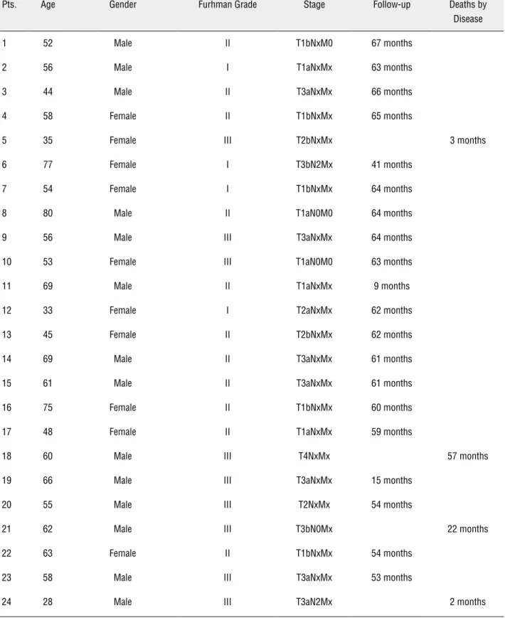
A total of 24 ccRCC samples and their ad-jacent normal renal tissues were collected after ra-dical nephrectomy at the Division of Urology of the National Institute of Cancer (Instituto Nacional do Câncer (INCA)) and Hospital Mário Kröeff (MK), Rio de Janeiro, Brazil, between 2005 and 2008 after approval by the INCA Ethical Committee with the number 38/05. Classification was according to the World Health Organization (WHO). Of the total of 24 patients 22 were operated in the Instituto Nacional do Cancer (INCA) and 2 at the Division of Urology in the Hospital Mário Kroeff. Patients included in the study were over 18 years, any gender, smokers and non-smokers. Patient with genetic syndromes, HIV infection, mental illness or previously treated for cancer were excluded. The cohort was composed of 14 males and 10 females. Their ages ranged from 28 to 80 years; mean age was 57 with standard devia-tion of 13.4. In our cohort of 24 tumors analysed, 14 (58%) tumors were confined to the kidney. These and other clinical data of patients are shown in Table-1.
Proteins extraction and quantification
Tumor and adjacent normal tissues were collected by a pathologist immediately after sur-gery. Approximately 100 mg of tissues were im-mersed in liquid nitrogen and macerated in the presence of 500µL lysis buffer, containing 7M urea, 2M thiourea, 4% w/v 3-[(3-cholamidopropyl) dimethylammonio]-1-propanesulfonate (CHAPS) and 1% w/v dithiothreitol (DTT). The mixture was agitated for 1 hour at room temperature and centrifuged at 16,000 g for 10 minutes. The su-pernatant containing proteins were collected and quantified using the kit 2D quant (GE Healthcare, Piscataway, USA).
Two-dimensional Gel (2DE)
After precipitation with trichloroace-tic acid, proteins were resuspended in reswelling buffer (4% w/v CHAPS, 8M urea, 1% w/v DTT, 0.002% w/v bromophenol blue). For isoeletric fo-cusing, a total of 1.5 mg of proteins was applied on 18 cm strips of linear pH 3-10 (10). Four repli-cates were made for the each pool of sample. The platform used for separation was Ettan IPGphorTM (GE Healthcare, Piscataway, USA) at 30 volts for 12 hours at 20°C, 100 volts for 1 hour, 200 volts for 1 hour, 500 volts for 1 hour, 1,000 volts for 1 hour, 1,000 to 8,000 volts for 0.5 hour and 8,000 volts up to 75,067 volts/hour.
After focusing, the strips were equilibra-ted and washed (10). The second dimension was performed in a SDS-PAGE gel 12.5%. The run was made in the EttanTM system (GE Healthcare) with tris(hydroxymethyl)aminomethane (Tris) - glyci-ne running buffer at 2.5 W/gel for 30 min and then 100 W until the end of the run. The gels were stained with Coomassie G-250 solution. Image and statistical analysis (Student’s t test, p-value < 0.05) of the stained gels were performed using Image Master 2D Platinum 5 (GE Healthcare, Pis-cataway, USA).
Mass spectrometry
concen-IBJU |DIFFERENCIAL PROTEOME OF CLEAR-CELL RENAL CELL CARCINOMA
85
Pts. Age Gender Furhman Grade Stage Follow-up Deaths by
Disease
1 52 Male II T1bNxM0 67 months
2 56 Male I T1aNxMx 63 months
3 44 Male II T3aNxMx 66 months
4 58 Female II T1bNxMx 65 months
5 35 Female III T2bNxMx 3 months
6 77 Female I T3bN2Mx 41 months
7 54 Female I T1bNxMx 64 months
8 80 Male II T1aN0M0 64 months
9 56 Male III T3aNxMx 64 months
10 53 Female III T1aN0M0 63 months
11 69 Male II T1aNxMx 9 months
12 33 Female I T2aNxMx 62 months
13 45 Female II T2bNxMx 62 months
14 69 Male II T3aNxMx 61 months
15 61 Male II T3aNxMx 61 months
16 75 Female II T1bNxMx 60 months
17 48 Female II T1aNxMx 59 months
18 60 Male III T4NxMx 57 months
19 66 Male III T3aNxMx 15 months
20 55 Male III T2NxMx 54 months
21 62 Male III T3bN0Mx 22 months
22 63 Female II T1bNxMx 54 months
23 58 Male III T3aNxMx 53 months
24 28 Male III T3aN2Mx 2 months
IBJU |DIFFERENCIAL PROTEOME OF CLEAR-CELL RENAL CELL CARCINOMA
86
trated to final volume of 5µL. MALDI analysis used 0.5µL of each peptide solution mixed with the same volume of a saturated matrix solution (α-cyano-4-hydroxycinnamic acid (Sigma Aldri-ch, Milwaukee, USA) at 0.05M in 50% acetonitri-le/ 0.1% trifluoroacetic acid) on the target plate and allowed to dry at room temperature. Data for protein identification were obtained on the 4700 Proteomics Analyzer (Applied Biosystems, Foster City, CA). Both MS and MS/MS data were acqui-red with a neodymium-doped yttrium aluminum garnet (Nd:YAG) laser with a 200-Hz repetition rate. Typically, 1600 shots were accumulated for spectra in MS mode while 2400 shots were accu-mulated for spectra in MS/MS mode. MS and MS/ MS mass spectra were acquired in reflector posi-tive ion mode after calibration with mixtures 1 or 2 (Sequazyme Peptide Mass Standard, Perseptive Biosystems, Foster City, CA). Proteins were identi-fied by correlation of tandem mass spectra to the NCBInr proteins database, using Mascot (Matrix Science, London, UK) with restricted taxonomy Homo sapiens. Search parameters were as follo-ws: MS and MS/MS tolerance of 0.6 Da, tryptic specificity allowing for one missed cleavage, fixed modification of carbamidomethylation of cysteine residues, and variable modification of oxidation of methionine, histidine and tryptophan residues; phosphorylation of tyrosine, serine and threonine residues and propionamide. Positive protein iden-tification was accepted with at least two peptides with a Mascot peptide score ≥ 35.
RESULTS
This study aimed to identify differentially expressed proteins in ccRCC tissues compared to their adjacent normal kidney tissues in 2DE gels. The patient cohort was composed of 14 males and 10 females. Their ages ranged from 28 to 80 years; mean age was 57 with standard deviation of 13.4.
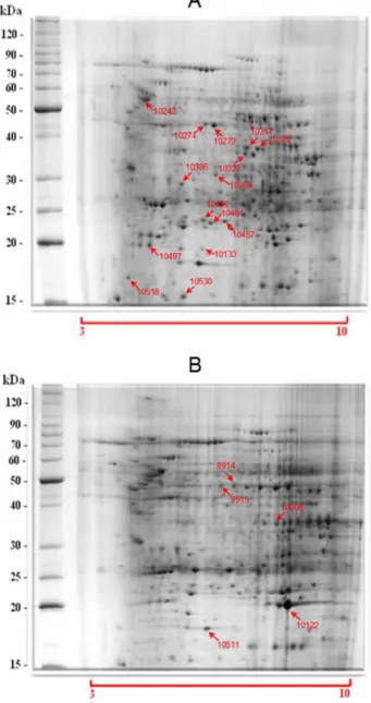
Figure-1 shows the representative 2DE gel profiles of the experimental replicates of the two protein pools (tumor and adjacent normal tissues). The gels were analyzed by Image Master 2D Platinum software. The volumes of the spots were compared between the gels. Twenty six spots were identified as differentially expressed between
Figure 1 - (A) Normal kidney tissue, (B) ccRCC tissue. The gels were stained with colloidal Coomassie. 1.5 mg of protein from pools of extracts was applied to the 2DE. First dimen-sion was carried out with 18 cm IPG strips, pH 3-10; Second dimension was carried out in 12.5% SDS-PAGE.
them, among which 20 were identified as 16 non--redundant proteins (Table-2).
IBJU
|
DIFFERENCIAL PRO
TEOME OF CLEAR-CELL RENAL CELL CARCINOMA
87
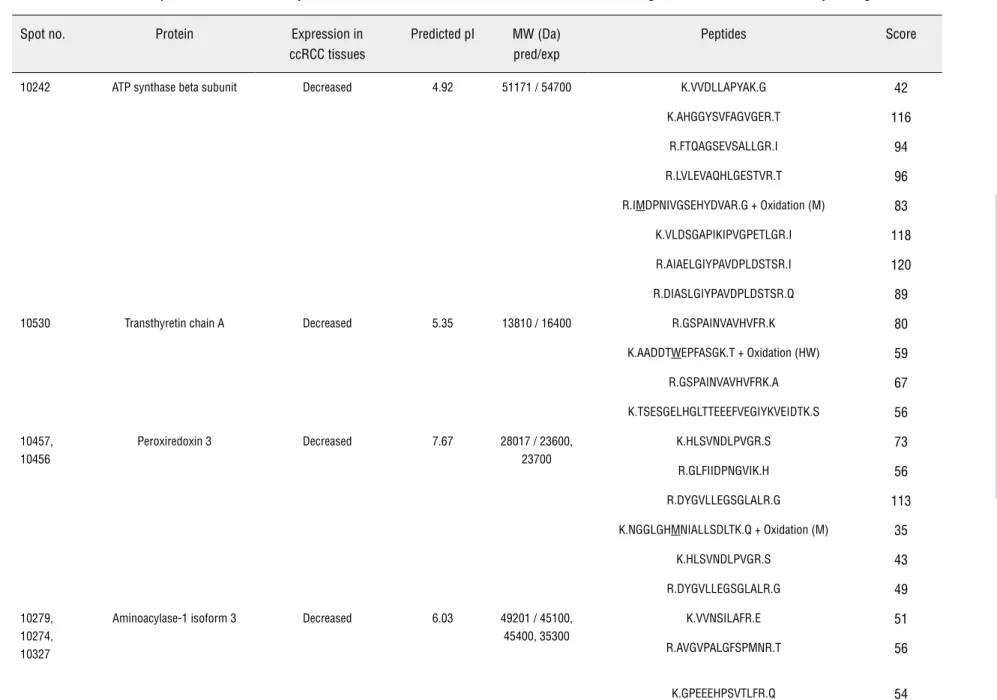
Table 2 - Identification of proteins with altered expression levels in RCC tissues. Proteins were identified using MALDI/TOF-TOF de novo sequencing.
Spot no. Protein Expression in ccRCC tissues
Predicted pI MW (Da) pred/exp
Peptides Score
10242 ATP synthase beta subunit Decreased 4.92 51171 / 54700 K.VVDLLAPYAK.G 42
K.AHGGYSVFAGVGER.T 116
R.FTQAGSEVSALLGR.I 94
R.LVLEVAQHLGESTVR.T 96
R.IMDPNIVGSEHYDVAR.G + Oxidation (M) 83
K.VLDSGAPIKIPVGPETLGR.I 118
R.AIAELGIYPAVDPLDSTSR.I 120
R.DIASLGIYPAVDPLDSTSR.Q 89
10530 Transthyretin chain A Decreased 5.35 13810 / 16400 R.GSPAINVAVHVFR.K 80
K.AADDTWEPFASGK.T + Oxidation (HW) 59
R.GSPAINVAVHVFRK.A 67
K.TSESGELHGLTTEEEFVEGIYKVEIDTK.S 56
10457, 10456
Peroxiredoxin 3 Decreased 7.67 28017 / 23600,
23700
K.HLSVNDLPVGR.S 73
R.GLFIIDPNGVIK.H 56
R.DYGVLLEGSGLALR.G 113
K.NGGLGHMNIALLSDLTK.Q + Oxidation (M) 35
K.HLSVNDLPVGR.S 43
R.DYGVLLEGSGLALR.G 49
10279, 10274, 10327
Aminoacylase-1 isoform 3 Decreased 6.03 49201 / 45100,
45400, 35300
K.VVNSILAFR.E 51
R.AVGVPALGFSPMNR.T 56
IBJU
|
DIFFERENCIAL PRO
TEOME OF CLEAR-CELL RENAL CELL CARCINOMA
88
R.TVQPKPDYGAAVAFFEETAR.Q 78
R.AGFALDEGIANPTDAFTVFYSER.S 149
K.GPEEEHPSVTLFR.Q 48
R.TVQPKPDYGAAVAFFEETAR.Q 75
R.AGFALDEGIANPTDAFTVFYSER.S 116
R.AGFALDEGIANPTDAFTVFYSER.S 59
R.LEHTHFFSR.H 41
10317, 10320
Aldehyde reductase Decreased 6.32 33365 / 37400,
36600
R.DAGHPLYPFNDPY.- 35
R.GLEVTAYSPLGSSDR.A 60
K.YALSVGYR.H 38
10386 Prohibitin Decreased 5.57 29843 / 29300 R.FDAGELITQR.E 89
K.DLQNVNITLR.I 63
R.ILFRPVASQLPR.I 54
R.IFTSIGEDYDER.V 78
R.KLEAAEDIAYQLSR.S 121
K.AAELIANSLATAGDGLIELR.K 92
R.NVPVITGSKDLQNVNITLR.I 56
R.IFTSIGEDYDERVLPSITTEILK.S 48
10133 Ferritin light chain Decreased 5.65 16441 / 18200 K.MGDHLTNLHR.L + Oxidation (M) 36
K.LNQALLDLHALGSAR.T 45
R.LGGPEAGLGEYLFER.L 117
K.KLNQALLDLHALGSAR.T 131
IBJU
|
DIFFERENCIAL PRO
TEOME OF CLEAR-CELL RENAL CELL CARCINOMA
89
10461 Abhydrolase domain
containing 14B
Decreased
5.94 22446 / 23000
R.FSVLLLHGIR.F 48
R.AVAIDLPGLGHSK.E 70
R.EGTIQVQGQALFFR.E 52
10518 Cytochrome b5 Decreased
4.82 15218 / 18400
K.STWLILHHK.V + Oxidation (HW) 50
K.FLEEHPGGEEVLR.E 112
K.TFIIGELHPDDRPK.L 84
R.EQAGGDATENFEDVGHSTDAR.E 104
10369 Ceto-hexokinase Undetected 5.99 25498 / 31200 K.WIHIEGR.N 46
K.LLHSDAFPPPR.V 50
K.HLGFQSAEEALR.G 53
R.VVDTLGAGDTFNASVIFSLSQGR.S 68
R.TIVLHDTSLPDVSATDFEKVDLTQFK.W 48
10497 Retinol binding protein Undetected
5.09 20532 / 19900
R.FSGTWYAMAK.K + Oxidation (HW); Oxidation (M) 36
R.LLNLDGTCADSYSFVFSR.D 95
K.GNDDHWIVDTDYDTYAVQYSCR.L + Oxidation (HW) 53
10006 Glycine amidinotransferase Increased 8.30 48869 / 35300 R.SIIKDYFHR.G 49
R.RPDPIDWSLK.Y + Oxidation (HW) 39
R.AENACVPPFTIEVK.A 53
K.TPDFESTGLYSAMPR.D + Oxidation (M) 42
K.FVTTEFEPCFDAADFIR.A 100
Spot no. Protein Expression in ccRCC tissues
Predicted pI MW (Da) pred/exp
IBJU
|
DIFFERENCIAL PRO
TEOME OF CLEAR-CELL RENAL CELL CARCINOMA
90
10122 Alpha B crystallin Increased
7.18 22435 / 20500
K.DRFSVNLDVK.H 55
R.RPFFPFHSPSR.L 63
R.APSWFDTGLSEMR.L 108
R.APSWFDTGLSEMR.L + Oxidation (HW) 75
K.HEERQDEHGFISR.E 59
K.VLGDVIEVHGQHEERQDEHGFISR.E 61
10511 Cu/Zn Superoxide dismutase Increased
8.76 16020 / 18600
K.GLTEGLHGFHVHEFGDNTAGCTSAGPHFNPLSR.K 60
R.HVGDLGNVTADKDGVADVSIEDSVISLSGDHCIIGR.T 55
K.GBGPVZGIIBFZZK.E 50
K.AVCVLKGBGPVZGIIBFZZK.E 41
K.GLTEGLHGFHVHEFGDNTAGCTSAGPHFNPLSR.K 60
9914 Glyceraldehyde 3-phosphate dehydrogenase
Exclusive 7.60
36910 / 47200
R.VPTANVSVVDLTCR.L 77
K.LVSWYDNEFGYSNR.V + Oxidation (HW) 63
K.FVTTEFEPCFDAADFIR.A 95
9915 Heat shock protein 27 Exclusive 5.98 22826 / 47200 R.LFDQAFGLPR.L 51
R.QDEHGYISR.C 40
IBJU |DIFFERENCIAL PROTEOME OF CLEAR-CELL RENAL CELL CARCINOMA
91
cytochrome b5, ceto-hexokinase and retinol bin-ding protein (Table-2).
On the other hand, five proteins were found to be either overexpressed or exclusively detected in the ccRCC extracts: glycine amidinotransferase, alpha B crystallin, Cu/Zn superoxide dismutase, glyceraldehyde 3-phosphate dehydrogenase and heat shock protein 27 (HSP27) (Table-2).
DISCUSSION
The search for the ideal set of protein ma-rkers of RCC tissues began when Sarto et al. (3) compared the tumor proteins from 10 patients with ccRCC with their respective adjacent normal tissue through 2DE gel. Since then, other studies were performed with the same goal under different conditions or different proteomics approaches (3-8). The method that was applied in this study is based on 2DE, the most traditional technology for separation of proteins. It is a very efficient me-thod, allowing good resolution of complex sam-ples, providing the molecular weight and pH of the analyzed protein that, in certain instances, generates information about post-translational modifications. This work is justified by the fact that no study with RCC Brazilian patients was ever produced.
Some of the proteins here identified are involved in cellular metabolism, such as the beta subunit of ATP synthase, glycine amidinotransfe-rase, cytochrome b5, glyceraldehyde 3-phosphate dehydrogenase, aminoacilase isoform 3, aldehyde reductase and ceto-reductase hexokinase.
The ATP synthase protein complex is di-rectly involved in ATP production through oxi-dative phosphorylation in mitochondria, and is a high-energy molecule used in several metabolic processes (11). Glycine amidinotransferase enzy-me participates in the enzy-metabolism of amino acids as well as in the urea cycle, and is found in all tissues of the body (11). Cytochrome b5 is a pro-tein that participates in the desaturation of fatty acids and was described to be underexpressed in RCC (8). Glyceraldehyde-3 phosphate dehydro-genase (GAPDH) participates in the oxidation of glyceraldehyde 3-phosphate, associated with the formation of ATP molecules in the electron
trans-port chain in mitochondria (11). GAPDH was ex-clusively detected in the ccRCC extract. GAPDH is a constitutive enzyme and therefore it is likely to be present in the normal extracts. Two hypothe-ses may explain our results: GAPDH is undergoing posttranslational modifications and/or degrada-tion in the normal tissue or GAPDH is modified and accumulates at detectable levels in the ccRCC tissue, whereas in the normal tissue it is present at undetectable levels. Previous works suggest that GAPDH is indeed target of modifications that can modulate its cellular level (12,13).
Aminoacylase-I is a homodimeric cytoso-lic enzyme that catalyzes the hydrolysis of a va-riety of N-acylated amino acids, generating free amino acids and acyl groups. The enzyme is found in many mammalian tissues, with highest activity in the kidney, and is involved in detoxification processes. Balabanov et al (11) and Raimondo et al (8) observed underexpression of aminoacylase-I when they analyzed the proteome of ccRCC tis-sues. An interesting fact is that the gene encon-ding for this enzyme is located on chromosome 3p21-1, and deletions in the short arm of chro-mosome 3 were observed in almost all renal cell carcinomas.
Ketohexokinase, also known as fructoki-nase, participates in the metabolism of fructose, catalyzing the conversion of fructose to fructo-se-1-phosphate. Although the ketohexokinase is mostly abundant in liver, it was also found in kid-ney, small intestine and pancreas (4). Hwa et al (4) found lower levels of ketohexokinase in ccRCC tissues, in accordance with our findings.
Aldehyde reductase is an oxidoreductase that catalyzes NADPH-dependent reduction of a variety of aliphatic and aromatic aldehydes. Howe-ver, little is known about its physiological role. We observed underexpression of aldehyde reductase in agreement with previous published data on RCC (4).
IBJU |DIFFERENCIAL PROTEOME OF CLEAR-CELL RENAL CELL CARCINOMA
92
possible that carcinogenesis by oxidative stress is mainly due to excessive production of reactive oxygen species (ROS) such as H2O2 and superoxide anion, leading to structural changes in DNA. Mo-reover, ROS can directly activate cytoplasmic and nuclear pathways of signal transduction that are associated with malignant transformation. The-refore, it is suggested that antioxidant enzymes, such as peroxiredoxins, play a functional role in carcinogenesis (15).
Soini et al (16) studied the expression of peroxiredoxins 1 to 6 in 138 RCC samples using immunohistochemistry and immunoblotting. The authors correlated the expression of peroxiredo-xins with tumor stage and patient survival rate. Non-malignant tubular cells were positive for all six Prxs. Most RCC were positive for Prxs 1 and 2, whereas only 15% of tumors showed expres-sion of Prxs 3 and 4. In this study, lower levels of peroxirredoxin-3 were observed in ccRCC tissues extracts, which is consistent with data previously published (16).
The superoxide dismutases (SOD) are an-tioxidant enzymes that catalyze the dismutation of superoxide radicals into hydrogen peroxide and oxygen (17). We found overexpression of SOD-1 in ccRCC extracts. This can be explained by the fact that the production of ROS is generally more active in malignant cells (17).
Transthyretin acts as a carrier of retinol (vitamin A), through its association with retinol--binding protein (RBP). Retinol is a natural pre-cursor of the retinoic acid, which regulates the transcription of specific genes through nuclear receptors of retinoic acid, having effects on cell growth and differentiation of normal cells, em-bryonic cells, as well as on neoplastic cells (18). We observed underexpression of transthyretin in the ccRCC extract, whereas RBP protein was not detected. Because the kidneys play a role in the metabolism of proteins of low molecular weight, such as RBP, it is possible that the disability of renal filtration that occurs in RCC leads to greater loss of circulating RBP. This, in turn, can result in decreasing levels of transthyretin.
Ferritin is the most important iron reser-ve protein and found in all cells. Under normal conditions, it can represent 25% of the total iron
found in the body (19). Anemia is the most com-mon hematologic abnormality associated with kidney cancer (1). This picture may be a conse-quence of bad function of iron storage proteins such as ferritin. Concurring with this, in the pre-sent study we observed lower levels of ferritin in ccRCC protein extracts.
The gene encoding prohibitin was first isolated due to its ability to down regulate rat hepatocytes cell proliferation (20). Its antiproli-ferative activity was further demonstrated when the micro-injection of synthetic mRNA in normal fibroblasts and HeLa cells blocked the entry of cells in S phase of the cell cycle (21). Furthermo-re, prohibitin is capable of inhibiting cell proli-feration by repressing the transcriptional activity mediated by E2F which regulates many genes in-volved in the transition G1 / S and DNA synthesis (22). The study of Fusaro et al (23) has also shown that in addition to its mitochondrial localization, prohibitin is also located in the nucleus, and is able to physically interact with p53 in vivo and in vitro, potentially modulating their regulatory pathways. These data suggested that prohibitin is able to act as tumor suppressor. However, the role of prohibitin as tumor suppressor has been de-bated. Prohibitin undergoes multiple post-trans-lational modifications, such as phosphorylation, glycosylation, palmitoylation, ubiquitination and others; therefore, it is reasonable to propose more than one function for prohibitin. In fact, the pre-sence of isoforms could explain the shift in mole-cular weight of the two prohibitin spots that were observed on RCC gels (24). In addition, prohibitin was recently found to combat oxidative stress, as well as to have a role in modulating mitochondrial stress-related autophagy (25). We found underex-pression of prohibitin in ccRCC extracts, which is consistent with its apparent tumor suppressor function and free radical protection.
compo-IBJU |DIFFERENCIAL PROTEOME OF CLEAR-CELL RENAL CELL CARCINOMA
93
nent of the vertebrate eye lens with chaperone--like functions. It is probable the up-expression of αβ-crystalin represents a response to stress.
Previous studies have shown higher expres-sion of HSP27 in ccRCC (27,29-32). We observed the same result. HSP27 down-regulates apoptosis through its ability to interact with key components of the apoptosis signaling pathway, in particular those involved in activation of caspases. Because of this physiologic effect, it is likely that HSP27 is involved in cell proliferation of ccRCC.
CONCLUSIONS
Our results present the first proteomics analysis of ccRCC tissues within a Brazilian popula-tion. Several proteins were found at differential le-vels when compared to their normal adjacent tissues and, moreover, many have been previously descri-bed in studies on RCC, when other populations were used. Therefore, this work accesses the robustness and validity of previously reported proteins and points out candidate biomarkers and/or targets for RCC that may be validated in future trials.
ACKNOWLEDGEMENTS
This work was supported by Programa de Oncobiologia (Rio de Janeiro, Brazil), the Carlos Chagas Foundation for the Support of Science (FAPERJ) and the Conselho Nacional de Desenvol-vimento Científico e Tecnológico (CNPq), Brazil.
CONFLICT OF INTEREST
None declared.
REFERENCES
1. Campbell SC, Novick AC. Renal Tumors. In: Walsh PC, Retik AB, Vaughan ED, Wein AJ (Editors), Kavoussi LR, Novick AC, Partin AW, Peters CA (ed.), Campbell’s Urology, Philadelphia, Elsevier, 2002; pp. 2672-86.
2. Ries LA, Melbert D, Krapcho M, Stinchcomb DG, Howlader N, Horner MJ, et al.: (ed.), SEER Cancer Statistics Review, 1975-2005, National Cancer Institute. Bethesda, MD, http:// seer.cancer.gov/csr/1975_2005/, based on November 2007 SEER data submission, posted to the SEER website, 2008.
3. Sarto C, Marocchi A, Sanchez JC, Giannone D, Frutiger S, Golaz O, et al.: Renal cell carcinoma and normal kidney pro-tein expression. Electrophoresis. 1997; 18: 599-604. 4. Hwa JS, Park HJ, Jung JH, Kam SC, Park HC, Kim CW, et
al.: Identification of proteins differentially expressed in the conventional renal cell carcinoma by proteomic analysis. J Korean Med Sci. 2005; 20: 450-5.
5. Perroud B, Lee J, Valkova N, Dhirapong A, Lin PY, Fiehn O, et al.: Pathway analysis of kidney cancer using proteomics and metabolic profiling. Mol Cancer. 2006; 5: 64.
6. Jones J, Otu HH, Grall F, Spentzos D, Can H, Aivado M, et al.: Proteomic identification of interleukin-2 therapy response in metastatic renal cell cancer. J Urol. 2008; 179: 730-6. 7. Siu KW, DeSouza LV, Scorilas A, Romaschin AD, Honey RJ,
Stewart R, et al.: Differential protein expressions in renal cell carcinoma: new biomarker discovery by mass spec-trometry. J Proteome Res. 2009; 8: 3797-807.
8. Raimondo F, Salemi C, Chinello C, Fumagalli D, Morosi L, Rocco F, et al.: Proteomic analysis in clear cell renal cell carcinoma: identification of differentially expressed protein by 2-D DIGE. Mol Biosyst. 2012; 8: 1040-51.
9. Engwegen JY, Mehra N, Haanen JB, Bonfrer JM, Schellens JH, Voest EE, e al.: Validation of SELDI-TOF MS serum pro-tein profiles for renal cell carcinoma in new populations. Lab Invest. 2007; 87: 161-72.
10. Görg A, Weiss W, Dunn MJ: Current two-dimensional elec-trophoresis technology for proteomics. Proteomics. 2004; 4: 3665-85. Erratum in: Proteomics. 2005; 5: 826-7. 11. Balabanov S, Zimmermann U, Protzel C, Scharf C, Klebingat KJ,
Walther R: Tumour-related enzyme alterations in the clear cell type of human renal cell carcinoma identified by two-dimen-sional gel electrophoresis. Eur J Biochem. 2001; 268: 5977-80. 12. Pierce A, Mirzaei H, Muller F, De Waal E, Taylor AB, Leonard
S, et al.: GAPDH is conformationally and functionally altered in association with oxidative stress in mouse models of amyo-trophic lateral sclerosis. J Mol Biol. 2008; 382: 1195-210. 13. Mazzola JL, Sirover MA: Alteration of intracellular structure
and function of glyceraldehyde-3-phosphate dehydroge-nase: a common phenotype of neurodegenerative disor-ders? Neurotoxicology. 2002; 23: 603-9.
14. Immenschuh S, Baumgart-Vogt E: Peroxiredoxins, oxida-tive stress, and cell proliferation. Antioxid Redox Signal. 2005; 7: 768-77.
15. Nonn L, Berggren M, Powis G: Increased expression of mitochondrial peroxiredoxin-3 (thioredoxin peroxidase-2) protects cancer cells against hypoxia and drug-induced hydrogen peroxide-dependent apoptosis. Mol Cancer Res. 2003; 1: 682-9.
IBJU |DIFFERENCIAL PROTEOME OF CLEAR-CELL RENAL CELL CARCINOMA
94
17. Hileman EO, Liu J, Albitar M, Keating MJ, Huang P:Intrin-sic oxidative stress in cancer cells: a biochemical basis for therapeutic selectivity. Cancer Chemother Pharmacol. 2004; 53: 209-19.
18. Soprano DR, Soprano KJ, Goodman DS: Retinol-binding protein messenger RNA levels in the liver and in extrahe-patic tissues of the rat. J Lipid Res. 1986; 27: 166-71. 19. Park KS, Kim H, Kim NG, Cho SY, Choi KH, Seong JK, et al.:
Proteomic analysis and molecular characterization of tis-sue ferritin light chain in hepatocellular carcinoma. Hepa-tology. 2002; 35: 1459-66.
20. McClung JK, Danner DB, Stewart DA, Smith JR, Schnei-der EL, Lumpkin CK, et al.: Isolation of a cDNA that hybrid selects antiproliferative mRNA from rat liver. Biochem Bio-phys Res Commun. 1989; 164: 1316-22.
21. Nuell MJ, Stewart DA, Walker L, Friedman V, Wood CM, Owens GA, et al.: Prohibitin, an evolutionarily conserved intracellular protein that blocks DNA synthesis in normal fi-broblasts and HeLa cells. Mol Cell Biol. 1991; 11: 1372-81. 22. Wang S, Nath N, Adlam M, Chellappan S: Prohibitin, a po-tential tumor suppressor, interacts with RB and regulates E2F function. Wang S, Nath N, Adlam M, Chellappan S. On-cogene. 1999; 18: 3501-10.
23. Fusaro G, Dasgupta P, Rastogi S, Joshi B, Chellappan S: Prohibitin induces the transcriptional activity of p53 and is exported from the nucleus upon apoptotic signaling. J Biol Chem. 2003; 278: 47853-61.
24. Junker H, Venz S, Zimmermann U, Thiele A, Scharf C, Wal-ther R: Stage-related alterations in renal cell carcinoma--comprehensive quantitative analysis by 2D-DIGE and pro-tein network analysis. PLoS One. 2011; 6: e21867. 25. Kathiria AS, Butcher LD, Feagins LA, Souza RF, Boland CR,
Theiss AL: Prohibitin 1 modulates mitochondrial stress-related autophagy in human colonic epithelial cells. PLoS One. 2012; 7: e31231.
26. Takashi M, Sakata T, Ohmura M, Kato K: Elevated concen-trations of the small stress protein HSP27 in rat renal tu-mors. Urol Res. 1997; 25: 173-7.
27. Shi T, Dong F, Liou LS, Duan ZH, Novick AC, DiDonato JÁ: Differential protein profiling in renal-cell carcinoma. Mol Carcinog. 2004; 40: 47-61.
28. Holcakova J, Hernychova L, Bouchal P, Brozkova K, Zalou-dik J, Valik D, et al.: Identification of alphaB-crystallin, a biomarker of renal cell carcinoma by SELDI-TOF MS. Int J Biol Markers. 2008; 23: 48-53.
29. Santarosa M, Favaro D, Quaia M, Galligioni E: Expression of heat shock protein 72 in renal cell carcinoma: possible role and prognostic implications in cancer patients. Eur J Cancer. 1997; 33: 873-7.
30. Erkizan O, Kirkali G, Yörükoğlu K, Kirkali Z: Significance of heat shock protein-27 expression in patients with renal cell carcinoma. Urology. 2004; 64: 474-8.
31. Sarto C, Valsecchi C, Magni F, Tremolada L, Arizzi C, Cor-dani N, et al.: Expression of heat shock protein 27 in human renal cell carcinoma. Proteomics. 2004: 2252-60.
32. Kim DS, Choi YP, Kang S, Gao MQ, Kim B, Park HR, et al.: Panel of candidate biomarkers for renal cell carcinoma. J Proteome Res. 2010; 9: 3710-9.