INTRODUCTION
It is known that renal cell carcinoma (RCC) is responsible for 3% of all malignancies. In Amer-ica, it is estimated that approximately 58,000 new cases were diagnosed and 13,000 patients died from RCC in 2010 (1). It is the seventh most mon malignancy in men and the ninth most com-mon in women, and occurs predominantly in the sixth and seventh decades of life (1).
Despite recent advances in the diagnosis and treatment of RCC, it is still a tumor of unpredictable presentation and clinical outcome. Currently, the only potentially curative treatment is surgery, through either radical nephrectomy or nephron sparing procedures. Systemic therapies such as anti-angiogenic agents show mixed results and often disappointing outcomes (2). Such drugs are available for patients with metastatic disease and its possible use as adjuvant or neoadjuvant agents Purpose: To analyze the immunohistochemical expression of the standard isoform
of CD44 (CD44s) adhesion molecule in clear cell renal cell carcinoma (CCRCC) and its impact on clinical outcomes.
Materials and Methods: Ninety-nine consecutive patients treated surgically for RCC between 1992 and 2009 were selected. A single pathologist reviewed all cases to effect a uniform reclassifi cation and determine the most representative tumor areas for construction of a tissue microarray. The same pathologist, who was blinded to the outcome of the cases, semi-quantitatively scored the staining intensity of CD44s in all specimens. The counting was done using the H-Score algorithm.
Results: Of the 99 immunostained RCC specimens, 57(57.7%) showed low expres-sion, and 42(42.4%) showed high expression levels of CD44s. The expression of CD44s was directly associated with tumor size (p = 0.03), clinical stage (p = 0.02) and Fuhrman grade (p = 0.02). Disease specifi c survival (DSS) rates for patients whose specimens expressed low and high levels of CD44s was 88.1% and 67.5%, respectively (p = 0.009). Progression free survival (PFS) rates in patients with low and high expression of CD44s were 78.8% and 61.7%, respectively (p = 0.05). Clas-sical features such as the presence of metastasis and clinical stage remained isolated predictors of survival.
Conclusions: Immunohistochemical expression of CD44s was associated with im-portant clinical variables such as stage and Fuhrman grade. However, it was not an independent predictor of survival. Therefore, we believe it has a limited role as a prognostic marker in patients with CCRCC.
Key words:
Carcinoma, Renal Cell; Antigens, CD44; Biological Markers; Prognosis
Int Braz J Urol. 2012; 38: 456-65
________________
Submitted for publication: March 06, 2012
________________
Accepted after revision: June 14, 2012
Immunohistochemical expression of CD44s in renal cell
carcinoma lacks independent prognostic signifi cance
_______________________________________________
Walter Henriques da Costa, Rafael Malagoli Rocha, Isabela Werneck da Cunha, Gustavo Cardoso
Guima-raes, Stenio de Cássio Zequi
A.C. Camargo Hospital, Sao Paulo, Brazil
ABSTRACT
ARTICLE
INFO
is still not well defined (3). Therefore, individual markers of tumor aggressiveness are needed (4-6).
CD44 and its variants constitute a protein family that is expressed in the cytoplasmic mem-brane and mediates cell-cell and cell-extracellular matrix interactions. It is encoded by a single gene and is subjected to post-translational modifications through alternative splicing. Its chief ligand is hy-aluronic acid, an important component of the ex-tracellular matrix (ECM) (7). As a molecule involved in mechanisms of cell motility, CD44 has been used as a marker of tumor aggressiveness in a number of malignancies such as stomach, liver and breast. However, in RCC there are contradictory results, with some series supporting the role of CD44 as a prognostic marker (8-10) while in other studies its importance as a predictor of survival was not con-firmed (11,12).
The purpose of this study was to analyze the immunohistochemical expression of the standard isoform of CD44 (CD44s) in clear cell RCC (CCRCC) and examine its association with pathological fea-tures and its impact on clinical outcomes.
MATERIALS AND METHODS
Patients
Ninety-nine consecutive patients submitted to radical nephrectomy or partial nephrectomy for RCC between 1992 and 2009 were selected from the medical records of our institution. The clinical data were obtained from medical charts in the hospital archives. A single pathologist (IWC) reviewed all cases to effect a uniform reclassification and deter-mined the most representative tumor areas for con-struction of a tissue microarray. Our internal review board approved this study.
The following variables were included in the data bank: age, gender, ECOG status, smoking status, time since diagnosis, type of surgery, stag-ing (TNM AJCC/UICC 2002), Furhman grade, histo-logical subtype (World Health Organization (WHO) 2004), microvascular invasion (MVI), lymph node involvement, presence of tumor necrosis, follow-up period, presence of metastases, clinical situation at the end of the study, storage time of the paraffin embedded samples and immunohistochemical ex-pression of CD44s.
Construction of tissue microarray
Two cylinders, measuring 2 mm in diam-eter, taken from different parts of the tumor were used to build a tissue microarray (TMA). Two cyl-inders from previously evaluated tumors--one that was positive for CD44s and one that was CD44s-negative--were included as internal con-trols. Sequential 4µm sections were generated and stained with hematoxylin and eosin (the first and last sections) to confirm the diagnosis, and the in-tervening sections were examined by immunohis-tochemistry.
Immunohistochemistry
The sections were mounted on positively charged glass slides, dried for 30 min. at 37° C, deparaffinized in xylene, and rehydrated in a se-ries of graded alcohols. The sections were then incubated with a primary antibody against CD44 standard isoform (Novocastra® clone F10-44-2, Newcastle, UK) at 1:40 for 30 minutes. All im-munohistochemistry procedures were performed automatically in a Link 48 autostainer (DAKO®) using the Flex Plus visualization system according to the supplier’s specifications. For control stain-ing of CD44s, 17 specimens of non-neoplastic re-nal parenchyma that were at least 1.0 cm from the tumor lesion were included. Both renal cortex and renal medulla were evaluated.
“low-” and “high-” expression levels were used based on the median value of H-Score algorithm.
Statistical Analysis
Analyses were performed using SPSS for Windows, version 17.0 (SPSS®, Chicago, Ill). To ver-ify the association between CD44s expression and other variables, Pearson chi-square test was per-formed. Fisher’s exact test was applied to cases in which the expected frequency was less than 5. The level of significance was 5%. Comparisons between the degrees of expression of CD44s in non-neoplas-tic renal parenchyma and CCRCC tissue were made by Kruskal-Wallis test and Mann-Whitney test.
Disease-specific survival (DSS) was defined as the interval between the primary surgery and the last follow-up visit or disease-related death. Progression-free survival (PFS) was defined as the interval between the primary surgery and the last follow-up visit if the patient showed no evi-dence of disease, recurrence, or metastasis of RCC. DSS and PFS were examined using Kaplan-Meier curves. To compare the estimated curves for each category within a given variable, log-rank test was used. The level of significance was 5%. Cox proportional hazards model was used to determine which variables influenced survival. Variables that significantly impacted survival in univariate analysis were included in multivariate analyzes. The confidence interval was 5%.
RESULTS
Characterization of patients
Of the total 99 patients, 71 (71.7%) patients underwent radical nephrectomy; 28 (28.3%) were treated with nephron-sparing procedures, all of which had negative margins. The patients ranged from age 27 to 79 years (average 55.5 years). The mean postoperative follow-up period was 43 months and ranged from 0 to 178 months. There was metastatic disease at the initial clinical pre-sentation in 13 (13.3%) patients; 50/99 (51.5%) presented with T1 stage disease. The cohort mean tumor size was 7.4 cm. Over 5 years, 21 (21.2%) pa-tients died of CCRCC. The clinical and pathological features of the patients are listed in Table-1.
Of the 99-immunostained RCC specimens, 57 (57.6%) showed low expression, and 42 (42.4%) showed high expression of CD44s, which was evenly distributed through the plasma membrane, cytoplasm and cell nucleus (Figure-1). In fact, it was observed an even distribution of the staining instead of the presence of high spots. Additionally, when divided into two groups according to storage time of the paraffin embedded samples (up to 10 years and above 10 years), we noted no significant impact on staining intensity (p = 0.42).
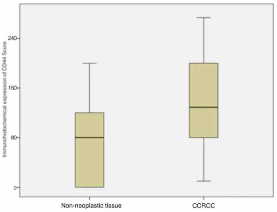
CCRCC cases expressed higher levels of CD44s compared with normal tissue (p = 0.01). The mean expression score for CD44s in non-neoplastic tissues was 88 (sd = 11.1, median = 81). In CCRCC samples, the mean score was 143 (sd = 12.9, me-dian = 149) (Figure-2).
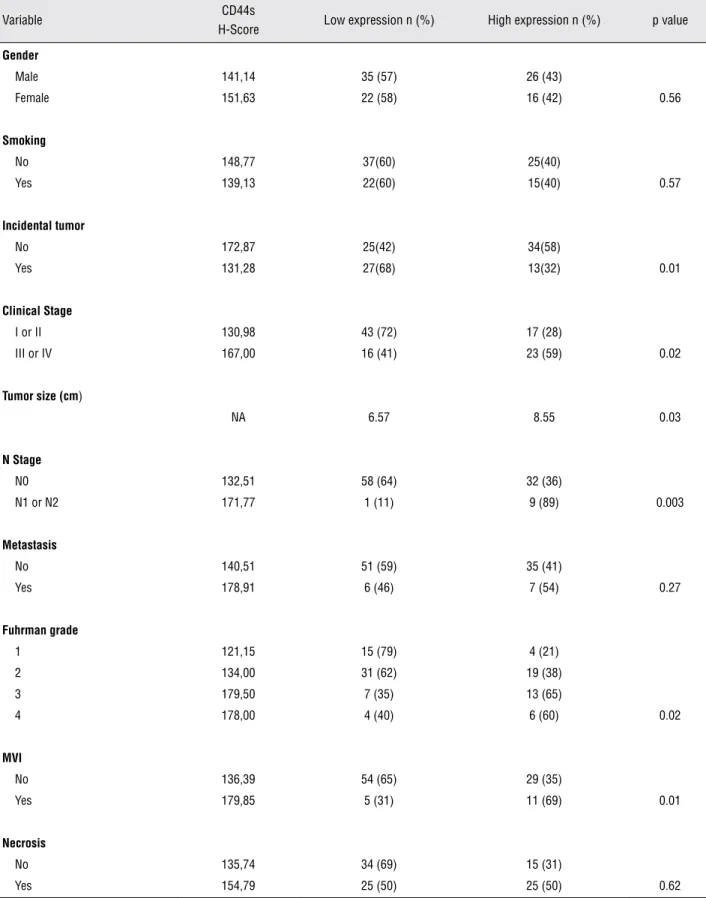
The expression of CD44s was directly asso-ciated with incidental tumors (p = 0.01), tumor size (p = 0.03), lymph node involvement (p = 0.003), microvascular invasion (MVI) (p = 0.01), clinical stage (p = 0.002) and Fuhrman grade (p = 0.02) as shown in Table-1.
Survival analysis
Disease-related death occurred in 21 pa-tients (21.2%) during follow-up. The following variables were associated with 5-year disease-spe-cific survival (DSS) rates in the univariate analysis: clinical stage (p < 0.001), MVI (p < 0.001), tumor size (p < 0.001), necrosis (p < 0.001), metastasis at presentation (p < 0.001), Fuhrman grade (p < 0.001), and ECOG PS (p = 0.009). The 5-year pro-gression-free survival (PFS) rate was 70.7%. Me-tastasis at diagnosis (p < 0.001), MVI (p = 0.007), clinical stage (p = 0.001), tumor size (p < 0.001), Fuhrman grade (p < 0.001), and ECOG PS (p = 0.01) were linked to 5-year PFS rates in the univariate analysis (Table-2).
DSS rates based on CD44s expression are shown in Figure-3A. The 5-year DSS for patients whose specimens expressed low and high levels of CD44s was 88.1% and 67.5%, respectively (p = 0.009). Five-year PFS rates in patients with low and high expression of CD44s were 78.8% and 61.7%, respectively (p = 0.05) (Figure-3B).
Variable CD44s
H-Score Low expression n (%) High expression n (%) p value
Gender
Male 141,14 35 (57) 26 (43)
Female 151,63 22 (58) 16 (42) 0.56
Smoking
No 148,77 37(60) 25(40)
Yes 139,13 22(60) 15(40) 0.57
Incidental tumor
No 172,87 25(42) 34(58)
Yes 131,28 27(68) 13(32) 0.01
Clinical Stage
I or II 130,98 43 (72) 17 (28)
III or IV 167,00 16 (41) 23 (59) 0.02
Tumor size (cm)
NA 6.57 8.55 0.03
N Stage
N0 132,51 58 (64) 32 (36)
N1 or N2 171,77 1 (11) 9 (89) 0.003
Metastasis
No 140,51 51 (59) 35 (41)
Yes 178,91 6 (46) 7 (54) 0.27
Fuhrman grade
1 121,15 15 (79) 4 (21)
2 134,00 31 (62) 19 (38)
3 179,50 7 (35) 13 (65)
4 178,00 4 (40) 6 (60) 0.02
MVI
No 136,39 54 (65) 29 (35)
Yes 179,85 5 (31) 11 (69) 0.01
Necrosis
No 135,74 34 (69) 15 (31)
Yes 154,79 25 (50) 25 (50) 0.62
460
Figure 1 - Photomicrographs of immunohistochemical expression of CD44s. A. Low expression of CD44s in non-neoplastic renal cortex. B. Low expression of CD44s in Fuhrman 2 CCRCC. C. High expression of CD44s in Fuhrman 4 CCRCC. Original magnification: 400 X.
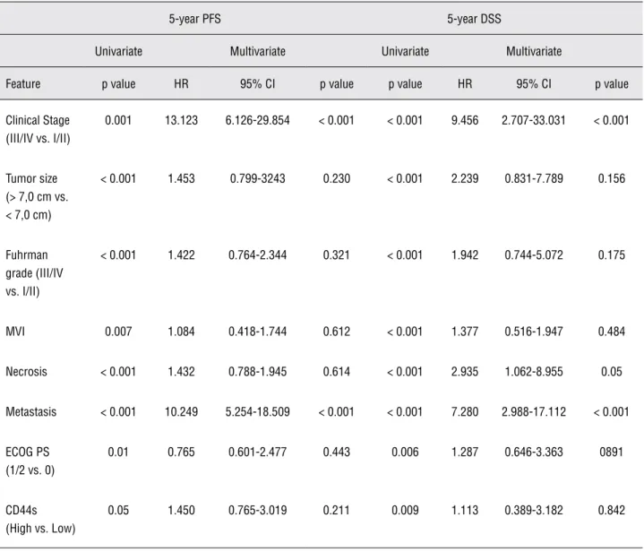
Table 2 - Cox regression analysis of DSS and PFS.
5-year PFS 5-year DSS
Univariate Multivariate Univariate Multivariate
Feature p value HR 95% CI p value p value HR 95% CI p value
Clinical Stage (III/IV vs. I/II)
0.001 13.123 6.126-29.854 < 0.001 < 0.001 9.456 2.707-33.031 < 0.001
Tumor size (> 7,0 cm vs. < 7,0 cm)
< 0.001 1.453 0.799-3243 0.230 < 0.001 2.239 0.831-7.789 0.156
Fuhrman grade (III/IV vs. I/II)
< 0.001 1.422 0.764-2.344 0.321 < 0.001 1.942 0.744-5.072 0.175
MVI 0.007 1.084 0.418-1.744 0.612 < 0.001 1.377 0.516-1.947 0.484
Necrosis < 0.001 1.432 0.788-1.945 0.614 < 0.001 2.935 1.062-8.955 0.05
Metastasis < 0.001 10.249 5.254-18.509 < 0.001 < 0.001 7.280 2.988-17.112 < 0.001
ECOG PS (1/2 vs. 0)
0.01 0.765 0.601-2.477 0.443 0.006 1.287 0.646-3.363 0891
CD44s (High vs. Low)
0.05 1.450 0.765-3.019 0.211 0.009 1.113 0.389-3.182 0.842
HR = Hazard ratio; CI = Confidence interval.
the presence of necrosis (p = 0.05) were indepen-dent predictors of DSS. However, with regard to PFS, only clinical stage (p < 0.001) and metastasis at diagnosis (p < 0.001) were predictive factors. CD44s tissue expression was not an independent predictor of DSS (p = 0.842) or PFS (p = 0.211) (Table-2).
DISCUSSION
Due to the increasing use of radiologi-cal procedures, particularly ultrasonography, the number of tumors that are diagnosed in the early
life expectancies or presenting comorbidities lim-iting surgical treatment (17). Therefore, we seek to identify molecular markers that have prognostic impact and may assist in therapeutic strategies.
The cell surface glycoprotein CD44 ranges in size from 80 to 200 Kd, as described by Naor et al. in 1997 (18). A gene that lies on the short arm of chromosome 11 and mediates cell-cell in-teractions, cell adhesion, and migration encodes it. CD44 is the principal cellular ligand of hyal-uronic acid, a glycosaminoglycan that abounds in the ECM. Described initially in mechanisms of lymphocyte activation and hematopoiesis, CD44 is an important molecule that regulates the biol-ogy of neoplastic cells, especially in interactions between cells and the ECM during tissue invasion and metastasis (11,19,20).
In our study, we observed that immunohis-tochemical expression of CD44s was significantly lower in non-neoplastic cells compared with RCC tissue (p = 0.01). Such observation only con-firms previous reports regarding the metabolism of CD44. It is known that during embryogenesis, the epithelium of the Wolffian or paramesoneph-ric duct expresses CD44, down regulating its ex-pression during differentiation of specialized col-lecting tubular epithelium (21). During malignant transformation, the up regulation of CD44 in the distal and proximal tubular epithelium might re-flect a more embryonic and dedifferentiated state of neoplastic renal cells. During embryogenesis, cells are highly invasive and assume many prop-erties of metastatic cells, such as high levels of adhesion molecules and proteases (22).
We noted that the expression of CD44s was distributed uniformly among cellular com-partments. In fact, CD44 is composed of extracel-lular, transmembrane, and cytoplasmic domains. The extracellular domain of CD44 interacts with ECM, and the intracellular domain is associated with the actin cytoskeleton via binding to ERM (ezrin, radixin, and moesin) proteins. Cleavage of the ectodomain is mediated by membrane-associ-ated matrix metalloproteinases such as MT1-MMP, ADAM10, and ADAM17. The release of the soluble ectodomain from the membrane is responsible for dynamic regulation of the interaction between CD44 and the ECM during cell migration on a HA-containing substratum. The ectodomain cleavage also triggers the intramembrane cleavage of CD44. The second cleavage releases the CD44 intracellu-lar domain into the cytoplasm, which migrates to the cell nucleus and through a feedback mecha-nism stimulates the production of new molecules of CD44 (23). Therefore, it is expected a uniform distribution of the staining of CD44 on the cell membrane, cytoplasm and cell nucleus as observed in our study.
The exact mechanism by which CD44 in-fluences tumor recurrence or progression is un-known. Active migration of tumor cells in ECM is a prerequisite for tumor cell invasion and metastasis. As the principal ligand of hyaluronan, it is pos-sible that CD44 mediates mechanisms of neoplastic cell adhesion (24) or signal transduction in tumor cells following receptor-mediated adhesion to ECM proteins (25). Still, it is known that CD44 molecule can undergo post-translational modifications such as alternative splicing. Previous studies describe CD44v5 and CD44v6 as variants presenting onco-genic potential (10,26).
In our study, we observed that the expres-sion levels of CD44s were associated with several clinical and pathological features such as tumor size (p = 0.03), N stage (p = 0.003), Fuhrman grade (p = 0.02) and clinical stage (p = 0.02). High ex-pression levels of CD44s were associated with larger tumors, high Fuhrman grade tumors and advanced disease. It is known that high Fuhrman grade tumors have a more aggressive phenotype and are associated with higher likelihood of local invasion and distant metastasis (5). Therefore, it
is expected that the metabolism of adhesion mol-ecules such as CD44s be increased in such tumors favoring the spread of tumor cells. It is also known that larger tumors are associated with advanced disease, which explains the lower survival rates observed in patients presenting high expression levels of CD44s (14).
There are few studies describing the impact of CD44s in RCC. And many of the results are con-tradictory, which stimulated the research for this marker in our series. Our findings confirm those of previous published series. Paradis et al. performed a seminal study on the prognostic role of CD44s in 91 cases of RCC who were treated surgically, report-ing a link between CD44s and tumor size, Fuhrman grade, and T stage (8). Lim et al. evaluated the prognostic value of CD44s in 125 cases of RCC of which 89 cases (71%) were composed of patients with metastatic disease. The authors reported great-er expression of CD44s in metastatic tumors and described such marker as an independent predictor of PFS in their series (9). We believe that the differ-ences between our series in which only 29% of pa-tients progressed to metastatic disease, and Lim et al. series might explain the fact that CD44s was not confirmed as a predictor of survival in our study. Previous series confirm our findings (11,12). Altinel and his colleagues from Turkey described increased expression of CD44s in tumors presenting venous thrombi, suggesting more aggressive behavior of tumors within this group (27). However, previous series have not reported an association of CD44 with pathological variables or survival rate (28,29).
Patients with higher levels of CD44s had lower DSS and PFS rates on univariate analysis. However, the pattern of expression of CD44s was not an independent predictor of survival in multi-variate analysis. In fact, classical parameters such as the presence of tumor necrosis, metastatic dis-ease and advanced clinical stage remained as sig-nificant predictors of DSS and PFS.
CONCLUSIONS
Although immunohistochemical expres-sion of CD44s was associated with important clin-ical variables such as stage and Fuhrman grade, its prognostic value was not confirmed in survival multivariate analysis. Therefore, we believe it has a limited role as a prognostic marker in patients with CCRCC.
CONFLICT OF INTEREST
None declared.
REFERENCES
1. Hernandez BY, Green MD, Cassel KD, Pobutsky AM, Vu V, Wilkens LR: 1-Preview of Hawaii Cancer Facts and Figures 2010. Hawaii Med J. 2010; 69: 223-4.
2. Sun M, Lughezzani G, Perrotte P, Karakiewicz PI: Treatment of metastatic renal cell carcinoma. Nat Rev Urol. 2010; 7: 327-38.
3. Molina AM, Motzer RJ: Clinical practice guidelines for the treatment of metastatic renal cell carcinoma: today and to-morrow. Oncologist. 2011; 16(Suppl 2): 45-50.
4. Gupta K, Miller JD, Li JZ, Russell MW, Charbonneau C: Epi-demiologic and socioeconomic burden of metastatic renal cell carcinoma (mRCC): a literature review. Cancer Treat Rev. 2008; 34: 193-205.
5. Kanao K, Mizuno R, Kikuchi E, Miyajima A, Nakagawa K, Ohi-gashi T, et al.: Preoperative prognostic nomogram (probability table) for renal cell carcinoma based on TNM classification. J Urol. 2009; 181: 480-5; discussion 485.
6. Eichelberg C, Junker K, Ljungberg B, Moch H: Diagnostic and prognostic molecular markers for renal cell carcinoma: a criti-cal appraisal of the current state of research and clinicriti-cal ap-plicability. Eur Urol. 2009; 55: 851-63.
7. Ponta H, Sherman L, Herrlich PA: CD44: from adhesion mol-ecules to signalling regulators. Nat Rev Mol Cell Biol. 2003; 4: 33-45.
8. Paradis V, Ferlicot S, Ghannam E, Zeimoura L, Blanchet P, Es-chwége P, et al.: CD44 is an independent prognostic factor in conventional renal cell carcinomas. J Urol. 1999; 161: 1984-7. 9. Lim SD, Young AN, Paner GP, Amin MB: Prognostic role of
CD44 cell adhesion molecule expression in primary and meta-static renal cell carcinoma: a clinicopathologic study of 125 cases. Virchows Arch. 2008; 452: 49-55.
10. Lucin K, Matusan K, Dordević G, Stipić D: Prognostic signifi-cance of CD44 molecule in renal cell carcinoma. Croat Med J. 2004; 45: 703-8.
11. Tawfik OW, Kramer B, Shideler B, Danley M, Kimler BF, Holz-beierlein J: Prognostic significance of CD44, platelet-derived growth factor receptor alpha, and cyclooxygenase 2 expres-sion in renal cell carcinoma. Arch Pathol Lab Med. 2007; 131: 261-7.
12. Yildiz E, Gokce G, Kilicarslan H, Ayan S, Goze OF, Gultekin EY: Prognostic value of the expression of Ki-67, CD44 and vascular endothelial growth factor, and microves-sel invasion, in renal cell carcinoma. BJU Int. 2004; 93: 1087-93.
13. Budwit-Novotny DA, McCarty KS, Cox EB, Soper JT, Mutch DG, Creasman WT, et al.: Immunohistochemical analyses of estrogen receptor in endometrial adenocarcinoma using a monoclonal antibody. Cancer Res. 1986; 46: 5419-25. 14. Lane BR, Babineau D, Kattan MW, Novick AC, Gill IS, Zhou
M, et al.: A preoperative prognostic nomogram for solid en-hancing renal tumors 7 cm or less amenable to partial ne-phrectomy. J Urol. 2007; 178: 429-34.
15. Eggener SE, Rubenstein JN, Smith ND, Nadler RB, Kontak J, Flanigan RC, et al.: Renal tumors in young adults. J Urol. 2004; 171: 106-10. Erratum in: J Urol. 2004; 171: 1639. Ru-benstein JR [corrected to RuRu-benstein JN].
16. Remzi M, Ozsoy M, Klingler HC, Susani M, Waldert M, Seitz C, et al.: Are small renal tumors harmless? Analysis of his-topathological features according to tumors 4 cm or less in diameter. J Urol. 2006; 176: 896-9.
17. Chawla SN, Crispen PL, Hanlon AL, Greenberg RE, Chen DY, Uzzo RG: The natural history of observed enhancing renal masses: meta-analysis and review of the world literature. J Urol. 2006; 175: 425-31.
18. Naor D, Sionov RV, Ish-Shalom D: CD44: structure, func-tion, and association with the malignant process. Adv Can-cer Res. 1997; 71: 241-319.
19. Goodison S, Urquidi V, Tarin D: CD44 cell adhesion mol-ecules. Mol Pathol. 1999; 52: 189-96.
20. Naor D, Wallach-Dayan SB, Zahalka MA, Sionov RV: Involve-ment of CD44, a molecule with a thousand faces, in cancer dissemination. Semin Cancer Biol. 2008; 18: 260-7. 21. Terpe HJ, Stark H, Prehm P, Günthert U: CD44 variant
iso-forms are preferentially expressed in basal epithelial of non-malignant human fetal and adult tissues. Histochemistry. 1994; 101: 79-89.
22. Ruiz P, Schwärzler C, Günthert U: CD44 isoforms during dif-ferentiation and development. Bioessays. 1995; 17: 17-24. 23. Nagano O, Saya H: Mechanism and biological significance of
CD44 cleavage. Cancer Sci. 2004; 95: 930-5.
24. Underhill C: CD44: the hyaluronan receptor. J Cell Sci. 1992; 103: 293-8.
26. Wu ST, Sun GH, Hsieh DS, Chen A, Chen HI, Chang SY, et al.: Correlation of CD44v5 expression with invasiveness and prognosis in renal cell carcinoma. J Formos Med Assoc. 2003; 102: 229-33.
27. Altinel M, Arpali E, Turhan N, Gonenc F, Yazicioglu A: In-creased expression of CD44s in conventional renal cell carcinomas with renal vein or vena cava thrombosis. a ret-rospective evaluation of the expression of CD44s by immu-nohistochemical analysis in conventional RCC patients and in conventional RCC patients with renal vein or vena cava thrombosis. Urol Int. 2008; 81: 452-5.
28. Bamias A, Chorti M, Deliveliotis C, Trakas N, Skolarikos A, Protogerou B, et al.: Prognostic significance of CA 125, CD44, and epithelial membrane antigen in renal cell carci-noma. Urology. 2003; 62: 368-73.
29. Matusan K, Dordevic G, Mozetic V, Lucin K: Expression of osteopontin and CD44 molecule in papillary renal cell tu-mors. Pathol Oncol Res. 2005; 11: 108-13.
______________________