Results in Malignant Phenotype of Hypertrophic
Cardiomyopathy
Yilu Wang1., Zhimin Wang2., Qi Yang3
, Yubao Zou1, Hongju Zhang2, Chaowu Yan4, Xinxing Feng5, Yi Chen6, Yin Zhang1, Jizheng Wang7, Xianliang Zhou8, Ferhaan Ahmad9, Rutai Hui1,7,8*", Lei Song1,8
*" 1Department of Cardiology, State Key Laboratory of Cardiovascular Disease, Fuwai Hospital, National Center for Cardiovascular Disease, Chinese Academy of Medical Sciences and Peking Union Medical College, Beijing, China,2Department of Ultrasound, Fuwai Hospital, Chinese Academy of Medical Sciences and Peking Union Medical College, Beijing, China,3Radiology Department, Xuanwu Hospital, Capital Medical University, Beijing, China, 4Department of Radiology, Fuwai Hospital, Chinese Academy of Medical Sciences and Peking Union Medical College, Beijing, China,5Endocrinology and Cardiovascular Center, Fuwai Hospital, Chinese Academy of Medical Sciences and Peking Union Medical College, Beijing, China,6Surgical ICU, Fuwai Hospital, Chinese Academy of Medical Sciences and Peking Union Medical College, Beijing, China,7Sino-German Laboratory for Molecular Medicine, State Key Laboratory of Cardiovascular Disease, Fuwai Hospital, National Center for Cardiovascular Disease, Chinese Academy of Medical Sciences and Peking Union Medical College, Beijing, China,8Hypertension Center, State Key Laboratory of Cardiovascular Disease, Fuwai Hospital, National Center for Cardiovascular Disease, Chinese Academy of Medical Sciences and Peking Union Medical College, Beijing, China,9Cardiovascular Genetics Center and Hypertrophic Cardiomyopathy Center, UPMC Heart and Vascular Institute, University of Pittsburgh, Pittsburgh, Pennsylvania, United States of America
Abstract
Background: Hypertrophic cardiomyopathy (HCM) due to mutations in genes encoding sarcomere proteins is most commonly inherited as an autosomal dominant trait. Since nearly 50% of HCM cases occur in the absence of a family history, a recessive inheritance pattern may be involved.
Methods:A pedigree was identified with suspected autosomal recessive transmission of HCM. Twenty-six HCM-related genes were comprehensively screened for mutations in the proband with targeted second generation sequencing, and the identified mutation was confirmed with bi-directional Sanger sequencing in all family members and 376 healthy controls.
Results:A novel missense mutation (c.1469G.T, p.Gly490Val) in exon 17 ofMYBPC3was identified. Two siblings with HCM
were homozygous for this mutation, whereas other family members were either heterozygous or wild type. Clinical evaluation showed that both homozygotes manifested a typical HCM presentation, but none of others, including 5 adult heterozygous mutation carriers up to 71 years of age, had any clinical evidence of HCM.
Conclusions:Our data identified aMYBPC3mutation in HCM, which appeared autosomal recessively inherited in this family.
The absence of a family history of clinical HCM may be due to not only a de novo mutation, but also recessive mutations that failed to produce a clinical phenotype in heterozygous family members. Therefore, consideration of recessive mutations leading to HCM is essential for risk stratification and genetic counseling.
Citation:Wang Y, Wang Z, Yang Q, Zou Y, Zhang H, et al. (2013) Autosomal Recessive Transmission ofMYBPC3Mutation Results in Malignant Phenotype of Hypertrophic Cardiomyopathy. PLoS ONE 8(6): e67087. doi:10.1371/journal.pone.0067087
Editor:Andreas R. Janecke, Innsbruck Medical University, Austria
ReceivedMarch 10, 2013;AcceptedMay 15, 2013;PublishedJune 28, 2013
Copyright:ß2013 Wang et al. This is an open-access article distributed under the terms of the Creative Commons Attribution License, which permits
unrestricted use, distribution, and reproduction in any medium, provided the original author and source are credited.
Funding:This work was supported by the Ministry of Science and Technology of China (2010CB732601 to Lei Song and 2009DFB30050 to Rutai Hui) and the National Natural Science Foundation of China (30971233 to Lei Song and 81070100 to Yubao Zou). The URL of the Ministry of Science and Technology of China is http://www.most.gov.cn/eng/, and the URL of the National Natural Science Foundation of China is http://www.nsfc.gov.cn/Portal0/default166.htm. The funders had no role in study design, data collection and analysis, decision to publish, or preparation of the manuscript.
Competing Interests:The authors have declared that no competing interests exist. * E-mail: [email protected] (RH); [email protected] (LS)
.These authors contributed equally to this work.
"These authors also contributed equally to this work.
Introduction
Hypertrophic cardiomyopathy (HCM) is the most common inherited heart disease and one of the common cause of sudden cardiac death (SCD) [1]. Most cases of HCM are caused by mutations in the genes encoding sarcomere proteins in a Mendelian autosomal dominant pattern [1–3]. Genetic testing of these genes in HCM patients has been recommended in the latest guidelines, because of its significant value in diagnosis and early
Methods
Subjects and Clinical Evaluation
The proband and his family were recruited at Beijing Fuwai Hospital, Chinese Academy of Medical Sciences. Physical examinations, resting and exercise stress M-mode, 2-D, and Doppler echocardiograms, 12-lead ECGs, 24-hour Holter ECGs, and cardiac magnetic resonance imaging (CMR) with late enhancement of gadolinium (LGE) were performed for thorough phenotype characterization of each family member. Three hundred and seventy six individuals with normal ECGs and echocardiograms were also included as healthy controls.
This study was performed in accordance with the principle of the Declaration of Helsinki and approved by the Ethics Committees of Fuwai Hospital. Written informed consents were provided by this family and the healthy controls.
Genetic Analysis
Genomic DNA was extracted from peripheral blood leukocytes [14]. In the proband, the entire coding sequence and the flanking regions of 26 HCM-related genes, including MYH7, MYBPC3, TNNT2, TNNI3, MYL2, MYL3, TPM1, ACTC1, MYH6, TNNC1, TTN, ACTN2, TCAP, VCL, ANKRD1, CAV3, CSPR3, LDB3,
MYOZ2, NEXN, JPH2, PLN, CASQ2, CALR3, PRKAG2 and
LAMP2, were enriched by using a custom designed library
(Agilent Technologies, Santa Clara, CA, USA), and subsequently sequenced on Genome Analyzer IIx (Illumina Inc, CA, USA). The variant was considered as disease-causing mutation if it was absent in the genetic database of 307 Chinese healthy controls, in which the 26 HCM-related genes were completely screened in the same manner as did in the proband. The identified mutation in the proband was then assessed in all family members and the other 376 healthy controls with bi-directional Sanger sequencing after PCR amplification of corresponding exon. Previous reports of the mutations in public polymorphism databases were determined by searching dbSNP and 1000 Genomes at http://www.ncbi.nlm. nih.gov/projects/SNP and http://www.1000genomes.org, respec-tively. The pathogenicity of the mutation was predicted with PolyPhen 2 and SIFT [15,16]. Protein sequence homology of mutation-affected regions among species was determined with Clustal W2 [17].
Results
Proband
The proband (III-2, 21 years old) was referred for cardiac evaluation after the SCD of his older brother (III-1) at 23 years of age, who had been diagnosed HCM in another hospital but had not been offered an implantable cardioverter defibrillator (ICD) because of the absence of clinical symptoms or family history (medical record was not available). The proband complained about mild chest pain after intense exertion over the past two years. His ECG showed diffuse repolarization changes with inverted T waves, transthoracic echocardiogram showed mid to distal interventricular septal hypertrophy and CMR showed hypertrophy of the mid to distal interventricular septum and the inferior ventricular wall (Table 1).
Family History
A detailed family history revealed that the proband’s paternal grandfather (I-3) had a 6-year history of coronary heart disease with chest pain. There was no cardiovascular symptoms or medical history identified in other family members. The proband’s
parents (II-1 and 2) were found to have a consanguineous relationship (Figure 1A).
Genotype
A total of 33 nonsynonymous variants were detected in the proband (Table S1). All the variants were present in the genetic database of 307 controls, except a homozygous mutation c.1469G.T within exon 17 of MYBPC3, which resulted in a replacement of glycine at the 490th amino acid by valine (p. Gly490Val). This novel mutation was absent from the other 376 healthy controls, and was not previously reported in the dbSNP and 1000 Genomes public polymorphism databases. Sequence comparisons revealed that the amino acid Gly490 is highly conserved among species (Figure 1B), localized in an immuno-globulin domain on MYBPC3 protein. Both PolyPhen 2 and SIFT predicted that this mutation was pathogenic.
Genetic screening of family members showed that the proband’s younger brother (III-3, 19 years old) was also homozygous and 6 other relatives (I-2 and 3, II-1 to 3, III-4), including the proband’s parents, were heterozygous for Gly490Val mutation. All other family members were normal at the 490th codon of MYBPC3 (Figure 1A).
Genotype-Phenotype Correlation
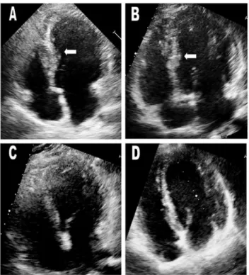
In order to confirm whether the Gly490Val mutation causes HCM as an autosomal recessive trait, family members underwent thorough clinical evaluations to detect the presence of HCM (Table 1). Only the two homozygotes exhibited a typical HCM phenotype, including inverted T waves on ECG (Figures 2A and 2B), hypertrophy of the mid to distal interventricular septum on echocardiography (Figures 3A and 3B). CMR showed hypertro-phy of the mid to distal interventricular septum and inferior ventricular wall in the proband, and isolated hypertrophic septum and inferior ventricular wall in his younger brother (Figures 4A and 4B). Both of the two homozygotes showed preserved cardiac function (left ventricular ejection fraction, 66% and 71%, respectively), normal atrial and ventricular chamber dimensions, no left ventricular outflow tract (LVOT) obstruction at rest and after exercise, and negative LGE.
All the heterozygous mutation carriers (I-2 and I-3, II-1 to II-3, III-4) showed no typical clinical manifestations of HCM (Table 1). Even the oldest heterozygous mutation carriers (I-2 and I-3), who were 71 and 62 years old, respectively, had no evidence of ECG abnormalities (Figures 2C and 2D) or left ventricular hypertrophy on echocardiogram (Figures 3C and 3D) and CMR. The proband’s parents (II-1 and II-2) and one of his paternal uncles (II-3) did not show any left ventricular hypertrophy nor LVOT obstruction even after exercise provocation, or any arrhythmias on 24-hour Holter. CMR of II-1 to 3 showed neither structural abnormalities nor cardiac fibrosis.
All family members without Gly490Val mutation had normal ECGs and echocardiograms (Table 1), except the maternal grandfather (I-1), who showed concentric hypertrophy on echo-cardiogram with 17 mm inter ventricular septum and 15 mm left ventricular posterior wall without abnormal T or Q waves or any arrhythmia. However, he had a history of uncontrolled hyperten-sion (170/100 mmHg at enrollment) for greater than 20 years. This type of left ventricular hypertrophy is a typical cardiac remodeling resulted from uncontrolled hypertension.
Discussion
Taking the advantage of second generation sequencing, we were able simultaneously to screen mutations in 26 known HCM
Autosomal Recessive Transmission Results in HCM
Table 1.Genotypes and clinical characteristics of all family members.
Echocardiogram CMR
Subject
Number Age (yr)
Mutation
Type Symptoms
Medical History
Blood Pressure
(mmHg) LVEDD(mm) IVS(mm) LVPW(mm) LVEF(%) LA Diameter
(mm) SAM
LVOT obstruction (mmHg)
ECG Findings (including
Holter) IVS(mm) LVPW(mm) Apex(mm) LGE
I-1 68 Wild Type No HT 20
years
170/100 43 17 15 64 36 No No Normal N.A.
I-2 71 Heterozygous No No 140/80 46 10 10 61 35 No No Normal 9 9 5 No
I-3 62 Heterozygous Chest pain
CAD 6 years
140/80 47 10 10 65 30 No No Normal 8 8 6 No
I-4 61 Wild Type No No 130/80 39 9 10 66 30 No No Normal N.A.
II-1 44 Heterozygous No No 120/75 46 9 8 62 32 No No Normal 8 5 No
II-2 42 Heterozygous No No 130/80 47 9 8 57 32 No No Normal 7 6 5 No
II-3 37 Heterozygous No No 140/100 52 11 10 67 34 No No Normal 8 7 5 No
II-4 29 Wild Type No No 120/86 52 10 10 64 36 No No Normal N.A.
III-2 21 Homozygous Chest pain
No 100/60 55 18 9 66 39 No No Diffuse repolarization changes with inverted T waves; premature ventricular contractions
17 10 8 No
III-3 19 Homozygous No No 100/60 46 15 9 71 34 No No Diffuse
repolarization changes with inverted T waves
13 9 12 No
III-4* 8 Heterozygous No No 95/60 38 5 6 67 38 No No Normal N.A.
III-5 4 Wild Type No No N.A. 31 6 6 68 21 No No Normal N.A.
LVEDD, left ventricular end-diastolic diameter; IVS, inter ventricular septum; LVPW, left ventricular posterior wall; LVEF, left ventricular ejection fraction; LA, left atrium; SAM, systolic anterior motion; LVOT, left ventricular outflow tract; CMR, cardiac magnetic resonance imaging; LGE, late enhancement of gadolinium; ECG, electrocardiographic; HT, hypertension; CAD, coronary artery disease; N.A., not applicable.
*LGE was not performed on III-4, because we considered that it was not necessary to perform an invasive examination at this young age. doi:10.1371/journal.pone.0067087.t001
Autosomal
Recessive
Transmissi
on
Results
in
ONE
|
www.ploson
e.org
3
June
2013
|
Volume
8
|
Issue
6
pathogenic genes, and identified a patient carrying a homozygous mutation in MYBPC3 without an apparent family history of clinical HCM. Autosomal recessive inheritance pattern of HCM due to this MYBPC3 mutation was supported by the following findings: (1) two clinically affected family members homozygous for the mutation were born to clinically unaffected parents; (2) the parents were consanguineous and heterozygous carriers of
MYBPC3 mutation; (3) all the adult family members who were
heterozygous for the mutation did not have a clinically apparent HCM phenotype, even into their 70 s; (4) the family members who harbored homozygous mutations expressed early-onset HCM. Therefore, in some patients with no apparent family history of HCM, an autosomal recessive pattern may be responsible for disease.
MYBPC3 is a crucial component of the sarcomere and an important regulator of muscle function. Among three different
Figure 1. Pedigree of the family with the mutation c.1469G.T (p.Gly490Val) inMYBPC3(A).Square, male; circle, female; empty, absent of
clinical findings; black, clinically affected; ‘‘w’’, wild-type allele; ‘m’, mutant allele; ?, no genetic testing performed; black arrow, proband. Protein sequence homology of mutation-affected regions among species (B), determined using Clustal W2. The Gly490Val substitution involves an amino acid that is highly conserved among species.
doi:10.1371/journal.pone.0067087.g001
Autosomal Recessive Transmission Results in HCM
MYBPC proteins, MYBPC3 is expressed exclusively in cardiac myocytes [18,19] and its HCM-causing mutations were first reported in 1995 [20]. Homozygous mutation in HCM was firstly reported by Ho CY et al in 2000. They described an homozygous Ser179Phe mutation inTNNT2gene which caused a severe form of HCM with striking morphological abnormalities and juvenile lethality [21]. From then on, more homozygous mutations were recognized either in case reports or in cohort studies. The inheritance traits were all autosomal dominant because the heterozygotes showed affirmatory but milder clinical evidence of HCM than the homozygotes [21–23]. In 2002, the first autosomal recessive transmission of HCM was reported in a family with Glu143Lys mutation in MYL2 gene. Abnormalities in echocar-diogram and ECG were only found in homozygous but not in heterozygous family members [24]. Recently Gray B et al reported a Arg162Trp mutation inTNNI3gene could also cause recessive HCM, but lack of clinical and genetic evaluation of old family members [25]. MYBPC3 is one of the most common disease-causing gene of HCM, accounting for 40–50% of known genetic causes of HCM patients, much higher than the frequency of mutation inMYL2(,3%) and TNNI3 (,6%). HCM caused by
MYBPC3mutations usually manifest lower penetrance, later onset
of disease and milder forms of disease progression in comparison to other gene mutations (i.e., MYH7) [26,27]. Patients with multiple mutations (i.e., compound or double heterozygotes) suffer
more severe phenotypes and increased risk of SCD [8,10,11,21]. Therefore, we postulated that some MYBPC3 mutations are functionally so mild that they do not lead to disease unless they are homozygous. In the present study, we screened theMYBPC3gene and identified a novel mutation which appeared autosomal recessive inheritance pattern, in a manner, supported the speculation above.
The present mutation (Gly490Val) we identified was a novel one in the domain C3 ofMYBPC3, with small change of side chain and kept the polarity neutral. Another mutation on the same amino acid residue (Gly490Arg) was reported to cause HCM in heterozygote in western population, substituting the small side chain for a bulky side chain and changing the polarity of amino acid residue into basic [8]. Therefore, the structural change of domain of MYBPC3 protein, which extended into the interfila-mental space in the motif binds to myosin S2 [28], due to mutation Gly490Arg much more prominent than that due to mutation Gly490Val. This might be the reason why these two kinds of mutations on the same position presented different inheritance patterns.
In the family described here, our documentation of inheritance of HCM as an autosomal recessive trait had clinical implications. The proband’s older brother had HCM in the absence of any other obvious heart diseases, and died of SCD at young age, suggesting that he was a highly suspicious homozygous mutation Figure 2. ECGs of proband (III-2) and his younger brother (III-3) (A&B), both of which show diffuse repolarization changes with
large negative T waves.ECGs of I-2, I-3 and II-1 to 3 (C to G), five heterozygous mutation carriers in the oldest generation, were normal. ECG of I-1
carrier. Therefore, the implantation of an implantable cardiover-ter defibrillator was recommended for the two surviving homo-zygotes in the family, the proband and his younger brother. Heterozygous family members were felt not to require long-term clinical follow-up.
Our results illustrate the complexity of genetic analysis for HCM. For example, ‘‘nonpathogenic’’ variants in HCM-related genes inherited from parents respectively may lead to HCM in the offspring as recessive mutations. Variants found in clinically unaffected individuals are often considered as benign polymor-phisms because almost HCM is most commonly inherited as an autosomal dominant trait. However, this strategy risks the missing of recessive disease-causing mutations. This may partly explain why disease-causing mutations were hard to be found in some
typical HCM patients and why more than half of the HCM patients do not have obvious family history.
In conclusion, our data identified a MYBPC3 mutation appeared to be an autosomal recessive transmission in HCM and suggest that the inheritance pattern may be more complex than previously thought. In clinical practice, the absence of a family history of clinical HCM may be due to not only a de novo mutation, but also recessive mutations that failed to produce a clinical phenotype in heterozygous family members. Therefore, consideration of recessive mutations leading to HCM is essential for risk stratification and genetic counseling.
Supporting Information
Table S1 The nonsynonymous variants found in the proband.
(XLS)
Acknowledgments
We are grateful to the family for their participation in the study.
Author Contributions
Conceived and designed the experiments: YW ZW JW RH LS. Performed the experiments: YW ZW QY Y.Zou HZ CY XF YC Y.Zhang JW XZ LS. Analyzed the data: YW JW FA RH LS. Contributed reagents/ materials/analysis tools: YW ZW QY Y.Zou HZ CY JW. Wrote the paper: YW JW FA RH LS.
References
1. Maron BJ (2002) Hypertrophic cardiomyopathy: a systematic review. JAMA 287(10): 1308–1320.
2. Bonne G, Carrier L, Richard P, Hainque B, Schwartz K (1998) Familial hypertrophic cardiomyopathy: from mutations to functional defects. Circ Res 83(6): 580–593.
3. Kimura A (2008) Molecular etiology and pathogenesis of hereditary cardiomy-opathy. Circ J 72 Suppl A(A38–48.
4. Ingles J, McGaughran J, Scuffham PA, Atherton J, Semsarian C (2012) A cost-effectiveness model of genetic testing for the evaluation of families with hypertrophic cardiomyopathy. Heart 98(8): 625–630.
5. Gersh BJ, Maron BJ, Bonow RO, Dearani JA, Fifer MA, et al. (2011) 2011 ACCF/AHA guideline for the diagnosis and treatment of hypertrophic cardiomyopathy: executive summary: a report of the American College of Cardiology Foundation/American Heart Association Task Force on Practice Guidelines. Circulation 124(24): 2761–2796.
6. Brito D, Richard P, Komajda M, Madeira H (2008) Familial and sporadic hypertrophic myopathy: differences and similarities in a genotyped population. A long follow-up study. Rev Port Cardiol 27(2): 147–173.
7. Brito D, Madeira H (2005) Malignant mutations in hypertrophic cardiomyop-athy: fact or fancy? Rev Port Cardiol 24(9): 1137–1146.
8. Van Driest SL, Vasile VC, Ommen SR, Will ML, Tajik AJ, et al. (2004) Myosin binding protein C mutations and compound heterozygosity in hypertrophic cardiomyopathy. J Am Coll Cardiol 44(9): 1903–1910.
9. Olivotto I, Girolami F, Ackerman MJ, Nistri S, Bos JM, et al. (2008) Myofilament protein gene mutation screening and outcome of patients with hypertrophic cardiomyopathy. Mayo Clin Proc 83(6): 630–638.
10. Ingles J, Doolan A, Chiu C, Seidman J, Seidman C, et al. (2005) Compound and double mutations in patients with hypertrophic cardiomyopathy: implications for genetic testing and counselling. J Med Genet 42(10): e59.
11. Maron BJ, Maron MS, Semsarian C (2012) Double or compound sarcomere mutations in hypertrophic cardiomyopathy: A potential link to sudden death in the absence of conventional risk factors. Heart Rhythm 9(1): 57–63. 12. Girolami F, Ho CY, Semsarian C, Baldi M, Will ML, et al. (2010) Clinical
features and outcome of hypertrophic cardiomyopathy associated with triple sarcomere protein gene mutations. J Am Coll Cardiol 55(14): 1444–1453. Figure 3. Echocardiograms of the proband (III-2) and his
younger brother (III-3) (A&B). White arrows indicate areas of
hypertrophy. Maximum wall thicknesses were 18 mm in the proband and 17 mm in his younger brother. Echocardiograms of heterozygous mutation carriers (I-2 and I-3) in the oldest generation were normal (C&D). White arrows indicate the interventricular septum.
doi:10.1371/journal.pone.0067087.g003
Figure 4. Cardiac magnetic resonance imaging (CMR) of the
proband (III-2) (A) and his younger brother (III-3) (B). White
arrows indicate areas of hypertrophy in the proband and asymmetri-cally hypertrophic and stiff ventricular wall in the younger brother (III-3). doi:10.1371/journal.pone.0067087.g004
Autosomal Recessive Transmission Results in HCM
13. Kubo T, Kitaoka H, Okawa M, Baba Y, Hirota T, et al. (2011)Genetic screening and double mutation in Japanese patients with hypertrophic cardiomyopathy. Circ J 75(11): 2654–2659.
14. Zou Y, Song L, Wang Z, Ma A, Liu T, et al. (2004) Prevalence of idiopathic hypertrophic cardiomyopathy in China: a population-based echocardiographic analysis of 8080 adults. Am J Med 116(1): 14–18.
15. Adzhubei IA, Schmidt S, Peshkin L, Ramensky VE, Gerasimova A, et al. (2010) A method and server for predicting damaging missense mutations. Nat Methods 7(4): 248–249.
16. Ng PC, Henikoff S (2001) Predicting deleterious amino acid substitutions. Genome Res 11(5): 863–874.
17. Larkin MA, Blackshields G, Brown NP, Chenna R, McGettigan PA, et al. (2007) Clustal W and Clustal X version 2.0. Bioinformatics 23(21): 2947–2948. 18. Furst DO, Vinkemeier U, Weber K (1992) Mammalian skeletal muscle
C-protein: purification from bovine muscle, binding to titin and the characteriza-tion of a full-length human cDNA. J Cell Sci 102 (Pt 4)(769–778.
19. Gautel M, Zuffardi O, Freiburg A, Labeit S (1995) Phosphorylation switches specific for the cardiac isoform of myosin binding protein-C: a modulator of cardiac contraction? EMBO J 14(9): 1952–1960.
20. Bonne G, Carrier L, Bercovici J, Cruaud C, Richard P, et al. (1995) Cardiac myosin binding protein-C gene splice acceptor site mutation is associated with familial hypertrophic cardiomyopathy. Nat Genet 11(4): 438–440.
21. Ho CY, Lever HM, DeSanctis R, Farver CF, Seidman JG, et al. (2000) Homozygous mutation in cardiac troponin T: implications for hypertrophic cardiomyopathy. Circulation 102(16): 1950–1955.
22. Ortiz MF, Rodriguez-Garcia MI, Hermida-Prieto M, Fernandez X, Veira E, et al. (2009) A homozygous MYBPC3 gene mutation associated with a severe phenotype and a high risk of sudden death in a family with hypertrophic cardiomyopathy. Rev Esp Cardiol 62(5): 572–575.
23. Zahka K, Kalidas K, Simpson MA, Cross H, Keller BB, et al. (2008) Homozygous mutation of MYBPC3 associated with severe infantile hypertro-phic cardiomyopathy at high frequency among the Amish. Heart 94(10): 1326– 1330.
24. Olson TM, Karst ML, Whitby FG, Driscoll DJ (2002) Myosin light chain mutation causes autosomal recessive cardiomyopathy with mid-cavitary hypertrophy and restrictive physiology. Circulation 105(20): 2337–2340. 25. Gray B, Yeates L, Medi C, Ingles J, Semsarian C (2012) Homozygous mutation
in the cardiac troponin I gene: Clinical heterogeneity in hypertrophic cardiomyopathy. Int J Cardiol.
26. Niimura H, Bachinski LL, Sangwatanaroj S, Watkins H, Chudley AE, et al. (1998) Mutations in the gene for cardiac myosin-binding protein C and late-onset familial hypertrophic cardiomyopathy. N Engl J Med 338(18): 1248–1257. 27. Wang S, Zou Y, Fu C, Xu X, Wang J, et al. (2008) Worse prognosis with gene mutations of beta-myosin heavy chain than myosin-binding protein C in Chinese patients with hypertrophic cardiomyopathy. Clin Cardiol 31(3): 114– 118.