Short Report
S
J. Braz. Chem. Soc., Vol. 23, No. 2, 349-354, 2012.Printed in Brazil - ©2012 Sociedade Brasileira de Química 0103 - 5053 $6.00+0.00
*e-mail: [email protected], [email protected].
Lycopodium
Alkaloids from
Palhinhaea cernua
Fu-Wei Zhao,a,b Qian-Yun Sun,c Fu-Mei Yang,c Ji-Feng Luo,a Guang-Wan Hu,a
Fang Liu,d Yue-Hu Wang*,a and Chun-Lin Long*,a,e
aKey Laboratory of Economic Plants and Biotechnology, Kunming Institute of Botany, Chinese Academy of Sciences, Kunming 650201, People’s Republic of China
bGraduate University of Chinese Academy of Sciences, Beijing 100049, People’s Republic of China
cKey Laboratory of Chemistry for Natural Products, Guizhou Province and Chinese Academy of Sciences, Guiyang 550002, People’s Republic of China
dCollege of Landscape and Horticulture, Yunnan Agricultural University, Kunming 650201, People’s Republic of China
eCollege of Life and Environmental Sciences, Minzu University of China, Beijing 100081, People’s Republic of China
Dois novos alcaloides tipo-licopódio, acetil-licoposerramina M e palcernina A foram isolados de extratos de plantas inteiras de Palhinhaea cernua L. juntamente com dez compostos
previamente identificados. As estruturas dos dois novos compostos foram elucidadas por métodos espectroscópicos e difratometria de Raios X de monocristal usando o parâmetro Flack.
Two new Lycopodium alkaloids, acetyllycoposerramine M and palcernine A were isolated from
whole plant extracts of Palhinhaea cernua L. together with ten previously identified compounds.
The structures of the new compounds were elucidated by spectroscopic methods and single-crystal X-ray diffraction analyses using the Flack parameter.
Keywords: Lycopodiaceae, Palhinhaea cernua, Lycopodium alkaloids
Introduction
Palhinhaea cernua L. (syn. Lycopodium cernuum (L.); Lepidotis cernua (L.) P. Beauv.; Lycopodiella cernua (L.) Pic.
Ser.), found in both the Flora of North America and the Flora of China,1 is a member of the Lycopodiaceae family. The
species is traditionally used to heal contusions, scalding and rheumatism by the Chinese people and is broadly distributed in Eastern and Southern China.2 The chemical constituents
of the genus Lycopodium (sensu lato) plants are mainly Lycopodium alkaloids which are quinolizine or pyridine and
α-pyridone type alkaloids.3-10 Some of these alkaloids inhibit
acetylcholinesterase (AChE) and provide a challenge for total synthesis.9-10 In our previous research on this plant,
palhinine A, a novel C16N-type alkaloid was isolated.9 We
now report the isolation and structural elucidation of two new Lycopodium alkaloids (1-2) along with ten previously
identified compounds (Figure 1), namely, lycoflexine
N-oxide (3)8, lycoflexine (4),11 lycodine (5),12α-obscurine
(6),12 dehydroisofawcettiine (7),13,14 lycopodine (8),14
5-acetyllycofoline (9),15 acetylfawcettiine (10),16,17 cernuine
(11)18 and lycocernuine (12).18
Results and Discussion
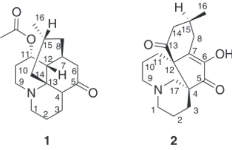
Acetyllycoposerramine M(1) was obtained as a colorless
columnar crystal (mp 197-198 °C). A molecular formula (C18H27NO3) was assigned by HREIMS (electrospray
N O
O O
1 2
H
1 4 2 3
5 6 7 15
14 8
9 11 10
12
13 16
H
N O
OH
1 2
3 4 5
6 7
8
9 101112
13 1415
16
17
H
O
ionization mass spectrometric) analysis (observed [M]+
at m/z 305.1995, calcd. [M]+: 305.1991), implying six
degrees of unsaturation. The IR (infrared) spectrum showed absorptions for ester (1695 cm−1) and keto (1733 cm−1)
carbonyl groups. One singlet (dH 2.09, 3H) and one doublet
(dH 0.88, 3H, J 6.3 Hz, H3-16) for two methyl groups
were observed in 1H NMR (nuclear magnetic resonance)
spectrum of 1 (Table 1). Meanwhile, signals of 18 carbons
were recorded in the 13C NMR and DEPT (distortionless
enhancement by polarization) spectra (Table 2), including the presence of one ester carbonyl (dC 170.3), one keto
carbonyl (dC 214.4, C-5), one sp3 quaternary, five sp3
methine, eight sp3 methylene and two methyl groups.
Specifically, the signals of an oxygenated methine group were recorded at dH 5.23 (ddd, J 2.9, 2.9, 2.9 Hz, H-11) and
dC 72.3 (C-11). The above mentioned analyses suggested
that 1 was a lyopodine alkaloid, and shared a similar
planar structure with the known Lycopodium alkaloid,
lycoposerramine M.19 Differentiations between the two
compounds were observed in that the additive signals (dH
2.09, 3H, s; dC 22.0 and 170.3) for an acetyl group appeared
in the 1H and 13C NMR spectra of 1, and the presence of
a chemical shift of H-11 in 1 was downshifted compared
with that of the known compound (dH 4.22).19 Therefore, it
could be speculated that 1 was the acetylated derivative of
lycoposerramine M. This was confirmed by the presence of the cross peak between H-11 and ester carbon in the HMBC (heteronuclear multiple bond correlation) spectrum of 1 (Figure 2).
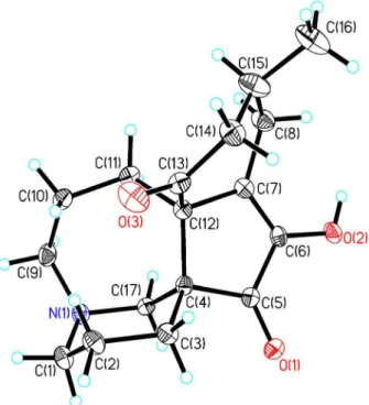
To elucidate the absolute configuration, the crystals of 1 were isolated from a mixed solution of petroleum
ether/Me2CO (9:1), and single-crystal X-ray crystallographic
analysis was performed (Figure 3). The results of the analysis revealed the absolute configuration of 1 to be 4R,
7R, 11R, 12R, 15R in light of the Flack parameter of 0.0(2),
using anomalous dispersion with copper radiation.20-22
Finally, 1 was elucidated as acetyllycoposerramine M.
Palcernine A (2) was obtained as a colorless block
crystal (mp 98-101 °C). Its molecular formula (C17H24NO3)
Table 1.1H NMR data of compounds 1-2 (d in ppm, J in Hz)
No. 1a 2b
1a 3.42 (dd, 14.2, 3.4) 3.09 (m)
1b 2.64 (m) 2.93 (m)
2a 1.89 (m) 2.51 (dd, 14.8, 4.0)
2b 1.51 (m) 1.34 (d, 13.4)
3a 2.13 (m) 2.29 (m)
3b 1.59 (ddd, 13.6, 13.6, 4.4) 1.99 (dd, 14.8, 9.4) 4 3.24 (dd, 12.0, 2.9)
6a 2.76 (dd, 16.6, 6.7)
6b 2.23 (d, 16.6)
7 2.33 (s)
8a 1.69 (br. d, 13.0) 2.90 (m; β)
8b 1.35 (ddd, 12.7, 12.7, 3.4) 2.66 (dd, 15.1, 8.0; α)
9a 3.37 (m) 2.99 (m)
9b 2.61 (m) 2.82 (m)
10a 2.10 (m) 2.15 (m)
10b 1.98 (m) 1.41 (m)
11a 5.23 (ddd, 2.9, 2.9, 2.9) 2.28 (m; α)
11b 1.65 (dd, 14.7, 11.8; β)
12 1.85 (m)
14a 2.66 (m) 2.49 (dd, 14.8, 4.0; α)
14b 1.09 (dd, 13.1,13.1) 1.97 (dd, 14.8, 9.4; β)
15 1.54 (m) 2.19 (m)
16 0.88 (3H, d, 6.3) 0.90 (3H, d, 6.7)
17a 3.10 (m)
17b 2.99 (m)
OCCH3 2.09 (3H, s)
6-OH 12.19 (br. s)
aRecorded at 500 MHz in CDCl
3; brecorded at 500 MHz in C5D5N.
Table 2.13C NMR data of compounds 1-2 (d in ppm)
No. 1a 2b
1 47.2 t 52.7 t
2 19.4 t 21.1 t
3 20.0 t 29.3 t
4 45.4 d 54.0 s
5 214.4 s 203.8 s
6 44.4 t 149.1 s c
7 35.1 d 145.0 s
8 44.3 t 28.4 t
9 42.1 t 57.7 t
10 31.0 t 26.1 t
11 72.3 d 34.0 t
12 46.8 d 61.7 s
13 58.9 s 210.0 s
14 43.8 t 48.9 t
15 25.5 d 27.4 d
16 22.8 q 21.7 q
17 57.8 t
OCCH3 170.3 s
OCCH3 22.0 q
aRecorded at 125 MHz in CDCl
3; brecorded at 125 MHz in C5D5N; cdetected by HMBC.
N O
O O
1H-1H COSY
HMBC
N O
O O
H
1 4 2 3
5 6 7 15
14 8
9 11 10
12
13 16
H
was established on the basis of HRESIMS for the [M + H]+
ion at m/z 290.1754 (calcd. 290.1756), implying seven
degrees of unsaturation. The IR spectrum showed the presence of hydroxy (3441 cm−1), ketone (1709 cm−1) and
α,β-unsaturated ketone (1631 cm−1) groups. A signal
dH 0.90 (d, 3H, J 6.7 Hz) for a methyl group was observed
in the 1H NMR spectrum of 2 (Table 1), and 17 carbon
signals for two ketone, one tetrasubstituted double bond, two sp3 quaternary, one methine, nine methylene and one
methyl groups were recorded in the 13C NMR and DEPT
of 2 (Table 2). Three fragments, a (C-1 to C-3), b (C-9
to C-12) and c (C-8–C-15–C-16) were elucidated by the
1H-1H COSY (correlation spectroscopy) experiments
(Figure 4). Based on the HMBC correlations (Figure 4) of H-2a/C-4, H-17a/C-2 & C-3, H-17a & H-3a/C-5, H-10b/C-12, H-11β/C-7 & C-13, H-8α/C-6, C-7 & C-12, H-15/C-13, and H3-16/C-14 & C-13, a fawcettimine
skeleton of 2 was determined, and the double bond was
Figure 3. ORTEP drawing of the asymmetric unit of compound 1.
Figure 4. Key 2D NMR correlations of 2.
located at C-6, and two ketone groups were located at C-5 and C-13, respectively. After analysis of the molecular formula of 2 the presence of a hydroxy functionality in 2
was confirmed, possibly being located at C-6.
The relative configuration of partial chiral center of 2
could be elucidated, integrating ROESY (rotating-frame Overhauser effect spectroscopy) experiment and analysis on coupling constant. In ROESY spectrum (Figure 4), the cross peaks of H-8α (dH 2.66, dd, J 15.1, 8.0 Hz)/H-11β (dH 1.65,
dd, J 14.7, 11.8 Hz), H-8α/H-14α (dH 2.49, dd, J 14.8,
4.0 Hz) and H-11β/H-14α were observed. H-14α and H-15 should be in cis configuration due to the coupling
constant of JH-14α/H-15 4.0 Hz (Table 1). Therefore, the
16-CH3 in 2 was β-oriented, which is rare because the 16-CH3
is α-oriented in most of the Lycopodium alkaloids.3-5 Based
on the single-crystal X-ray crystallographic analysis (Figure 5), the absolute configuration of 2 was determined
to be 4S, 12S, 15S in light of the Flack parameter of 0.1(2),
using anomalous dispersion with copper radiation.20-22
Accordingly, 2 was elucidated as shown in Figure 2 and
named palcernine A.
Compounds 1-3 were tested for their inhibition activities
against acetylcholinesterase, butyrylcholinesterase and human chronic myelogenous leukemia K562 cells, respectively by the improved Ellman’s method,23-27 and the
MTT method.28 However, using these methods no inhibitory
activities for the compounds were detected.
Conclusions
Two new Lycopodium alkaloids, acetyllycoposerramine M
and palcernine A were isolated from whole plant extracts of Palhinhaea cernua L. together with ten
previously identified compounds. The structures of the new compounds were elucidated by spectroscopic methods and single-crystal X-ray diffraction analyses using the Flack parameter. The two new Lycopodium
alkaloids along with lycoflexine N-oxide were tested for
their inhibition activities against acetylcholinesterase, butyrylcholinesterase and human chronic myelogenous leukemia K562 cells, respectively by the improved Ellman’s method, and the MTT method. However, using these methods, none of the inhibitive activities of the compounds were detected.
Experimental
General experimental procedures
Melting points (mp) were determined using an X-4 melting point apparatus (Yingyu Yuhua Apparatus Factory, Gongyi, P. R. China), and was not adjusted. Bruker SMART APEX-II and Bruker APEX DUO diffractometers using graphite-monochromated Cu Kα radiations were employed for the intensity data collection, and the structures of compounds were solved by direct methods (SHELXS97). Optical rotations were determined on a JASCO DIP-370 automatic digital polarimeter. UV spectra were recorded on a Shimadzu double-beam 210A spectrometer. IR spectra were recorded on a Bio-Rad FTS-135 infrared spectrophotometer. 1D and 2D NMR spectra were recorded on Bruker AM-400, DRX-500 and Bruker Avance 600 spectrometers with chemical shifts in ppm relative to TMS (tetramethylsilane) as internal standard. ESIMS and HRESIMS were measured using an API QSTAR Pulsar 1 spectrometer, as well as EIMS and HREIMS using a VG Autospec Premier spectrometer. Column chromatography was performed over Al2O3 (200-300 mesh, Wusi Lt. Co., Shanghai),
silica gel G (80-100 and 300-400 mesh), silica gel H (10-40 µm) and Sephadex LH-20 (40-70 µm, Amersham Pharmacia Biotech AB, Uppsala, Sweden). HPLC (high-performance liquid chromatographic) separations were performed using an Agilent 1200 series pump equipped with a diode array detector and a semi-preparative Zorbax SB-C18 (5 µm, φ 9.4 × 250 mm) column. TLC (thin layer
chromatography) was conducted on precoated GF254 silica
gel plates (Qingdao).
Plant material
Whole plant samples of P. cernua, a nodding
club-moss was collected from Liping County of Guizhou Province, P. R. China and identified by Dr. Guang-Wan Hu at the Kunming Institute of Botany, Chinese Academy of Sciences. A voucher specimen (No. SZ09048) was deposited in the Key Laboratory of Economic Plants and Biotechnology, Kunming Institute of Botany.
Extraction and isolation
The air-dried and powdered sample (16.5 kg) was three times extracted with MeOH (4, 4, and 3 h) at 70 °C. The extracts were partitioned between EtOAc and 1% HCl/H2O.
Water-soluble materials were adjusted to pH 10 with 17% NaOH/H2O solution and extracted with CHCl3, producing
an alkaloid-rich extract (82.0 g). The latter was submitted to a silica gel column (CHCl3/MeOH, 1:0 to 0:1) to produce
four fractions: Fr. 1-Fr. 4.
Fr. 1 was partitioned over C18 column eluting with
MeOH/H2O (20% to 100%, v/v). The fraction by 50%
MeOH/H2O was chromatographed over Sephadex LH-20
(MeOH), Al2O3 [petroleum ether/Me2CO/NH3•H2O, 80:10:1
(v/v/v)] and silica gel [petroleum ether/Me2CO/NH3•H2O,
80:10:1 (v/v/v)] columns to obtain two sub-fractions: Fr. 1-50-1 and Fr. 1-50-2. Fr. 1-50-1 was deposited at room temperature, and compound 1 (246.2 mg) was
crystallized out [petroleum ether/Me2CO, 9:1 (v/v)].
The mother solution was evaporated and the residue was subjected to silica gel with petroleum ether/Me2CO
(8:1, v/v) to yield compound 7 (26.5 mg). Fr. 1-50-2 was
submitted to Al2O3 and silica gel columns and preparative
TLC [petroleum ether/Me2CO/NH3•H2O, 80:10:1 (v/v/v)]
to yield compound 3 (11.0 mg). The fraction by 60%
MeOH/H2O was purified by Sephadex LH-20 (MeOH) and
preparative TLC [petroleum ether/Me2CO/NH3•H2O,
80:10:1 (v/v/v)] to yield compound 5 (14.1 mg).
Fr. 2 was chromatographed over C18 column eluting
with MeOH/H2O (30% to 100%, v/v), and the fraction
by 50% MeOH/H2O was subjected to Sephadex LH-20
(MeOH) and silica gel [petroleum ether/(C2H5)2NH,
100:1 (v/v)] columns to give compounds 4 (98.6 mg) and 8 (16.5 mg).
Fr. 3 was chromatographed over C18 column eluting
with MeOH/H2O (30% to 100%, v/v). The fraction
by 50% MeOH/H2O was separated into two
sub-fractions (Fr. 3-50-1 and Fr. 3-50-2). Fr. 3-50-1 was successively subjected to Sephadex LH-20 (MeOH) and Al2O3 [CHCl3/MeOH, 30:1 (v/v)] columns to afford
Sephadex LH-20 (MeOH) and silica gel [CHCl3/MeOH/
NH3•H2O, 300:20:1 (v/v/v)] columns to give compound 9
(26.4 mg). The fraction by 60% MeOH/H2O was subjected
Sephadex LH-20 (MeOH) and silica gel [CHCl3/MeOH,
30:1 (v/v/v)] columns to obtain compounds 6 (138.6 mg) and 10 (407.3 mg).
Fr. 4 was chromatographed over C18 column eluting
with MeOH/H2O (30% to 100%, v/v). The fraction
by 40% MeOH/H2O was purified by Sephadex LH-20
(MeOH), and compound 12 (7.4 mg) was obtained in light
of recrystallization [petroleum ether/MeOH, 9:1 (v/v)]. The fraction by 50% MeOH/H2O was separated on Sephadex
LH-20 (MeOH) and silica gel [CHCl3/MeOH, 20:1 (v/v)]
columns to yield compound 11 (16.9 mg).
Acetyllycoposerramine M (1)
Colorless columnar crystal; mp. 197-198 °C (from petroleum ether/Me2CO, 9:1); [α]D23.6 + 45.9 (c 0.35,
CHCl3); UV (CHCl3) λmax/nm (log e) 241 (2.94); IR (KBr)
vmax/cm−1 1733, 1695, 1235 and 1196; 1H and 13C NMR
data, see in Table 1 and Table 2; EIMS m/z (%) 305 (M+,
83), 262 (38), 246 (95); HREIMS m/z (%) 305.1995 (M+,
C18H27NO3+, calcd. 305.1991).
Crystal data for 1
C18H27NO3, MW = 305.41; colorless columnar, size
0.58 × 0.42 × 0.36 mm3, orthorhombic, space group P21 21 21; a = 8.43070 (10) Å, b = 10.9756 (2) Å, c = 17.8783 (3) Å, a = b = γ = 90.00°, V = 1655.31 (5) Å3, T = 296(2) K, Z = 4,
rcalcd = 1.226 Mg m−3, µ(Cu Kα) = 0.7 mm−1, F(000) = 664, 10967 reflections in h(−10/10), k(−13/12), l(−21/21), measured in the range 4.73° ≤q≤ 66.50°, completeness qmax = 99.1%, 2843 independent reflections, Rint = 0.0386,
2878 reflections with |F|2 ≥ 2s |F|2, 202 parameters,
0 restraints, GOF = 1.116. Final R index: R1 = 0.0424,
wR2 = 0.1089. R index (all data): R1 = 0.0425, wR2 = 0.1092.
Flack parameter 0.0(2), largest difference peak and hole = 0.222 and −0.277 e Å−3. The intensity data for 1 were
collected on a Bruker APEX DUO diffractometer with a graphite monochromater, Cu Kα radiation. The structure of 1 was solved by direct methods (SHELXS97), expanded
using difference Fourier techniques, and refined by the program and full-matrix least-squares calculations. The non-hydrogen atoms were anisotropically refined, and hydrogen atoms were fixed at calculated positions. Crystallographic data for the structure of 1 were
deposited in the Cambridge Crystallographic Data Center (deposition No. CCDC 817948). Copies of the data can be obtained free of charge from the CCDC via www.ccdc.cam.ac.uk.
Palcernine A (2)
Colorless block crystal; mp. 98-101 °C (from Me2CO/MeOH, 5:1); [α]D21.7 – 21.0 (c 0.06, MeOH);
UV (MeOH) λmax/nm (log e) 270 (3.12), 205 (3.44); IR (KBr) vmax/cm−1 3441, 1709, 1631, 1384 and 1102; 1H and 13C NMR data, see in Table 1 and Table 2; ESIMS m/z 290
[M + H]+, 601 [2M + Na]+, 890 [3M + Na]+; HRESIMS m/z 290.1754 [M + H]+ (C17H24NO3, calcd. 290.1756).
Crystal data for 2
C17H23NO3, MW = 289.36; colorless blocks, size
0.22 × 0.12 × 0.08 mm3, orthorhombic, space group P21 21 21; a = 6.35940 (10) Å, b = 13.3290 (2) Å, c = 17.5422 (3) Å, a = b = γ = 90.00°, V = 1486.96 4) Å3, T = 296(2) K, Z = 4, rcalcd = 1.293 Mg m−3,
µ(Cu Kα) = 0.7 mm−1, F(000) = 624, 2774 reflections in h(-6/6), k(−14/13), l(−19/15), measured in the range 4.17° ≤ q ≤ 64.73°, completeness qmax = 71.1%, 1617
independent reflections, Rint = 0.0131, 1597 reflections with
|F|2≥ 2s |F|2, 190 parameters, 0 restraints, GOF = 1.075.
Final R index: R1 = 0.0395, wR2 = 0.1191. R index (all
data): R1 = 0.0400, wR2 = 0.1203. Flack parameter 0.1(2),
largest difference peak and hole = 0.318 and −0.201 e Å−3.
The intensity data for 2 were collected on a Bruker SMART
APEX-II diffractometer with a graphite monochromater using Cu Kα radiation. The structure of 2 was solved by direct methods (SHELXS97), expanded using difference Fourier techniques, and refined by the program and full-matrix least-squares calculations. The non-hydrogen atoms were refined anisotropically, and hydrogen atoms were fixed at calculated positions. Crystallographic data for the structure of 2 were deposited in the Cambridge
Crystallographic Data Center (deposition No. CCDC 831841). Copies of the data can be obtained free of charge from the CCDC via www.ccdc.cam.ac.uk.
Bioactivity test in vitro
Acetylcholinesterase and tacrine were purchased from Sigma-Aldrich Corporation, S-butyrylthiocholine
iodide and 5,5-dithiobis-2-nitrobenzoic acid (DTNB) were purchased from Fluka Chemie GmbH and Acros Organics, respectively. Inhibitory activity of all new compounds against acetylcholinesterase was performed by the improved Ellman’s method, and tacrine was used as positive (IC50 = 0.20 µM).
DTNB were purchased from Acros Organics. Inhibitory activity of all new compounds against butyrylcholinesterase was performed by the improved Ellman’s method, and tetraisopropylpyrophosphoramide was treated as positive (IC50 = 1.35 µM).
K562 human chronic myelogenous leukemia cell line was purchased from the China Center for Type Culture Collection, Hubei Province, P. R. China. K562 cells were maintained at 37 °C in RPMI-1640 medium (Hyclone) containing 10% fetal bovine serum (Hangzhou Sijiqing Biological Engineering Materials Co., Ltd., P. R. China) in an atmosphere of humidified 5% CO2. The cytotoxic assay
was carried out in quintuplicate in 96-well microplates (Corning), and the amount of viable cells at the end of the incubation period was determined by using MTT [3-(4,5-dimethylthiazol-2-yl)-2,5-diphenyltetrazolium bromide] assay. K562 cells were exposed for 24 h to the solution of all new compounds. Non-treated culture cells were used as a negative control. IC50 was calculated by
the GWBASIC software as the concentration (µM) of the compounds causing a 50% inhibition of cell viability. Adriamycin was treated as positive control (IC50 = 0.96 µM).
Supplementary Information
Supplementary information is available free of charge at http://jbcs.sbq.org.br, as PDF file.
Acknowledgements
This work was sponsored by the National Natural Science Foundation of China (No. 20972166), the National Basic Research Program of China (973 Program) (No. 2009CB 526512), the Ministry of Education of China through its 111 and 985 Projects (B08044 and MUC 985-9), and the Knowledge Innovation Project of the Chinese Academy of Sciences. We appreciate Dr. Jia-Liang Zhong (at the Shanghai Institute of Pharmaceutical Industry) and Xiao-Nian Li (at the Kunming Institute of Botany) who measured and elucidated the crystal structure. We are also grateful to Adam Nerin (Ph.D. candidate at the City University of New York, USA) and Jeffrey R. Boutain (Ph.D. candidate at the University of Hawaii at Manoa, USA) for editing the English.
References
1. Zhang, X. C.; Zhang, L. B.; Flora Reipublicae Popularis Sinicae; Science Press: Beijing, China, 2004, ch. 6(3). 2. Chinese Herbs Edition Board; Chinese Herbs, Shanghai
Science and Technology Press: Shanghai, China, 2005, ch. 2.
3. Hirasawa, Y.; Kobayashi, J.; Morita, H.; Heterocycles 2009, 77, 679.
4. Ayer, W. A.; Nat. Prod. Rep. 1991, 8, 455.
5. Ma, X.; Gang, D. R.; Nat. Prod. Rep. 2004, 21, 752.
6. Ishiuchi, K.i.; Kubota, T.; Ishiyama, H.; Hayashi, S.; Shibata, T.; Kobayashi, J.; Tetrahedron Lett.2011, 52, 289.
7. Ishiuchi, K.i.; Kubota, T.; Ishiyama, H.; Hayashi, S.; Shibata, T.; Mori, K.; Obara, Y.; Nakahata, N.; Kobayashi, J.; Bioorg. Med. Chem.2011, 19, 749.
8. Katakawa, K.; Mito, H.; Kogure, N.; Kitajima, M.; Wongseripipatana, S.; Arisawa, M.; Takayama, H.; Tetrahedron
2011, 67, 6561.
9. Zhao, F. W.; Sun, Q. Y.; Yang, F. M.; Hu, G. W.; Luo, J. F.; Tang, G. H.; Wang, Y. H.; Long, C. L.; Org. Lett. 2010, 12, 3922.
10. Kitajima, M.; Takayama, H.; Topics in Current Chemistry; Springer-Verlag: Heidelberg, 2011, vol. 1.
11. Takayama, H.; Katakawa, K.; Kitajima, M.; Yamaguchi, K.; Aimi, N.; Tetrahedron Lett. 2002, 43, 8307.
12. Nakashima, T. T.; Singer, P. P.; Browne, L. M.; Ayer, W. A.;
Can. J. Chem. 1975, 53, 1936.
13. Burnell, R. H.; Taylor, D. R.; Tetrahedron 1961, 15, 173. 14. MacLean, D. B.; Can. J. Chem. 1963, 41, 2654.
15. Anet, F. A. L.; Ahmad, M.; Khan, N. H.; Can. J. Chem. 1962,
40, 236.
16. Burnell, R. H.; Mootoo, B. S.; Taylor, D. R.; Can. J. Chem.
1960, 38, 1927.
17. Laemmerhold, K. M.; Breit, H.; Angew. Chem., Int. Ed. 2010,
49, 2367.
18. Braekman, J. C.; Nyembo, L.; Bourdoux, P.; Kahindo, N.; Hootele, C.; Phytochemistry 1974, 13, 2519.
19. Takayama, H.; Katakawa, K.; Kitajima, M.; Yamaguchi, K.; Aimi, N.; Chem. Pharm. Bull. 2003, 51, 1163.
20. Flack, H.; Acta Crystallogr., Sect. A: Found. Crystallogr.1983, 39, 876.
21. Flack, H. D.; Bernardinelli, G.; J. Appl. Crystallogr. 2000, 33, 1143.
22. Sheldrick, G. M; SHELXS-97, Program for Crystal Structure Solution; University of Gottingen: Gottingen, Germany, 1997.
23. Ellman, G. L.; Courtney, K. D.; Andres, V. J.; Featherstone, R. M.;
Biochem. Pharmacol. 1961, 7, 88.
24. Rhee, I. K.; Appels, N.; Hofte, B.; Karabatak, B.; Erkelens, C.; Stark, L. M.; Flippin, L. A.; Verpoorte, R.; Biol. Pharm. Bull.
2004, 27, 1804.
25. Sun, Q. Y.; Yang, F. M.; Chin. Pharm. Bull. 2008, 24, 1387.
26. Vogel, H. G.; Vogel, W. H.; Drug Discovery and Evaluation: Pharmacological Assays; Springer: New York, 2008.
27. Yang, F. M.; Sun, Q. Y.; Chin. Pharm. Bull. 2009, 25, 690. 28. Mosmann, T.; J. Immunol. Methods 1983, 65, 55.