Recebido em 16.03.2000. / Received in March, 16thof 2000.
Aprovado pelo Conselho Consultivo e aceito para publicação em 22.08.2002. / Approved by the Consultive Council and accepted for publication in August, 22thof 2002. * Trabalho realizado para defesa de tese de Mestrado em Anatomia Patológica da Universidade Federal de Pernambuco / Work done for Masters thesis in Anatomic Pathology, Federal University of Pernambuco.
1 Prof. Adjunto de Dermatologia da Universidade Federal de Pernambuco ( UFPE) e Mestra em Anatomia Patológica da UFPE / Adjunct Prof. of Dermatology, Federal University of Pernambuco (UFPE) and Masters degree in Anatomic Pathology, UFPE
2 Prof. Adjunto de Anatomia Patológica da UFPE. e Universidade de Pernambuco / Adjunct Prof. of Anatomic Pathology, UFPE and Pernambuco University 3 Prof. Titular de Patologia Geral da UFPE / Titular Prof. of General Pathology UFPE
4 Prof. Assistente do Departamento de Estatística da UFPE / Assistant Prof., Dept. of Statistics, UFPE
©2003 by Anais Brasileiros de Dermatologia
Carcinomas basocelulares: estudo clínico
e anatomopatológico de 704 tumores
*Basal cell carcinomas: anatomopathological and
clinical study of 704 tumors
*Aurilene Monteiro Bandeira
1Valdir Bandeira
2José Figueredo da Silva
3Edmilson Mazza
4Resumo: FUNDAMENTOS- Este é um estudo retrospectivo e anatomopatológico de 704 carcinomas basocelulares de 623 pacientes,
diagnos-ticados no período de 1991 a 1996, no setor de Dermatopatologia da Clínica Dermatológica do Hospital das Clínicas da UFPE e em um laboratório privado de dermatopatologia da cidade do Recife.
OBJETIVOS- Caracterizar aspectos clínicos e anatomopatológicos dos carcinomas basocelulares diagnosticados nos dois serviços da região de Pernambuco.
MÉTODOS- Para o estudo clínico, os dados foram retirados dos prontuários e para o estudo anatomopatológico, macro e microscópicos,
foram feitas revisão dos preparados histológicos. Para o crescimento vertical empregaram-se métodos baseados nas técnicas histoprognós-ticas de Clark e Breslow, aplicadas ao melanoma maligno.
RESULTADOS- Clínicos: a maior incidência foi no sexo feminino (55,7%), faixa etária de 55 a 72 anos, tempo de evolução com variabilidade eleva-díssima, desde um mês a 40 anos, e a cabeça foi a região topográfica mais freqüente (73,8%), principalmente nasal (21,1%) e zigomática (18,5%). A forma nodular pigmentada (47,4%) foi a mais encontrada e o tamanho das lesões independeu do tempo evolutivo. Histologicamente os padrões considerados baseados apenas nos arranjos parenquimais, foram os adenóide, compacto, fibroepitelial de Pinkus plexiforme, pseudocístico, reti-culado, superficial e tricoepitelial, predominando o adenóide (28,3%). A média de crescimento foi em 2/3 da derme reticular (32,4%), e os tumo-res que mais se aprofundaram mostraram fibroplasia intensa. Houve concomitância de vários tipos celulatumo-res em um mesmo tumor e o pigmen-to melânico esteve mais presente nos tricoepitelioides.
CONCLUSÃO- A fundamental importância da caracterização clínica e anatomopatológica dos carcinomas basocelulares destes serviços, sem
diferenças muito significativas para os grupos e atenção para definições comportamentais e proposições ao relatório histopatológico. Palavras-chave: carcinoma basocelular.
Sum m a ry: BACKGROUND- A retrospective and anatom opathological study was perform ed on 704 basal carcinom as of 623 patients, diag-nosed from 1991 to 1996, at the Derm atopathology section of the Derm atology Clinic of the Hospital das Clinicas, UFPE and at a priva-te derm atopathology laboratory in the city of Recife.
OBJECTIVE- To characterize the clinical and anatom opathological aspects of the basal cell carcinom as diagnosed by the two services of
Pernam buco region.
METHODS- For the clinical study, the data were collected from the patient files and for the anatom opathological, m acro and m icroscopic study a revision was m ade of the histological specim ens. For determ ination of vertical growth, m ethods were used based on Clark and Breslow's histoprognostic techniques applied to m alignant m elanom a.
RESULTS- Clinical: the highest incidence was in the fem inine sex (55.7%) and in the 55 to 72-year-old age group. Disease duration was highly
variable, ranging from one m onth to 40 years, and the head was the m ost frequent topographical area (73.8%), m ainly nasal (21.1%) and zygom atic (18.5%). The nodular pigm ented form (47.4%) was found m ost frequently and the size of the lesions did not depend on the disease duration. Histologically the patterns considered based only on the parenchym al arrangem ents, were the adenoid, com pact, ple-xiform Pinkus fibroepitheliom a, pseudocystic, reticulated, superficial and trichoepithelioid, though predom inantly the adenoid form (28.3%). The m ean growth involved 2/3 of the reticular derm is (32.4%), and the deepest tum ors presented intense fibroplasia. There was concom itance of several cellular types within a single tum or and m elanin pigm ent was found m ostly in the trichoepithelioid type. CONCLUSION- The clinical and anatom opathological characterization of the basal cell carcinom as is of fundam ental im portance at these services, where there is no m ajor difference between groups, calling attention to behavioral definitions and propositions for the histo-pathological report of these tum ors.
Key words: carcinom a, basal cell.
INTRODUÇÃO
As primeiras descrições clínicas do carcinoma baso-celular foram feitas por Jacob, em 1827,1quando mostrou
tratar-se de ulceração diferente do cancróide, sem determi-nar adenopatias regionais. Propôs a denominação ulcus rodens, que Darier2descreveu como lesão especial, clínica
e estrutural.
Desde os primeiros estudos da histogênese aos dias atuais, tem-se procurado creditar a origem dos carcinomas basocelulares a partir das células basais da epiderme e dos anexos cutâneos.
Krompecher, em 1900,3reconheceu certa
semelhan-ça entre as células basais da epiderme e dos anexos, e as células periféricas dos arranjos do parêquima tumoral. Baseado nessas observações, admitiu sua origem nessas estruturas e, em 1903, denominou-o epitelioma basocelular. Lever, em 1948,1considerou o carcinoma
basocelu-lar tumor de origem embrionária derivado de estruturas vinculadas às células do germe epitelial primário, denomi-nando-o, por isso, tumor nevóide ou hamartoma organóide.
Entretanto, Abulafia,1 com base em 12 anos de
observações, mostrou a existência de continuidade hística, porém não citológica, dos brotos epiteliomatosos com a epiderme e anexos. Mostrou, ainda, que havia diferencia-ção anexial e arquitetural organóide, com hiperplasia de melanócitos em alto índice de mecanismo embrionário de indução, semelhante ao que possui o folículo piloso na fase anágena. Diante do exposto, sugeriu a denominação de epi-teliomas anexiais fetais.
O desenvolvimento do estroma na constituição e interação com os carcinomas basocelulares foi visto por
Pinkus em 1962 e em 1967.2 Naquele momento, mostrou
tratar-se de tecido mesodérmico organizado, sem, contudo, acompanhar, o desenvolvimento parenquimal. Apesar da invasão local, acreditou não serem tumores totalmente malignos. Posteriormente, entretanto, acompanhou algu-mas metástases.
O que motivou a realização deste trabalho foi a necessidade de conhecer o comportamento clínico e histo-patológico dos carcinomas basocelulares, em serviços de dermatopatologia, na região de Pernambuco, em face da alta incidência e, conseqüentemente, disponibilidade de material biológico, além do fato de ele representar uma contribuição de importância, tendo em vista a falta de refe-rências bibliográficas regionais sobre o assunto. Mediante análise estatística, verifica-se a existência ou não de dife-renças e associações significativas entre as variáveis da pesquisa.
MATERIAL E MÉTODOS
Foram estudadas a clínica e a anatomopatologia de 704 casos de carcinomas basocelulares entre 1991 e 1996. Desses, 189 pertenciam ao Laboratório de Dermato-patologia da Clínica Dermatológica do Hospital das Clínicas (HC), da Universidade Federal de Pernambuco
-INTRODUCTION
The first clinical descriptions of basal cell carcino-ma were carcino-made by Jacob in 18271when he showed that the ulceration differed from cancroid, without determining regional adenopathy. He proposed the designation ulcus rodens, which Darier2described as a special lesion in both clinical and structural aspects.
From the first studies of its histogenesis up to the present time, the tendency has been to credit the origin of basal cell carcinomas as having started from the basal cells of the epidermis and cutaneous annexes.
Krompecher (1900)3noted certain similarities bet-ween the basal cells of the epidermis and the cutaneous annexes, and the peripheral cells in the arrangements of parenchymal tumors. Based upon his observations, which showed their origin to be within these structures, he named it basal cell epithelioma in 1903.
Lever (1948)1thought that the basal cell carcinoma tumor was of embryonic origin derived from structures lin-ked to the cells of the primary epithelial germ, therefore he denominated it a nevoid tumor or organoid hamartoma.
However, Abulafia,1based on 12 years of observa-tions, showed the existence of a histological rather than a cytological continuity of the epitheliomatous growths within the epidermis and cutaneous annexes. He also reported that there was organoid annexal and architectural differentiation, with hyperplasia in the melanocytes and a high degree of embryonic inductive activity similar to that of the pilar follicle in the anagen phase. In view of this, he suggested the term fetal adnexal epitheliomas.
The development of stroma in its constitution and interaction with basal cell carcinomas was noted by Pinkus in 1962 and in 1967.2At that time, he showed that this involved organized mesodermic tissue, without, howe-ver, accompanying the parenchymal development. Despite the local invasion, he considered that these tumors were not totally malignant. Later, however, he observed some metastases.
The objective of the present work was to clarify the clinical and histopathological behavior of basal cell carci-nomas, especially in the dermatopathology services of the Pernambuco region. This area is subject to a high inciden-ce and consequent ready availability of biological mate-rial. Furthermore, this work offers an important contribu-tion in view of the lack of regional bibliographical referen-ces on the subject. Statistical analysis was used to verify the existence or nonexistence of significant differences and associations in the variables under research.
MATERIAL AND METHODS
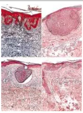
Figu ra 1: Carcin oma basocelu lar com variações n o p adrão su p erficial
(a, b, c, d). Esp aço de retração p arên qu imo-estron al (b, d). H&E x 40 (a, b, d) TM x 40 (c)
Figu re 1: Ba sal cell carcin om a w ith va ria tion s in the su perficial form (a, b, c, d ). Spa ce of paren chym a -strom a l retra ction (b, d ). H&E x 40 (a, b, d ) TM x 40 (c)
Federal University of Pernambuco (HC-UFPE) and a further 515 by a private laboratory of dermatopathology in Recife. These were cases involving primary tumors that had not been submitted to any prior therapeutics but had been removed by complete surgical excision. Some individuals presented more than one tumor, accounting for the total of only 623 patients. The cases were divided into two groups, the first (Group 1) included the patients of the Dermatology Clinic, HC-UFPE, and the second (Group 2), those of the private laboratory.
In the clinical study, data collected from records of each laboratory were considered (gender, age, origin and patients' color; duration of the disease and the topography of the lesions).
The anatomopathological study was an evaluation of the macro and microscopic morphological aspects. The mor-phological macroscopic data were extracted from patient files, registering the form, color and measurements of the tumors. The morphological microscopic data was gathered by a revision of the histological preparations of each tumor, and from a diagnosis by optical microscopy of epidermal, parenchymal aspects, stromal alterations and tumor exten-sion. The histological preparations, when necessary, were submitted to depigmentation by potassium permanganate and, for the study of fibrosis, to Masson's trichrome stain.4
To measure tumoral extension, a millimetric reference lens of MCM 1 1 100 MF 33F B KR - 207 21mmwas used in the ocular lens, in conjunction with a 4/0.12 objective lens gau-ged with an Olympus OB-M-1/100micrometric objective lens.
The histopathological aspects were evaluated by the author and then rechecked by a pathologist on the basis of predetermined parameters. In this manner, the histological pattern was established only according to the arrangements of the tumor cells, and the cellular type in each tumor, rela-tive to the predominant aspects.
The palisade and spaces of parenchymal-stromal (Figure 1) retraction were observed and classi-fied according to their presence and amount of involvement, into one third (slight degree); two thirds (moderate degree) or three thirds (accentuated degree) of the tumor.
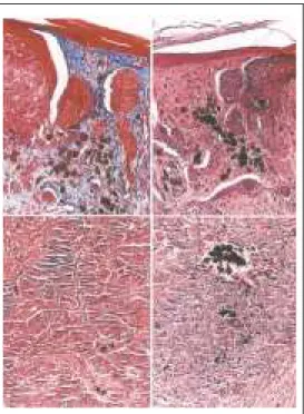
Pigment with focal distribu-tion in the parenchyma or in the stro-ma (Figure 2) was evaluated, for area and quantity, into slight (rare and scarce), moderate (some and more condensed) and accentuated (seve-ral, but considerably condensed).
UFPE, e 515, a um laboratório privado de dermatopatologia, em Recife. Eram tumores primários que não haviam sido submetidos a quaisquer métodos terapêuticos, retirados por excisão cirúrgica total, alguns pertencentes a um mesmo indivíduo, correspondendo ao total de 623 pacientes. Formados dois grupos, o primeiro (Grupo 1) incluiu os pacientes da Clínica Dermatológica do HC, da UFPE, e o segundo (Grupo 2), os do laboratório privado.
No estudo clínico, foram considerados dados refe-renciais recolhidos de prontuários de cada laboratório (sexo, idade, procedência e cor dos pacientes; tempo de evolução e topografia das lesões).
No estudo anatomopatológico avaliaram-se os aspectos morfológicos macro e microscópicos. Os dados morfológicos macroscópicos foram extraídos dos prontuá-rios, registrando-se a forma, a cor e as medidas dos tumo-res; os morfológicos microscópicos resultaram da revisão dos preparados histológicos de cada tumor, desde o diag-nóstico aos aspectos epidérmicos, parenquimais, alterações estromais e extensão tumoral à microscopia óptica. Os pre-parados histológicos, quando necessários, foram submeti-dos à despigmentação pelo permanganato de potássio e, para o estudo da fibrose, à coloração pelo tricrômico de Masson.4
Para medir a extensão tumoral, utilizou-se lente milimétrica de referência MCM 1 1 100 MF 33F B KR - 207 21mm, na ocular, com a objetiva 4/0.12, aferida pela objeti-va micrométrica Olympus OB-M-1/100.
Os aspectos histopatológicos foram avaliados levan-do-se em conta a acuidade visual do autor, e conferidos por um patologista, com base em parâmetros predeterminados. Dessa forma, o padrão histológico estabeleceu-se apenas segundo os arranjos das células tumorais, e o tipo celular em cada tumor, de acordo com sua predominância.
Observaram-se a paliçada e os espaços de retração parênquimo-estromal (Figura 1) segundo a pre-sença e o comprometimento, em um terço (grau leve); dois terços (grau moderado) ou três terços (grau acentuado) do tumor.
Figu ra 2: Carcin oma basocelu lar com grau s de in ten sidade do
p igmen to n o p arên qu ima e estroma: leve (d), moderado (a, b)
e acen tu ado (c) H&E x 40 (a), H&E x 100 (b,d), TM x 40 (c)
Interpretou-se a fibroplasia como leve (fibras colágenas delica-das, poucos fibroblastos, e o parên-quima predominando sobre o estro-ma), moderada (fibras colágenas mais compactas, regular presença de fibroblastos, e o estroma tenden-do ao equilíbrio) e acentuada (colá-geno compacto, riqueza em fibro-blastos, por vezes esclero-hialiniza-do, predominando o estroma sobre o parênquima).
O infiltrado inflamatório foi considerado tendo em vista a distri-buição focal e difusa. Na
distribui-ção focal, em leve (raros focos com poucas células inflama-tórias); moderado (vários focos com regular número de células inflamatórias) e acentuado (numerosos focos, por vezes confluentes, ou extenso). Na distribuição difusa, ocu-pando o estroma peri e intraparenquimal, com os mesmos graus de intensidade já mencionados.
A extensão tumoral baseou-se nos métodos histo-prognósticos para melanomas malignos (Clark e Breslow), com níveis de invasão considerados dérmicos (papilar, um, dois e três terços reticular); hipodérmicos e musculares. A espessura tumoral foi tomada em profundidade a partir da camada basal epidérmica, mas, nas lesões ulceradas, a par-tir de área vizinha com epitélio íntegro.
O nível de significância utilizado nas decisões de todos os testes estatísticos foi de 5%, e o software para a obtenção dos cálculos estatísticos, o Statistical Analysis System (SAS/ STAT).5
RESULTADOS Clínico
Dos 704 tumores estudados, 189 (26,8%) eram do Grupo 1, e 515 (73,2%), do Grupo 2. A Tabela 1 apresenta a distribuição dos casos por ano e por grupo, mostrando fre-qüência irregular, que se acentua nos últimos anos.
Na distribuição dos pacientes por número de tumo-res, a maioria foi expressiva para apenas um tumor (89,2%). Quanto ao sexo, o feminino contribuiu com 55,7%, e o mas-culino, com 44,3%, mostrando diferença não significativa (!2= 0,198 e P = 0,657).
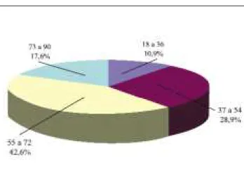
Quanto à idade dos pacientes (Gráfico 1), houve variação entre 18 e 90 anos, com predominância entre 55 e 72 anos (42,6%). No Grupo 1, a média de idade foi de 60,57 anos, e o desvio padrão, de 13,77. No Grupo 2, 56,45 anos e 15,52, respectivamente, comprovando-se a diferença
signi-Fibroplasia was interpreted as slight (delicate collagen fibers, few fibroblasts, and the parenchyma predominant over the stroma); moderate (collagen fibers more compact, the presence of regular fibroblasts and the stroma tending to be balanced); and accentuated (compacted collagen, rich in fibro-blasts, sometimes with sclerotic hyaline, stroma predominant over the parenchyma).
Inflammatory infiltration was considered in terms of focal and diffuse distribution. Focal distribu-tion was classified as: slight (rare focuses with few inflam-matory cells); moderate (several focuses with a number of regular inflammatory cells) and accentuated (numerous focuses, sometimes confluent, or extensive). In the diffuse distribution, occupying the peri- and intra-parenchymal stroma, the same degrees of intensity mentioned above were applied.
Quantification of the tumor extension was based on the histoprognostic methods for malignant melanomas (Clark and Breslow), with invasion levels considered dermal (papillary with one, two, or three thirds reticular); hypodermic and mus-cular. The depth of tumor thickness was measured starting from the epidermal basal layer, but in ulcerated lesions, starting from a neighboring area presenting a complete epithelium.
The significance level adopted in statistical tests was 5%. The software used for statistical calculations was Statistical Analysis System (SAS / STAT).5
RESULTS Clin ica l
Of the 704 tumors studied, 189 (26.8%) were in Group 1, and 515 (73.2%), Group 2. Table 1 presents the distribution of the cases according to year and group, showing demonstrating an irregular frequency that has increased in the last few years.
In the distribution of patients according to the number of tumors, it was significant that the majority had only one tumor (89.2%). As for gender, females constitu-ted 55.7% of the total, and males 44.3%, this did not show a significant difference (!2= 0.198 and P = 0.657).
Regarding patients' age (Graph 1), this ranged from 18 to 90 years, with a predominance between 55 and 72 years (42.6%). In Group 1, the mean age was 60.57 years, and the standard deviation, 13.77 against Group 2, 56.45
Figu re 2: Ba sal cell carcin om a w ith
years and 15.52, respectively. Mann-Whitney test indicated a significant difference (Z = 2.32 and P = 0.00199).
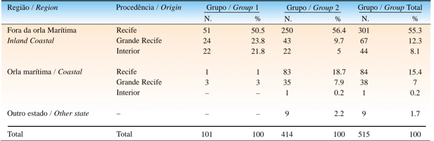
Table 2 shows the patients' distribution according to their regional origin: Recife, Greater Recife (both coastal) and inland cities, the majority being inhabitants of inland areas (55.3%). Of those living along the coast, Group 2 contributed a larger percentile (18.75%).
Most of the patients had leukoderma (78.4%), though there was a higher frequency in Group 2 (81.7%) than in Group 1 (70.2%), revealing a significant difference
(!2= 8.99 and P = 0.0031).
The duration of the lesions varied from one month to 40 years for all the patients, with a mean of 2.49 years, and a standard deviation of 3.81, indicating high variability, according to the coeffi-cient of variation (C.V. = 153.01%). For Group 1, the mean was 2.75 years, the standard deviation, 4.68, and C.V., 170.18%. In Group 2, t was 2.29 years, 2.98 and 130.13%, respectively. In about 54.1% of ficativa entre eles por meio do teste de Mann-Whitney
(Z=2,32 e P=0,00199).
A Tabela 2 mostra a distribuição de pacientes segun-do a procedência: Recife, Grande Recife e cidades segun-do inte-rior, predominando habitantes de fora da orla marítima (55,3%). Dos habitantes da orla marítima, o Grupo 2 contri-buiu com maior percentual (18,75%).
A maioria dos pacientes era leucoderma (78,4%), mais no Grupo 2 (81,7%) do que no Grupo 1 (70,2%), verificando-se diferença significativa entre eles (!2= 8,99 e P = 0,0031).
O tempo de evolução das lesões variou de um mês a 40 anos para todos os pacientes, com média de 2,49 anos, e desvio padrão de 3,81, indicando variabilidade elevadíssi-ma, segundo o coeficiente de variação (C.V.= 153,01%). Para o Grupo 1, a média foi de 2,75 anos, o desvio padrão, de 4,68, e o C.V., de 170,18%, e, no Grupo 2, 2,29 anos, 2,98 e 130,13%, respectivamente. Em cerca de 54,1% dos
Tabela 1: Distribu ição dos casos estu dados p or an o e gru p o / Ta ble 1: Distribu tion of the ca ses accord in g to year an d grou p
Ano / Year Grupo / Group 1
N. %
30 23 23 8 61 44
189
15.9 12.2 12.2 4.2 32.3 23.3
100,0
45 25 66 99 133 147
515
8.7 4.9 12.8 19.2 25.8 28.5
100.0
75 48 89 107 194 191
704
10.7 6.8 12.6 15.2 27.6 27.1
100.0 91
92 93 94 95 96
Total
Grupo / Group 2 N. %
Grupo / Group Total N. %
Fon te: Gru p o 1 – Casos registrados n a Clín ica Dermatológica do H.C. da UFPE. - Gru p o 2 – Casos registrados em laboratório p rivado de dermatop atologia, Recife, PE
Sou rce: Grou p 1 – Cases registered a t the d erm a tology clin ic HC-UFPE.- Grou p 2 – Ca ses registered at the private d erm a topa thology clin ic, Recife, PE
Tabela 2: Distribu ição dos p acien tes segu n do a p rocedên cia p or gru p o / Table 2: Distribu tion of the cases a ccord in g to region of origin per grou p
Região / Region Procedência / Origin Grupo / Group 1
N. %
51 24 22
1 3 –
–
101
50.5 23.8 21.8
1 3 –
–
100 250
43 22
83 35 1
9
414
56.4 9.7
5
18.7 7.9 0.2
2.2
100 301
67 44
84 38 1
9
515
55.3 12.3 8.1
15.4 7 0.2
1.7
100 Fora da orla Marítima
Inland Coastal
Orla marítima / Coastal
Outro estado / Other state
Total
Recife Grande Recife Interior
Recife Grande Recife Interior
–
Total
Grupo / Group 2 N. %
Grupo / Group Total N. %
Fon te: Gru p o 1 – Casos registrados n a Clín ica Dermatológica do H.C. da UFPE - Gru p o 2 – Casos registrados em laboratório p rivad o de dermatop atologia, Recife, PE
casos registrados, a evolução foi de um ano, e, em 20,7%, de dois anos.
Anato m o pato ló gico
- Morfológico macroscópico Observa-se bastante similaridade entre os grupos 1 e 2. Quanto à coloração dos tumores, os pigmentados (47,4%) e os brancacentos (45,8%) foram os mais repre-sentativos.
Na mensuração longi-tudinal, os valores medianos
diferiram de 0,10mm a favor do Grupo 1, com variabilidade de razoável à elevada, indicando diferença significativa entre os dois grupos (Z=2,0511 e P = 0,0403). Na mensuração transver-sal, a variabilidade também foi de razoável a elevada, forte-mente significativa entre os grupos, indicada através dos resul-tados do teste Mann-Whitney (Z=3,0100 e P = 0,0026), sendo a média (0,65) e a mediana (0,6) mais elevadas no grupo 1, que no grupo 2 (0,61 e 0,5) respectivamente.
- Morfológico microscópico
Houve bastante similaridade no comportamento tumo-ral para ambos os grupos. Em razão disso, algumas análises foram realizadas conjuntamente.
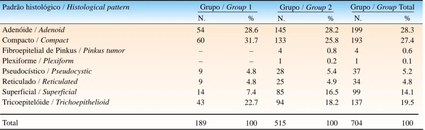
A Tabela 3 apresenta resultados dos padrões histológi-cos encontrados em todos os tumores, com tipos diversifica-dos, segundo a literatura consultada. Dessa forma, foi conside-rado apenas o padrão principal em cada tumor, sendo o adenói-de (28,3%) e o compacto (27,4%) os mais vistos.
Na associação entre o padrão histológico e o tipo celu-lar foi possível mostrar a predominância das células basalóides nos seis padrões analisados; entretanto, os padrões reticulado, tricoepitelióide e superficial tiveram também, de forma signi-ficativa, a presença de células fusiformes (32,4%, 25,5% e 8,1%); no adenóide, as células claras (17,1%); no compacto, as transicionais (15,5%); e no pseudocístico, as células escamói-des (10,8%).
A paliçada esteve presente em 90,5% dos tumores, apresentando-se mais no Grupo 1 (44,4%) do que no Grupo 2 (39%), com intensidade acentuada (94,7% e 88,9%, respecti-vamente). Associando-se o padrão histológico à paliçada, o resultado mostrou baixíssima probabilidade de significância P do teste Qui-quadrado de independência ou associação (!2=
39,323 e P = 0,001).
Os espaços de retração parênquimo-estromal (fenda) foram freqüentemente encontrados (93,2%) no grupo total, predominando a intensidade acentuada (37,6% e 47,4%). Na
the cases registered, the course of the disease was one year, and, in 20.7%, two years.
Ana tom op a thologica l
- Macroscopic morphology Considerable simila-rity can be observed between Groups 1 and 2. As for colora-tion of the tumors, the pigmen-ted (47.4%) and the whitish (45.8%) were the most repre-sentative.
In the longitudinal dimension, the median values differ by 0.10mm in favor of Group 1, with reasonable to high variability, indicating a significant difference between the two groups (Z=2.0511 and P = 0.0403). In the transversal measure-ment, the variability was also from reasonable to elevated and strongly significant between the groups, as shown by the results of the Mann-Whitney test (Z=3.0100 and P = 0.0026), while in Group 1 the mean (0.65) and median (0.6) was higher than Group 2 (0.61 and 0.5, respectively).
- Microscopic morphology
There was a notable similarity in tumoral behavior in both groups. For which reason, some analyses were performed simultaneously.
Table 3 presents results of the histological patterns found in all of the tumors, of various types, classified according to the consulted literature. Thus only the main pattern in each tumor was considered, of these the adenoid (28.3%) and the compact (27.4%) were the most frequently observed.
In the relationship between the histological pattern and the type of cell, it was possible to show the predominance of basaloid cells in the six analyzed patterns. At the same time, in the reticulated, trichoepithelioid and superficial specimens there was also, significantly, the presence of fusiform cells (32.4%, 25.5% and 8.1%); in the adenoid, clear cells (17.1%); compact, transitional cells (15.5%); and pseudocystic, squa-mous cells (10.8%).
Palisade cells were present in 90.5% of the tumors, occurring more in Group 1 (44.4%) than in Group 2 (39%), with accentuated intensity (94.7% and 88.9%, respectively). Relating the histological pattern to the palisade, the result showed a very low significance probability P of the Chi-square test of independence or association (!2= 39.323 and P
= 0.001).
The spaces of parenchymal-stromal retraction (-cracks) were frequently found (93.2%) in the total group, predominantly in areas of accentuated intensity (37.6% and Gráfico 1: Distribu ição dos p acien tes p esqu isados
qu an to a idade p or gru p o total
47.4%). In relation to the histological pattern, the reticula-ted pattern stood out because it showed moderate (35.3%) and slight (29.4%) intensities, suggesting a very significant association (!2= 45.492 and P <0.0001).
Pigment in the parenchyma appeared in only 20.3% of the tumors with slight intensity (11.1%); in the stroma, in 22.1%, also with slight intensity, mainly in Group 1 (32.3%). The asso-ciation of the histological pattern with the pigment in the parenchyma and/or stroma was seen more in the trichoepithe-lioid pattern and especially in those with accentuated intensity. In both groups fibroplasia was present in 95% of the cases, being predominant in areas of slight intensity (39.9%) mainly associated with the reticulated pattern (29.4%). There is a very strong significance in this associa-tion (!2= 85.030 and P <0.0001).
In measuring the extension of the tumors, it was found that Group 2 reached a vertical growth of 0.05 to 0.45mm (38.5%) and Group 1, 0.46 to 0.85mm (39.3%). The median for the patients as a whole was 0.49mm; in Group 1, 0.71mm and in Group 2, 0.49mm, demonstrating a significant difference bet-ween the two groups (Z = 8.043 and P <0.0001). In the analysis of the levels of involvement of the tumors, most tumors (31.4%) involved two thirds of the reticular dermis.
In the study of the relationship between the histological pattern and the level of dermal and hypodermic involvement (Table 4), the pseudocystic pattern stands out (48.7%) having a greater depth, involving three thirds of the reticular layer and revealing a very significant association (!2 = 354.413 and P
<0.001). However, in the comparison of vertical growth with
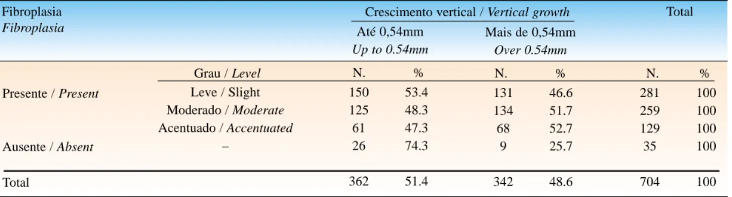
fibroplasia (Table 5), the greater the tumors' depth, the more accentuated was the fibroplasia, according to the level of signi-ficance adopted (!2 = 9.674 and P = 0.022).
In relating disease duration to the measurements of tumoral growth, using the Pearson ® coefficient of correlation, none were significantly above zero.
associação com o padrão histológico, o padrão reticulado des-tacou-se por mostrar intensidades moderada (35,3%) e leve (29,4%) com associação fortemente significativa ( !2= 45,492 e
P < 0,0001).
O pigmento no parênquima apareceu em apenas 20,3% dos tumores com intensidade leve (11,1%); no estroma, em 22,1%, também com intensidade leve, principalmente no Grupo 1 (32,3%). A associação do padrão histológico com o pigmento no parênquima e ou no estroma foi mais vista no padrão tricoe-pitelióide, sobretudo com na intensidade acentuada.
Em ambos os grupos a fibroplasia esteve presente em 95%, sendo a intensidade leve predominante (39,9%) principal-mente associada ao padrão reticulado (29,4%), existindo fortís-sima significância nessa associação (!2=85,030 e P<0,0001).
Medindo-se a extensão tumoral, verificou-se que o Grupo 2 atingiu crescimento vertical de 0,05 a 0,45mm (38,5%) e o Grupo 1, de 0,46 a 0,85mm (39,3%). A mediana, para o total de pacientes, foi de 0,49mm; no Grupo 1, de 0,71mm e no Grupo 2, de 0.49mm, comprovando-se diferen-ça significativa entre os grupos (Z = 8,043 e P < 0,0001). Na aná-lise dos níveis de comprometimento dos tumores, a maioria (31,4%) comprometeu dois terços da derme reticular.
No estudo da associação entre padrão histológico e os níveis de comprometimento dérmico e hipodérmico (Tabela 4), destaca-se o padrão pseudocístico (48,7%) com maior profundidade, comprometendo três terços da camada reticular, com forte associação significativa (!2= 354,413 e P
< 0,001). Entretanto, na associação do crescimento vertical
com a fibroplasia, os tumores que mais se aprofundaram mostraram fibroplasia (Tabela 5) mais acentuada, conforme o nível de significância considerado. (!2= 9,674 e P = 0,022).
Associando-se o tempo de evolução com as medidas de crescimento tumoral, por meio do coeficiente de correla-ção de Pearson ®, nenhuma delas foi significativamente diferente de zero.
Tabela 3: Distribu ição dos casos segu n do o p adrão histológico p or gru p o / Ta ble 3: Distribu tion of the ca ses a ccord in g to histological pattern per grou p
Padrão histológico / Histological pattern Grupo / Group 1
N. %
54 60 – – 9 9 14 43
189
28.6 31.7 – – 4.8 4.8 7.4 22.7
100 145 133 4 1 28 25 85 94
515
28.2 25.8 0.8 0.2 5.4 4.9 16.5 18.2
100 199 193 4 1 37 34 99 137
704
28.3 27.4 0.6 0.1 5.2 4.8 14.1 19.5
100 Adenóide / Adenoid
Compacto / Compact
Fibroepitelial de Pinkus / Pinkus tumor Plexiforme / Plexiform
Pseudocístico / Pseudocystic Reticulado / Reticulated Superficial / Superficial
Tricoepitelóide / Trichoepithelioid
Total
Grupo / Group 2 N. %
Grupo / Group Total N. %
Fon te: Gru p o 1 – Casos registrados n a Clín ica Dermatológica do H.C. da UFPE - Gru p o 2 – Casos registrados em laboratório p rivad o de dermatop atologia, Recife, PE
O infiltrado inflamatório no grupo total mostrou mais freqüentemente distribuição focal e intensidade leve (38,8%), porém, quando essa distribuição se mostrou difusa, a intensi-dade predominante foi a moderada (16,5%).
Das alterações epidérmicas, como revestimento tumo-ral, a atrofia foi a mais observada em ambos os grupos (71,6%).
DISCUSSÃO
Os carcinomas basocelulares são os tumores malig-nos cutâneos mais freqüentes, e, a cada ano, vem aumentan-do sua incidência, principalmente nos países situaaumentan-dos em baixa latitude e/ou com forte radiação solar,6-10apesar de os
registros mostrarem que alguns surgem em conseqüência de outros fatores mais raros.11,12
Inflammatory Infiltrate in the total group more fre-quently showed a focal distribution and slight intensity (38.8%), however, it was seen that when the distribution was diffused, the predominant intensity was moderate (16.5%).
Of the epidermal alterations, such as the tumor covering, atrophy was the most frequently observed in both groups (71.6%).
DISCUSSION
Basal cell carcinomas are the most frequent cuta-neous malignant tumors. Every year the incidence of these tumors is increasing, mainly in countries located in the low latitudes or with strong solar radiation.6-10Although there are records that show that some appear as a consequence of other less frequent factors.11,12
Tabela 4: Distribuição dos casos segundo o p adrão histológico e o nível de comp rometimento dérmico Ta ble 4: Distribu tion of the ca ses a ccord in g to histological pattern an d level of d erm a l in volvem en t
Padrão histológico Histological pattern
Adenóide / Adenoid Compacto / Compact Pseudocístico / Pseudocystic Reticulado / Reticulated Superficial / Superficial Tricoepitelóide / Trichoepithelioid
Total
Total
Fon te: Gru p o 1 – Casos registrados n a Clín ica Dermatológica do H.C. da UFPE - Gru p o 2 – Casos registrados em laboratório p rivad o de dermatop atologia, Recife, PE
Sou rce: Grou p 1 – Cases registered at the d erm a tology clin ic HC-UFPE.- Grou p 2 – Ca ses registered a t the private d erm a topa thology clin ic, Recife, PE
Nível de comprometimento / Level of involvement
Papilar Papillary
1/3 camada reticular 1/3 reticular layer
2/3 camada reticular 2/3 reticular layer
3/3 camada reticular 3/3 reticular layer
N. % N. % N. % N. % N. %
12 9 2 3 79 13 118 6.1 4.8 5.4 8.8 83.1 9.5 17.2 41 41 5 8 10 37 142 20.8 21.7 13.5 23.5 10.5 27.2 20.6 80 74 12 12 5 36 219 40.6 39.1 32.4 35.3 5.3 26.5 31.8 64 65 18 11 1 50 209 32.5 34.4 48.7 32.4 1.1 36.8 30.4 197 189 37 34 95 136 688 100 100 100 100 100 100 100
Tabela 5: Distribu ição dos casos qu an to a fibrop lasia e crescimen to vertical Ta ble 5: Distribu tion of the ca ses accord in g to fibroplasia an d vertical grow th
Fibroplasia Fibroplasia
Presente / Present
Ausente / Absent
Total
Total
Fon te: Gru p o 1 – Casos registrados n a Clín ica Dermatológica do H.C. da UFPE - Gru p o 2 – Casos registrados em laboratório p rivad o de dermatop atologia, Recife, PE
Sou rce: Grou p 1 – Cases registered at the d erm atology clin ic HC-UFPE.- Grou p 2 – Ca ses registered a t the private d erm a topa thology clin ic, Recife, PE
Crescimento vertical / Vertical growth Até 0,54mm
Up to 0.54mm
Mais de 0,54mm Over 0.54mm
Grau / Level N. % N. %
131 134 68 9 342 46.6 51.7 52.7 25.7 48.6 N. %
150 125 61 26 362 53.4 48.3 47.3 74.3 51.4 281 259 129 35 704 100 100 100 100 100 Leve / Slight
Moderado / Moderate Acentuado / Accentuated
The findings in the 704 cases of basal cell carcino-mas registered in the two services coincide with those in national and international literature regarding factors of irregular frequency, age group and increase in incidence in recent years.7,10,13Although it has been mentioned above that several tumors may belong to the same patient, most patients presented only a single lesion. These observations are similar to those of Kikuchi and col.,14who studying 243 Japanese patients with basal cell carcinomas, reported multiple lesions in only four patients.
In spite of the majority being of the female gender, the difference was not significant, corroborating the results obtained in other populations (Asian, African, Mexican and Spanish).14,15,16
The tumor is more frequently found to occur between the third and fifth decades of life,17 although, in this work, the youngest patients had been registered at the age of 18 years and the oldest at 90 years of age, this being similar to that reported in other work.9,18Some authors consider such extreme ages to be less frequent.14,15
Goldberg19demonstrated that, when individuals sub-mit themselves to solar exposure at around 20 years of age, a carcinogenic process begins that results in a delayed manifestation eventually seen in the period from about 40 to 60 years of age, thus explaining the source of the tardive cumulative effects.
In evaluating the relative importance of the coastal lifestyle, it was found that it was not a significant factor for greater incidence of basal cell carcinomas, but attention was called to the continuous solar exposure of those who do not live on the coast 20,6but that habitually spend much of their lives out-of-doors.13
The disease duration of the tumors presented great variation, from one month to 40 years. Genetic studies jus-tify this length of time, showing replication of DNAwith a prolonged phase of synthesis. Clinically, the tumor takes years to double its size due to the small percentile of active proliferative tumoral cells.21
In this analysis, the main locations of the tumors were: head (73%), anterior (9.9%) and posterior (9%) areas of the trunk, superior (5.5%) and inferior (1.7%) members. In the head, they were mainly on the scalp (6.2%) and forehead (10.1%), orbital (6.3%), auricular (2.1%), mastoid (1.8%), zygomatic (18.5), nasal (21.2%), buccal (2.6%), mental (1.2%) and cervical (3.8%) areas. Such results are similar those reported by Grosshans.22
As for unusual locations, there is a report of a case in in the urogenital (vulvar) area, similar to that found at Harvard University.23Also there was one in a gluteal loca-tion, an area that was not mentioned in reports in the con-sulted literature. It is important to call attention to the reports of tumors in areas not limited to the face.6
Os achados dos 704 casos de carcinomas basocelu-lares registrados nos dois serviços coincidem com as da lite-ratura nacional e internacional sobre freqüência irregular, faixa etária e crescimento, nos últimos anos.7,10,13Embora se
tivesse observado que vários tumores pertenciam a um mesmo paciente, a maioria apresentou apenas uma lesão. Essas observações são semelhantes às de Kikuchi e col.,14
que, estudando 243 pacientes japoneses com carcinomas basocelulares, verificaram multiplicidade de lesões em ape-nas quatro pacientes.
Apesar da predominância no sexo feminino, a dife-rença não foi significativa, coincidindo com os resultados obtidos em outras populações (asiática, africana, mexicana e espanhola).14,15,16
O tumor é mais freqüente entre a terceira e a quinta década de vida,17embora, neste trabalho, tenham sido
regis-trados os pacientes mais jovens com 18 anos e o mais idoso com 90 anos, assemelhando-se a dados da literatura.9,18
Alguns autores consideram esses extremos idades de baixa freqüência.14,15
Goldberg19evidenciou que, quando os indivíduos se
submetem a exposições solares por volta dos 20 anos, ini-cia-se um processo de carcinogênese que se manifesta tar-diamente, por volta da faixa de 40 a 60 anos, justificando, dessa forma, os efeitos cumulativos mais tardios.
Procurando associar a importância ou não da habita-ção em orla marítima, esse não foi um dado significativo de maior incidência para os carcinomas basocelulares, mas chama-se a atenção, para a exposição solar continuada daqueles que não moram em orla marítima6,20 e que têm
hábitos de vida ao ar livre.13
O tempo de evolução dos tumores apresentou grande variabilidade, de um mês a 40 anos. Os estudos genéticos justificam esse tempo, mostrando a replicação do DNAcom fase de síntese prolongada. Clinicamente, o tumor leva anos para duplicar seu tamanho, além de ser pequeno o percentual de células tumorais proliferativa-mente ativas.21
Nesta análise, as principais localizações dos tumores foram: região da cabeça (73% ), regiões anterior (9,9%) e posterior (9%) do tronco, membros superiores (5,5% ) e inferiores (1,7%). Na cabeça, destacam-se o couro cabeludo (6,2%) e as regiões frontal (10,1%), orbital (6,3%), auricular (2,1%), mastoídea (1,8%), zigomática (18,5), nasal (21,2%), bucal (2,6%), mental (1,2%) e cervical (3,8%). Tais resulta-dos assemelham-se aos apresentaresulta-dos por Grosshans.22
Quanto às localizações não usuais, tem-se o registro de um caso na região urogenital (vulvar), semelhante ao encontrado na Universidade de Harward,23e um de
Ana tom op a thologica l Com m e nts
As in the clinical forms, the histological forms also presented variations,11,24,25most of which were in the small tumors, coinciding with the information in the literature.26 Few tumors present a single histological pattern.27The defi-nition adopted here was based only on the form of presen-tation of these arrangements, considering that stromal alte-rations should not be included in the nomenclature of basal cell carcinomas, but that these alterations provide data relevant to a conclusive diagnosis of tumoral behavior. While remaining in accordance with these parameters, it should be noted that some authors exclude from the litera-ture several histological patterns of basal cell carcinomas that are included here; these are, however, based on the consulted literature. For example, the Pinkus type which has a reticulated pattern was considered special according to the observations of Grinspan & Abulafia.28
The variation in histological patterns was justified by Petersen in 1902, Madsen in 1941 and Foot in 1947.29Using a three-dimensional study of basal cell carcinomas they showed the impossibility of an exact image - in that what one sees is only an approximation of the true histological pattern. Others consider that there are architectural modifications taking place resulting from the development and deepening, and that these hinder the imaging in the histological sections.18,29,30,31 This being the case, the superficial pattern seen presented a highly varied architecture, everything from compact, adenoid, reticulated and trichoepithelioid arrangements, with single or multiple growths connected to the epidermis.29,32,33
The cellular variations were grouped into five types, these were seen to be in a complex arrangement in each tumor and rarely presented as pure.1,27,34,35,36
There was agreement with the literature in reference to the palisade, as a constant characteristic of basal cell carcinomas. On the other hand, its absence and the varia-tion of intensity found in the reticulated pattern (23.5%) were not considered in the literature consulted.25,27,37
The spaces of parenchymal-stromal retraction were always very much in evidence, seeming to corroborate the observations of Miller.21This apparently indicates that they result from desmosome alterations and a decrease in colla-gen from the collacolla-genase synthesis.
Some tumors presented more squamous cells as an epidermoid differentiation, which is an important factor in their aggressive behavior38 and their greater growth at depth,31,32,33,37Most of the time (31.4%), they did not exceed 1mm, in spite of the fact that tumors present more intense fibroplasia if they penetrate deeper.37,39,40
The inflammatory infiltration, besides the lympho-cytic elements, was occasionally composed of plasm cells and mast cells, mainly in ulcerated areas representing the local immunity.40,41It has been demonstrated that the pres-ence of the mast cells participates in the synthesis of gly-cosaminoglycan in the fibroblastic proliferation and in the tumoral aggressiveness.21,40
Co m e ntário s Anato m o pato ló gico s
Assim como as formas clínicas, as formas histológi-cas também apresentaram variações.11,24,25A maioria era de
pequenos tumores, coincidindo com as informações da lite-ratura.26 Poucos tumores apresentaram padrão histológico
único.27A denominação aqui adotada foi baseada apenas na
forma de apresentação desses arranjos, considerando que as alterações estromais não devam ser incluídas na nomencla-tura dos carcinomas basocelulares, mas que sejam dados relevantes a um diagnóstico conclusivo do comportamento tumoral. Seguindo essas determinações, verifica-se que alguns autores excluem da literatura vários padrões histoló-gicos dos carcinomas basocelulares; os aqui relatados, entretanto, tiveram embasamento na literatura consultada. O tipo de Pinkus com padrão reticulado foi considerado especial, segundo observações de Grinspan & Abulafia.28
A variação dos padrões histológicos foi justificada por Petersen em 1902, Madsen, em 1941 e Foot, em 1947,29
mediante estudo tridimensional dos carcinomas basocelula-res, em que mostraram a impossibilidade de uma visão exata - o que se vê é uma aproximação do verdadeiro padrão histológico. Outros acreditam que haja modificações arquiteturais, decorrentes de seu desenvolvimento e apro-fundamento, dificultando as imagens nas secções
histológi-cas.18,29,30,31Assim sendo, o padrão superficial visto
apresen-tou arquitetura muito variada, desde arranjos compactos, adenóides, reticulados e tricoepitelióides, com únicos ou múltiplos brotos, mas conectados à epiderme.29,32,33
As variações celulares foram agrupadas em cinco tipos, vistos de maneira complexa em cada tumor e que raramente se mostravam puros.1,27,34,35,36
Houve concordância com a literatura quanto à pali-çada, como característica constante nos carcinomas basoce-lulares. Entretanto, a ausência e a variação de intensidade encontradas no padrão reticulado (23,5%) não foram consi-deradas na literatura consultada.25,27,37
Os espaços de retração parênquimo-estromal estive-ram sempre muito presentes, parecendo colaborar com as observações de Miller,21as quais indicam serem resultantes
de alterações desmossômicas e diminuição do colágeno pela síntese de colagenase.
Alguns tumores apresentaram mais células escamói-des como diferenciação epidermóide, o que não deixou de ser dado de importância para o comportamento agressivo38
e de maior crescimento em profundidade,31,32,33,37o que, na
maioria das vezes (31,4%), não ultrapassou 1mm, apesar de os tumores que apresentaram fibroplasia mais intensa se terem aprofundado mais.37,39,40
O infiltrado inflamatório, além dos elementos lin-focitários, ocasionalmente compunha-se de células plas-máticas e mastócitos, principalmente em áreas ulceradas representando a imunidade local.40,41 A presença dos
CONCLUSIONS
Although the present work was performed with a sepa-ration into two socially different groups, the social inequali-ties did not produce sufficient significant data to allow speci-fic conclusions to be applied to each group.
Clin ica l
1 - Basal cell carcinomas are occurring with greater and abnormal frequency mainly in the last few years. There is a slight bias in the female gender. The age group having the greatest probability is from 55 to 72 years, pointing to the gro-wing emergence in a younger population.
2 - In tropical areas, living on the coast in itself is not a significant factor for higher registrations of incidence of basal cell carcinomas, but rather continuous solar exposure arising from peoples' life styles.
3 - They are tumors that develop slowly in the first two years. Their size does not depend on the disease duration.
4 - The most common locations were the head (nasal and zygomatic) and (though with few registrations) superior and inferior members.
Ana tom op a thologica l
1 - The nomenclature of these histological patterns should be based only on the arrangements of the parenchymal cells, thus making the language more uniform. The patterns researched were: adenoid, compact, plexiform, pseudocystic, reticulated, trichoepithelioid, Pinkus fibroepithelial and super-ficial. The superficial has similar arrangements to several other patterns, showing it to be but a stage of their development. Of them, the most frequent were the adenoid and the compact.
2 - Basaloid cells, as prototypes of basal cell carcino-mas, were found more often, followed by the fusiform, transi-tional, clear and squamous; squamous cells were seen more in the pseudocystic pattern, and fusiform in the reticulated pattern. These are considered to be the more aggressive tumors.
3 - Palisade cells were always very present with accen-tuated intensity, but generally seen more in the compact and reticulated patterns.
4 - The spaces of parenchymal-stromal retraction and the melanin pigment (parenchyma and stroma), in any distri-bution, were more frequent in the trichoepithelioid pattern.
5 - Most of the tumors had little deep growth, but the cellular type of the tumoral parenchyma and fibroplasia inter-fered in that growth.
6 - The inflammatory infiltrate of basal cell carcino-mas was usually focal and of mild intensity, and rarely dif-fuse.
7 - The histological behavior of the tumors was simi-lar in both groups from the public and private
dermatopatho-logical services. !
CONCLUSÕES
Embora o presente trabalho tenha sido realizado com a formação de dois grupos socialmente diferentes, essas desigualdades não representaram dados significativos para conclusões específicas a cada grupo.
Clínicas
1 - Os carcinomas basocelulares têm freqüência ele-vada, irregular, principalmente nos últimos anos, com dis-creta predominância no sexo feminino. A faixa etária de maior freqüência é a de 55 a 72 anos, ressaltando-se o cres-cente aparecimento na população jovem.
2 - Nas regiões tropicais, morar em orla marítima não é um dado significativo para maiores registros de incidência dos carcinomas basocelulares, mas sim a exposição solar continuada, decorrente do comportamento populacional.
3 - São tumores que evoluem lentamente nos dois pri-meiros anos, e seu tamanho independe do tempo de evolução.
4 - As localizações preferenciais foram a cabeça (nasal e zigomática) e (com poucos registros) membros superiores e inferiores.
Anato m o pato ló gicas
1 - A nomenclatura dos padrões histológicos deverá ser baseada apenas nos arranjos das células parenquimais, uniformizando-se a linguagem. Os padrões registrados foram: adenóide, compacto, plexiforme, pseudocístico, reti-culado, tricoepitelióide, fibroepitelial de Pinkus e superfi-cial. O superficial tem arranjos similares a vários padrões, demonstrando ser um momento de seu desenvolvimento. Deles, os mais freqüentes foram o adenóide e o compacto.
2 - As células basalóides, como protótipo dos carci-nomas basocelulares, foram as mais encontradas, seguidas das fusiformes, transicionais, claras e escamóides; as célu-las escamóides foram mais vistas no padrão pseudocístico, e as fusiformes, no padrão reticulado, considerados tumores mais agressivos.
3 - A paliçada esteve sempre muito presente com intensidade acentuada, em geral mais vista nos padrões compacto e reticulado.
4 - Os espaços de retração parênquimo-estromal e o pigmento melânico (parênquima e estroma), em qualquer dis-tribuição, foram mais freqüentes no padrão tricoepitelióide.
5 - A maioria dos tumores teve crescimento pouco profundo, mas o tipo celular do parênquima tumoral e a fibroplasia interferiram nesse crescimento.
6 - O infiltrado inflamatório dos carcinomas basoce-lulares foi, geralmente, focal e de intensidade leve, e rara-mente difuso.
7 - O comportamento histológico dos tumores foi semelhante nos grupos dos serviços de dermatopatologia
REFERÊNCIAS / REFERENCES
1. Abuláfia J. Epiteliomas cutâneos. Separata de An. Bras. Dermatol. 1962;38(3/4):14-31.
2. Darier J. Epitheliomas. In:_.La pratique dermatologique. Paris; Masson, 1907;2:395-422.
3. Degos R, Civatte, Belaich S. Tumeurs malignes; epiteliomas basocelulareis. In:_.Dermatologie. Paris, Flamarion Medicine Sciences, 1981:2;843-8.
4. Beçak W, Paulete J. Técnicas de citologia e histologia. Rio de Janeiro: Livros Técnicos e Científicos, 1976:2;359.
5. Statistical analysis system (SAS/SAT): procedures guide for per-sonal computers. Versão 6. Ed. Cay: SAS Institute Inc., 1985: 378. 6. Reizner GT. et al. Basal cell carcinoma in Kauai, Hawaii, the highestdocumented incidence in the United States. J. Am. Acad. Dermatol. Aug. 1993:29(n.2,pt.1);184-9.
7. Matsumura Y. et al. Characterization of p53 gene mutations in basal cell carcinomas: comparison between sun-exposed and less exposed skin areas. Int. J. Cancer, 1996:65;778-80.
8. Bastos AF, Fonseca A, de Faria JL. Acerca do carcinoma da pele (a propósito de uma estatística). Med. Cut. I.L.A. 1980:8;109-12. 9. Healy E, Collins P, Barnes L. Non melanoma skin cancer in a Irish population: an appraisal of risk factor. Ir. Med. J. Mar./Apr. 1995:88(2);58-9.
10. Walberg P, Skog E. The incidence of basal cell carcinoma in an area of Stockholm county during the period 1971-1980. Acta. Dermatol. Venereol. 1991:71;134-7.
11. Goldberg L. H. Basal - cell carcinoma as predictor for the can-cers. The Lancet, Mar. 1996:349;664-5.
12. Grob J. Cancers cutanés epithéliaux. La Rev. Practicien, 1995:45;645-51.
13. Porto JA. Câncer da pele; prevenção primária. Atual. Dermatol. abr./jun.1995:1(1);20-3.
14. Kikuchi A, Shimizu H, Nishikawa T. Clinical and histopatho-logical characteristics of basal cell carcinoma in Japanese patients. Arch. Dermatol., 1996:132;320-4.
15. N Jimenez, S Navarro, LM Delgado, Velasquez B. Estudio epi-demiologico del cancer cutáneo en el Estado de Oaxaca, Mexico de diez años (1982-1992). Med. Cut. I.L.A.1995:23(5);258-62. 16. Perez ON, Cañizo FD, Quiñones PA. Epiteliomas basocelu-lares sobre nevisebáceos. Med. Cut. I.L.A. 1991:(19);171-5. 17. Mahmoud SF, Azadeh B. Basal cell carcinoma in Qatar. Int. J. Dermatol. Oct 1996:35(10);704-6.
18. Labareda JM, Silva LG. e. Carcinomas basocelulares no couro cabeludo; revisão de 77 doentes com 81 tumores. Med. Cut. I. L. A. 1988:16;367-72.
19. Goldberg LH. Basal - cell carcinoma. The Lancet, Mar 1996:(347)663-7.
20. Gallargher, R. P. et al. Trends in basal cell carcinoma, squamous cell carcinoma, and melanoma of the skin from 1973 through 1987. J. Am. Acad. Dermatol. Sept. 1990:23(3pt.1);413-21.
21. Miller S J. Biology of basal cell carcinoma (part I ). J. Am. Acad. Dermatol. Jan. 1991:24(1);1-13.
22. Grosshans, E. Les epithéliomas cutanés. In:_. Encyclopédie medico-chirurgicale. Comitê Científico J. F. Bach [e outros]. Paris: Ed. Techniques, 1989:3.
23. Halder RM, Bridgeman - Shah, S. Skin cancer in African Americans. Cancer, Jan. 1995:75(Supl 2);667-73.
ENDEREÇO PARA CORRESPONDÊNCIA: / MAILINGADDRESS: Au rilen e Mon teiro Ba n d eira
Ru a Pad re Rom a , 688 - a pto. 2002 Recife PE 52060-060
Tel: (81) 3267-6155 / 9975-7764 Fax : (81) 3327-1396
E-m ail: au rilen e@elogica .com .br
24. Maloney ME. et al. Pigmented basal cell carcinoma: investIgations of 70 cases. J. Am. Acad. Dermatol. July 1992:27(1)74-8.
25. Bleehen S S. Pigmented basal cell epithelioma; light and elec-tron microscopic studies on tumours and cell cultures. Br. J. Dermatol. 1975:93; 361-70.
26. Dahl E. et al. Basal cell carcinoma; an epidemiologic study in a defined population. Cancer, July 1992:70;(1)104-8.
27. Garcia Prats, M. D. et al. Granular cell basal, cell carcinoma; light microscopy, immunohistochemical and ultrastrurctural study. Virchows Arch. A: Pathol. Anat. 1993:422;173-7. 28. Grinspan D, Abuláfia J. Tumor fibroepitelial de Pinkus. Separata de Arch. Arg. Dermatol., Buenos Aires, Mar.1963:23(1):23-44. 29. Sanderson, K. V. The architecture of basal-cell carcinoma. Br. J. Dermatol. 1961:73;455-74.
30 Goldberg LH, Fachin-Viso, R. Extenso carcinoma basocelular multifocal en un hombre joven. Med. Cut. I. L. A.,1983:(11);19-194, 31. Hendrix JR., J. D., Parlette, H. L. Micronodular basal cell car-cinoma. Arch. Dermatol. Mar 1996:(132)295-8.
32. Sloane JP. The value of typing basal cell carcinomas in pre-dicting recurrence after surgical excision. Br. J. Dermatol., 1977:96;127-32.
33. Lazaro SR, Sanchez RM, Contreras R F. Carcinoma basocelu-lar: fatores predictivos da recidiva. Med. Cut. I.L.A., 1995:23-(4);183-6.
34. Rossen K. et al. Bcl - 2 over expression in basaloid prolifera-tions overling dermatofibromas and basal cell carcinomas. APMIS, 1997:105;35-40.
35. Reck A. et al. Clear cell basal carcinoma: an unusual variant. Histopathology, Oct.1996:29(4);390-1.
36. Elder D, et al. Tumors and Cysts of the Epidermis. In:_. Lever’s Histopathology of the skin. Philadelphia: Lippincott-Raven Publishers, 1997:(Cap. 30);719-746.
37. De Rosa G. et al. Comparative morphometric analysis of aggressive and ordinary basal cell carcinoma of the skin. Cancer, Feb.1990:65;544 -9.
38. Santos OLR. et al Carcinoma basoescamoso pigmentado. An. Bras. Dermatol. jan./fev.1994:69(1);22-6.
39. Kunrath SO, Alchorne MMA. Pereira JPM. Estudo das fibras do sistema elástico no carcinoma basocelular e esclerodermiforme da pele humana. An. Bras. Dermatol. set./out.1997:72(5); 441-445.