Relationship of Tumor Thickness with Neck Node
Metastasis in Buccal Squamous Cell Carcinoma:
An Experience at a Tertiary Care Hospital
Sadaf Qadeer Ahmed
1Montasir Junaid
2Sohail Awan
3Moaz M. Choudhary
4Maliha Kazi
5Aria Masoom
6Hareem Usman Khan
71Department of Otorhinolaryngology, Sir Syed College of Medical
Sciences for Girls Ringgold Standard Institution, Karachi, Pakistan
2Department of Otorhinolaryngology, Jinnah Medical and Dental
College Ringgold Standard Institution, Karachi, Pakistan
3Department of Otorhinolaryngology, Aga Khan University Ringgold
Standard Institution, Karachi, Pakistan
4Department of Medicine, Aga Khan University Ringgold Standard
Institution, Karachi, Pakistan
5Department of Otorhinolaryngology, Manchester Royal Infirmary,
Manchester, United Kingdom
6Department of Otorhinolaryngology, Bolan Medical College
Ringgold Standard Institution, Quetta, Pakistan
7Department of Medicine, Shifa College of Medicine Ringgold
Standard Institution, Islamabad, Pakistan
Int Arch Otorhinolaryngol 2017;21:265–269.
Address for correspondence Sadaf Qadeer Ahmed, FCPS, Assistant Professor, Department of Otorhinolaryngology, Sir Syed College of Medical Sciences for Girls, Korangi Road, Karachi 74800, Pakistan (e-mail: [email protected]; [email protected]).
Keywords
►
buccal mucosa
►
squamous cell
carcinoma
►
metastasis
Abstract
Introduction
Squamous cell carcinoma is the most common malignancy of the head
and neck, with the buccal mucosa being the most common site involved. Early
locoregional metastasis is a hallmark of this disease, and early stage tumors may
harbor metastatic nodes that are occult. Certain parameters can help identify high-risk
patients for whom the pattern of occult nodal metastasis can be predicted. Tumor
thickness is one such objective parameter.
Objective
To determine the relationship of tumor thickness with neck node
metas-tasis in squamous cell carcinoma of the buccal mucosa.
Methods
A retrospective chart review of 102 patients with biopsy-proven squamous
cell carcinoma of the buccal mucosa with N
0Necks was performed. All patients
underwent tumor resection with neck dissection, and the tumor thickness was
measured. Univariate and multivariate analyses were performed.
Results
A total of 102 patients, of which 73.53% were males and 26.47% were
females. The mean age of the patients was 49.3
11.1 years. It was found that the risk
of neck node metastasis in buccal squamous cell carcinoma increases 35.5 times for a
tumor thickness
2 mm, and the risk of neck node metastasis in buccal squamous cell
carcinoma decreases by 0.58 times for each centimeter decrease in tumor size, while
the rate of occult neck lymph node metastasis was found to be 37%.
Conclusion
We conclude that tumor thickness is signi
fi
cantly related with neck nodal
metastasis in buccal squamous cell carcinoma, considering the age of the patient and
the size of the tumor.
received July 23, 2016 accepted January 15, 2017 published online February 21, 2012
DOIhttps://doi.org/ 10.1055/s-0037-1599061. ISSN 1809-9777.
Copyright © 2017 by Thieme Revinter Publicações Ltda, Rio de Janeiro, Brazil
Introduction
Oral cancer is the most frequent head and neck malignancy worldwide. According to the World Health Organization (WHO), oral cancer rates are expected to increase from 10
million cases in 2000 to 15 million cases in 2020.1There is
global and regional variation in the incidence of oral cancers due to local geographical, biological, dietary and
environ-mental factors.2 Needless to say, the majority of these
cancers are squamous cell carcinomas. Among the subsites of the oral cavity, the buccal mucosa is one of the most
common areas involved, accounting for 50% of all oral
cavity tumors. Buccal mucosa involvement is due to people’s
frequent consumption of pan, betel nut, tobacco, and
niswaar.2
Squamous cell carcinoma of the buccal mucosa is an aggressive malignancy, with a greater propensity for inva-sion into the surrounding tissues and metastasis to the
cervical lymph nodes.3 The involvement of the cervical
lymph nodes greatly impacts the treatment protocol and the prognosis of squamous cell carcinoma of the buccal
mucosa.4
Advanced stage tumors with clinical or radiological
evidence of neck nodes warrant a definitive neck dissection.5
It is the early stage tumors without any clinical or
radiolo-gical evidence whose management remains controversial.6–8
They may harbor disease (36–42%), as reported in the
literature;9–11 however, at the same time, performing an
unnecessary neck dissection can lead to avoidable complica-tions and gruesome outcomes.
Certain parameters can help in identifying high-risk
patients, for whom an elective neck dissection is justifi
-able.12–15Tumor thickness is one such objective parameter,
and it is assessed in various studies. The increasing depth of invasion and the microvascular proliferation caused by the neoplastic growth might determine the proximity to blood
vessels and lymphatics, thus facilitating the tumor’s ability to
metastasize.4,5,16
Our experience in using tumor thickness as a predictor for identifying occult neck nodal metastases in clinically and radiologically negative necks in squamous cell carcinoma of the buccal mucosa is described as follows.
Objective
To determine the relationship of the tumor thickness with the neck node metastasis in squamous cell carcinoma of the buccal mucosa.
Materials and Methods
After taking approval from the ethical review committee of the Aga Khan University & Hospital, the study was initiated. This study was a retrospective chart review of patients treated for squamous cell carcinoma of the buccal mucosa
at the Department of Otolaryngology – Head and Neck
Surgery of the Aga Khan University & Hospital from May 1st 2008 to May 31st 2013. All patients, irrespective of their
age, gender, race or tumor stage were recruited. A total of 102 patients were recruited in the study. Data were recorded using a structured questionnaire. Patients with a biopsy-proven squamous cell carcinoma of the buccal mucosa with-out any clinical or radiological signs of cervical node metas-tasis were included in the study. Patients who had previously received treatment and patients who had recurrence were excluded. All the patients involved in the study underwent wide local excisions of the buccal mucosa lesion along with ipsilateral functional neck dissection. The excised specimens were sent routinely to the Histopathology Department of the Aga Khan University & Hospital for assessment.
Data were collected from the assessment of patient charts and the review of histopathology reports. The variables assessed were age, gender, addiction, T-stage of the tumor and tumor thickness. All tumor margins were negative for
tumor involvement in thefinal histopathology. The presence
or absence of occult cervical lymph node metastasis was also
extracted from thefinal histopathology report.
Tumor thickness, which is the objective parameter of the depth of invasion, was measured by a senior histopathologist from the surface of the tumor to the deepest point of the
tumor invasion, as proposed by Moore et al17(►Fig. 1).
All the variables mentioned before were analyzed for their association with the presence of occult cervical lymph nodes
metastasis on thefinal histopathology.
Statistical Analysis
Data were analyzed using the Statistical Package for Social Sciences (SPSS, SPSS Inc., Chicago, IL, USA) software, version
19.0. Continuous variables were expressed as mean
nodes metastasis adjusting for other variables. The indepen-dent variables included were tumor thickness, age and
tumor size. Model adequacy was checked by Hosmer–
Leme-show test.
Results
Total 102 patients (n¼102) according to the eligibility
criteria were included in the study; 73.53% (75) of them were male, and 26.47% (27) were female. The mean age of the
patients was 49.311.1 years; the mean size of the tumors
was 3.51.8 cm; 92.15% (94) had a history of addiction
(alcohol use, cigarette smoking or chewing tobacco). Patients were categorized according to tumor thickness
<2 mm, or>2 mm. Thickness<2 mm was found in
91.17% (93) of the patients, and less than 9% had a
thickness>2 mm. The majority of the patients had early
stage tumors, that is, 34% had stage I, and 35% had stage II tumors; and 23% and 8% of them had stage III and IV tumors respectively. From all patients, 25.50% (26) had well-differ-entiated tumors, and 67.65% (69) and 6.85% (7) had
moder-ately, and poorly differentiated tumors respectively.
Cervical lymph node metastasis was found in 37% of the patients, and 63% of them had no signs of cervical node metastasis on the histopathology report.
The patients’ age, tumor thickness and sizer were
sig-nificant predictors of cervical lymph node metastasis on the
univariate analysis, with apvalue<0.05 (►Table 1).
When these variables were analyzed using the multi-variate regression, it was noted that the associations among age, tumor thickness and tumor size were statistically
sig-nificant with cervical node metastasis (►Table 2). It was
further identified that the risk of neck node metastasis in
buccal squamous cell carcinoma increases 35.5 times for a
tumor thickness of2 mm, provided the confounding
variables remain constant.
With every 10 years increase in age, the risk of neck node metastasis in buccal squamous cell carcinoma increases by 1.84 times, adjusting for other variables. The risk of neck node metastasis in buccal squamous cell carcinoma decreases by 0.58 times for each centimeter decrease in
tumor size. The Hosmer–Lemeshow test showed model
adequacy atp¼0.417.
Discussion
The WHO estimates the incidence of cancer is bound to
increase globally, with head and neck cancers being thefifth
most common malignancy.1However, the situation in South
Asia is completely different, with head and neck cancers being ranked as the most common malignancies in males, and the
second most common in females.18Maintaining consistency
with the data from the western Europe and North America, the oral cavity remains the most commonly involved site; however, the buccal mucosa seems to be the most frequent subsite in our part of the world, compared with the oral
tongue in the West.19This difference in the subsite is quite
expected when we take into consideration the prevalent cultural practice of chewing tobacco in South Asia. Similarly, recent local studies have also shown that squamous cell carcinoma of the buccal mucosa appears to be more
aggres-sive compared with the other subsites of the oral cavity,20–22
which contradicts recent reports from other parts of the
world.23In our study, we also found that 92% of the patients
had a history of addiction that was comparable.
It is an established fact that cervical lymph node metas-tasis is the most critical prognostic factor for head and neck
cancers, and that has been shown in multiple studies;3,24–26
thus, the treatment must not only address the primary site, but should also address the cervical lymph nodes. For advanced stage tumors with clinically or radiologically po-sitive neck nodes, the decision to perform a neck dissection is not debatable. However, the treatment of early stage tumors with no clinical or radiological evidence of metastasis to the
cervical lymph nodes remains controversial.7,8Various
stu-dies have shown the presence of occult cervical lymph node
metastasis to be as high as 45%.9,11With the use of advanced
immunohistochemical and molecular analyses of neck speci-mens instead of the traditional hematoxylin & eosin (H&E) and light microscopy, the exact incidence of occult cervical lymph nodes metastasis is believed to be higher than actually
reported.27–29Our study showed 63% of patients had cervical
lymph node metastasis in thefinal histopathology report.
Furthermore, despite the great advances in technology, none of the currently available imaging modalities is reliably able
to detect the presence of occult lymph node metastasis.30,31
There have been multiple studies that identify the tumor
thickness as a predictor of cervical lymph metastasis,12,13,25
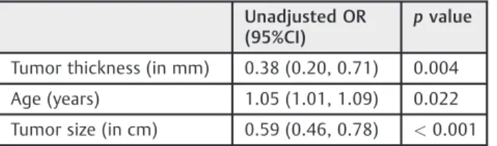
but none of them showed the similar cutoff point of tumor thickness on which neck dissection should be performed. For instance, Mishra et al, in their study of 176 patients with buccal squamous cell carcinoma, found 4 mm tumor Table 1 Univariate analysis (n¼102)
Unadjusted OR (95%CI)
pvalue
Tumor thickness (in mm) 0.38 (0.20, 0.71) 0.004
Age (years) 1.05 (1.01, 1.09) 0.022
Tumor size (in cm) 0.59 (0.46, 0.78) <0.001
Abbreviations: 95%CI, 95% confidence interval; cm, centimeters; mm, millimeters; OR, odds ratio.
Table 2 Multivariate analysis (n¼102)
Adjusted OR (95%CI) pvalue
Tumor thickness (T2 mm) (Ref: T<2 mm)
35.52 (2.39, 527.26) 0.009
Age (years) 1.06 (1.01, 1.12) 0.027
Tumor size (in cm) 0.58 (0.40, 0.82) 0.002
thickness to be a significant predictor of lymph node
me-tastasis.32On the other hand, Urist et al described a series of
89 patients with squamous cell carcinoma of the buccal mucosa and showed that greater than 6 mm tumor thickness
was a worse prognostic factor.33Both of these observations
are in contrast with ourfindings, in which a tumor thickness
of even 2 mm proves to be significantly associated with
lymph node metastasis. So, the relationship between tumor thickness and neck nodal metastasis should not be consid-ered questionable; rather, the size of tumor thickness is the subject of debate.
While considering the outcomes of patients, Janot et al34
and Kantola et al35 found an association between the
pa-tients’age and poor outcomes, which was something also
observed in our study. Further on, it was also noted that an increase in age is associated with an increased risk of cervical lymph node metastasis.
Very little has been done to explore the predictors of cervical neck node metastasis, with respect to tumor thick-ness, from our part of the world. There is limited local literature regarding the subject. With buccal mucosa tumors forming a very high proportion of oral cavity squamous cell carcinoma in our region, and having an unexpectedly high aggressive behavior, we believed that the predictors of occult cervical lymph node metastasis would be worthy of research.
Our study showed that tumor thickness is significantly
associated with the presence of lymph node metastasis in the
univariate and the multivariate analyses (p<0.05). The
association between tumor thickness and lymph node metastasis has been well demonstrated. However, there has been a lack of homogeneity in terms of study population and site of the primary tumor. We were also able to demon-strate that a tumor thickness of 2 mm or more favors an elective neck dissection where squamous cell carcinoma of the buccal mucosa is concerned.
Tumor thickness significantly influences the survival of
the patients, as reported in Gonzalez-Moles et al, Brown et al
and Spiro et al.36–38 As an increase in tumor thickness
reduces the survival rates among the patients, the causal relationship between tumor thickness and neck node me-tastasis in buccal squamous cell carcinoma needs to be explored, and further prospective studies with larger sample sizes and longer follow-up (up to 10 years) periods are required to unify the criteria for the measurement of this important prognostic factor.
Conclusion
We conclude that tumor thickness is significantly related to
neck nodal metastasis in buccal squamous cell carcinoma, considering the age of the patient and the size of the tumor.
References
1 Mignogna MD, Fedele S, Lo Russo L. The World Cancer Report and the burden of oral cancer. Eur J Cancer Prev 2004;13(02):139–142 2 Musani MA, Jawed I, Marfani S, Khambaty Y, Jalisi M, Khan SA. Carcinoma cheek: regional pattern and management. J Ayub Med Coll Abbottabad 2009;21(03):87–91
3 Mamelle G, Pampurik J, Luboinski B, Lancar R, Lusinchi A, Bosq J. Lymph node prognostic factors in head and neck squamous cell carcinomas. Am J Surg 1994;168(05):494–498
4 Huang SH, Hwang D, Lockwood G, Goldstein DP, O’Sullivan B. Predictive value of tumor thickness for cervical lymph-node in-volvement in squamous cell carcinoma of the oral cavity: a meta-analysis of reported studies. Cancer 2009;115(07):1489–1497 5 Pentenero M, Gandolfo S, Carrozzo M. Importance of tumor
thickness and depth of invasion in nodal involvement and prog-nosis of oral squamous cell carcinoma: a review of the literature. Head Neck 2005;27(12):1080–1091
6 Cheng A, Schmidt BL. Management of the N0 neck in oral squamous cell carcinoma. Oral Maxillofac Surg Clin North Am 2008;20(03):477–497
7 Dünne AA, Folz BJ, Kuropkat C, Werner JA. Extent of surgical intervention in case of N0 neck in head and neck cancer patients: an analysis of data collection of 39 hospitals. Eur Arch Otorhino-laryngol 2004;261(06):295–303
8 Werning JW, Heard D, Pagano C, Khuder S. Elective management of the clinically negative neck by otolaryngologists in patients with oral tongue cancer. Arch Otolaryngol Head Neck Surg 2003; 129(01):83–88
9 Capote A, Escorial V, Muñoz-Guerra MF, Rodríguez-Campo FJ, Gamallo C, Naval L. Elective neck dissection in early-stage oral squamous cell carcinoma–does it influence recurrence and sur-vival? Head Neck 2007;29(01):3–11
10 Okada Y, Mataga I, Katagiri M, Ishii K. An analysis of cervical lymph nodes metastasis in oral squamous cell carcinoma. Rela-tionship between grade of histopathological malignancy and lymph nodes metastasis. Int J Oral Maxillofac Surg 2003;32-(03):284–288
11 Pimenta Amaral TM, Da Silva Freire AR, Carvalho AL, Pinto CA, Kowalski LP. Predictive factors of occult metastasis and prognosis of clinical stages I and II squamous cell carcinoma of the tongue andfloor of the mouth. Oral Oncol 2004;40(08):780–786 12 Alkureishi LW, Ross GL, Shoaib T, et al. Does tumor depth affect
nodal upstaging in squamous cell carcinoma of the head and neck? Laryngoscope 2008;118(04):629–634
13 Clark JR, Naranjo N, Franklin JH, de Almeida J, Gullane PJ. Estab-lished prognostic variables in N0 oral carcinoma. Otolaryngol Head Neck Surg 2006;135(05):748–753
14 Jing J, Li L, He W, Sun G. Prognostic predictors of squamous cell carcinoma of the buccal mucosa with negative surgical margins. J Oral Maxillofac Surg 2006;64(06):896–901
15 Sheahan P, O’Keane C, Sheahan JN, O’Dwyer TP. Effect of tumour thickness and other factors on the risk of regional disease and treatment of the N0 neck in early oral squamous carcinoma. Clin Otolaryngol Allied Sci 2003;28(05):461–471
16 Kane SV, Gupta M, Kakade AC, D’Cruz A. Depth of invasion is the most significant histological predictor of subclinical cervical lymph node metastasis in early squamous carcinomas of the oral cavity. Eur J Surg Oncol 2006;32(07):795–803
17 Moore C, Kuhns JG, Greenberg RA. Thickness as prognostic aid in upper aerodigestive tract cancer. Arch Surg 1986;121(12): 1410–1414
18 Hanif M, Zaidi P, Kamal S, Hameed A. Institution-based cancer incidence in a local population in Pakistan: nine year data analysis. Asian Pac J Cancer Prev 2009;10(02):227–230 19 Bhurgri Y, Bhurgri A, Usman A, et al. Epidemiological review of
head and neck cancers in Karachi. Asian Pac J Cancer Prev 2006; 7(02):195–200
20 Diaz EM Jr, Holsinger FC, Zuniga ER, Roberts DB, Sorensen DM. Squamous cell carcinoma of the buccal mucosa: one institution’s experience with 119 previously untreated patients. Head Neck 2003;25(04):267–273
22 Sieczka E, Datta R, Singh A, et al. Cancer of the buccal mucosa: are margins and T-stage accurate predictors of local control? Am J Otolaryngol 2001;22(06):395–399
23 Shaw RJ, McGlashan G, Woolgar JA, et al. Prognostic importance of site in squamous cell carcinoma of the buccal mucosa. Br J Oral Maxillofac Surg 2009;47(05):356–359
24 Ferlito A, Rinaldo A, Devaney KO, et al. Prognostic significance of microscopic and macroscopic extracapsular spread from meta-static tumor in the cervical lymph nodes. Oral Oncol 2002;38(08): 747–751
25 Larsen SR, Johansen J, Sørensen JA, Krogdahl A. The prognostic significance of histological features in oral squamous cell carci-noma. J Oral Pathol Med 2009;38(08):657–662
26 Warburton G, Nikitakis NG, Roberson P, et al. Histopathological and lymphangiogenic parameters in relation to lymph node metastasis in early stage oral squamous cell carcinoma. J Oral Maxillofac Surg 2007;65(03):475–484
27 Barrera JE, Miller ME, Said S, Jafek BW, Campana JP, Shroyer KR. Detection of occult cervical micrometastases in patients with head and neck squamous cell cancer. Laryngoscope 2003;113-(05):892–896
28 Enepekides DJ, Sultanem K, Nguyen C, Shenouda G, Black MJ, Rochon L. Occult cervical metastases: immunoperoxidase analy-sis of the pathologically negative neck. Otolaryngol Head Neck Surg 1999;120(05):713–717
29 Yoshida K, Kashima K, Suenaga S, Nomi N, Shuto J, Suzuki M. Immunohistochemical detection of cervical lymph node micro-metastases from T2N0 tongue cancer. Acta Otolaryngol 2005; 125(06):654–658
30 Akoğlu E, Dutipek M, BekişR, Değirmenci B, Ada E, Güneri A. Assessment of cervical lymph node metastasis with different imaging methods in patients with head and neck squamous cell carcinoma. J Otolaryngol 2005;34(06):384–394
31 Merritt RM, Williams MF, James TH, Porubsky ES. Detection of cervical metastasis. A meta-analysis comparing computed tomo-graphy with physical examination. Arch Otolaryngol Head Neck Surg 1997;123(02):149–152
32 Mishra RC, Parida G, Mishra TK, Mohanty S. Tumour thickness and relationship to locoregional failure in cancer of the buccal mu-cosa. Eur J Surg Oncol 1999;25(02):186–189
33 Urist MM, O’Brien CJ, Soong SJ, Visscher DW, Maddox WA. Squamous cell carcinoma of the buccal mucosa: analysis of prognostic factors. Am J Surg 1987;154(04):411–414
34 Janot F, Klijanienko J, Russo A, et al. Prognostic value of clinico-pathological parameters in head and neck squamous cell carci-noma: a prospective analysis. Br J Cancer 1996;73(04):531–538 35 Kantola S, Parikka M, Jokinen K, et al. Prognostic factors in tongue cancer - relative importance of demographic, clinical and histo-pathological factors. Br J Cancer 2000;83(05):614–619
36 Gonzalez-Moles MA, Esteban F, Rodriguez-Archilla A, Ruiz-Avila I, Gonzalez-Moles S. Importance of tumour thickness measurement in prognosis of tongue cancer. Oral Oncol 2002;38(04):394–397 37 Brown B, Barnes L, Mazariegos J, Taylor F, Johnson J, Wagner RL. Prognostic factors in mobile tongue andfloor of mouth carci-noma. Cancer 1989;64(06):1195–1202