J of Evolution of Med and Dent Sci/ eISSN- 2278-4802, pISSN- 2278-4748/ Vol. 3/ Issue 22/June 02, 2014 Page 5997
UTILITY OF HEMATOLOGICAL AND LABORATORY DATA IN ASSESSMENT
OF IMMEDIATE PROGNOSIS IN MULTIPLE MYELOMA
Kalaranjini K. V1, Vinukumar V2, Sankar S3, Sheela Vasudevan4
HOW TO CITE THIS ARTICLE:
Kalaranjini K. V, Vinukumar V, Sankar S, Sheela Vasudevan. Utility of Hematological and Laboratory Data in Assessment of Immediate Prognosis in Multiple Myeloma . Journal of Evolution of Medical and Dental Sciences 2014; Vol. 3, Issue 22, June 02; Page: 5997-6002, DOI: 10.14260/jemds/2014/2695
ABSTRACT: BACKGROUND: Plasma cell myeloma comprises about 1% of malignant tumors, 10-15% of hematopoietic neoplasms and causes 20% of deaths from hematologic malignancies. Several clinical, laboratory and histological/cytological variables help us in determining the prognosis of the disease.There have been very few studies from India evaluating bone marrow histology of plasma cells in myeloma. The present study is aimed at analyzing and studying the various clinical presentations, biochemical, hematologic and histological parameters determining immediate prognosis [mortality] in myeloma. MATERIAL AND METHODS: Prospective study was conducted from April 1st 2008 to March 2010. 48 patients were diagnosed with multiple myeloma during study
period; 41 were available for follow-up.ie for a minimum 6 months from the time of diagnosis. Among these seven cases expired i.e. in the period of 6 months from the respective time of diagnosis. Biochemical and hematological variables of those expired [seven] and those alive available for follow-up [thirty-Four] were compared. Results of biochemical investigations were collected from the case sheet. Peripheral smear, Bone marrow aspiration and biopsy were done for all cases. STATISTICAL ANALYSIS USED: student’s t test and chi square test. RESULTS: Among the variables considered mean hemoglobin level in expired vs. alive was 4.2 vs. 7.5 g/dl. Mean ESR was 133 vs. 102 mm/hr. Mean blood urea was 97.3 vs. 38.5 mg/dl. A/G reversal was present in 85.7% of expired and in 50% of alive patients. Average plasma cell percentage in the bone marrow aspirate in expired vs. alive was 74.7% and 43.1%.Plasmablastic variety was present in 71.4% expired and 17.6% of alive patients. Diffuse pattern of infiltration was present in 85.7% of expired and 44.1% of alive patients. Grade III fibrosis was present in 85.7% of expired and 20.6% of alive patients. All variables except A/G reversal had p <.005. CONCLUSION: Immediate Prognostication of Multiple myeloma requires both hematological and laboratory data. Low hemoglobin, raised blood urea, and elevated ESR, plasma cell percentage, plasmablastic variety, diffuse pattern of infiltration and grade III fibrosis were proved to have a direct link to immediate mortality.
KEYWORDS: Multiple myeloma, immediate mortality, prognosis, bone marrow, plasmablastic
INTRODUCTION: Plasma cell myeloma is a bone marrow based multifocal plasma cell neoplasm associated with an M protein in serum and/or urine. In most cases there is disseminated bone marrow involvement. The disease spans a clinical spectrum from asymptomatic to aggressive forms and disorders due to deposition of abnormal immunoglobulin in tissues.
Plasma cell myeloma comprises about 1% of malignant tumors, 10-15% of hematopoietic neoplasms1 and causes 20% of deaths from hematologic malignancies. Geographic and familial
clustering2 of cases can occur.
J of Evolution of Med and Dent Sci/ eISSN- 2278-4802, pISSN- 2278-4748/ Vol. 3/ Issue 22/June 02, 2014 Page 5998 substances or radiation.3,4,5 But most patients have no identifiable toxic exposure or known chronic
antigenic stimulation. Several studies show an increase in incidence of myeloma during the last decade; this may be due to increased awareness, improved diagnostic approaches and access to many effective treatments.
The current WHO Classificationof Tumors of Hematopoietic and lymphoid Tissues (2007) has put forward a new diagnostic criteria1 and according to this the most important criteria for
symptomatic myeloma are manifestations of end organ damage (CRAB) hypercalcemia, renal insufficiency, anemia, and bone lesions.
Bone marrow examination continues to be the cornerstone for establishing a diagnosis in association with other clinical and laboratory parameters. Several clinical, laboratory and histological/cytological variables help us in determining the prognosis of the disease. The first histological classification and staging of multiple myeloma, based on the bone marrow trephine biopsy, was put forwarded by Bartl et al.6 in 1987.
The Durie and Salmon7 clinical staging system, proposed in 1977, is still being used today,
though it has been replaced by the ISS staging at many places. Prognostic markers like beta2 microglobulin and Interleukin-6 (IL-6) are not available in most centres in developing countries like India.
Histological parameters that have a definitive prognostic significance in multiple myeloma are the percentage of myeloma cells in the marrow, pattern of infiltration, degree of plasma cell atypia, marrow fibrosis and mitotic index.There have been very few studies from India evaluating bone marrow histology of plasma cells in myeloma.
The present study is aimed at analyzing and studying the various clinical presentations, biochemical, hematologic and histological parameters in myeloma with relation to immediate prognosis.
MATERIALS AND METHODS: The study was conducted in Department of Pathology, Medical college Kottayam from April 1st 2008 to March 2010. All patients who satisfied the criteria for diagnosis of
plasma cell myeloma included in the study. Of the 48 patients diagnosed 41 were available for follow-up. i.e. for a minimum of 6 months from the time of diagnosis. Of them seven cases expired i.e. in the period of 6 months from the respective time of diagnosis. Variables of those expired [seven] and those alive available for follow-up [thirty-four] were compared.
Peripheral blood taken for routine investigations (Hemoglobin, TC, DC, ESR) and blood urea. Bone marrow aspirates and bone marrow trephine biopsy sections were also taken.
Bone marrow aspirate smears are dried and stained with Leishman’s stain. The adequacy, cellularity, hematopoiesis of all three cell lines and percentage of plasma cells estimated by a 500 cell differential count. The criteria put forward by the German myeloma task force was used to type the plasma cell morphology into well differentiated, intermediately differentiated or poorly differentiated plasma cell.8
J of Evolution of Med and Dent Sci/ eISSN- 2278-4802, pISSN- 2278-4748/ Vol. 3/ Issue 22/June 02, 2014 Page 5999
STATISTICAL ANALYSIS: Data collected were analyzed using SPSS v 13. Categorical variables analyzed for statistical significance by student’s t test and chi square test. Statistical significance assumed to be present if p value <0.05.
RESULTS: Of the 48 patients diagnosed 41 were available for follow-up. i.e. for a minimum of 6 months from the time of diagnosis. Of them seven cases expired i.e. in the period of 6 months from the respective time of diagnosis. Variables of those expired [seven] and those alive available for follow-up [thirty-four] were compared.
Hematological and biochemical variables compared included hemoglobin, blood urea, ESR and A/G reversal.
The results with the corresponding p values are tabulated below:
Mean Hb g% Mean ESR mm/hr.
Mean urea. mg/dl
A/G reversal Present
Expired 4.68 114 100.87 6 cases
Alive 7.2 110 60 17 cases
P value .003 .003 .001 .301
Table 1
Low Hemoglobin, raised blood urea, raised ESR at time of presentation correlated with immediate prognosis i.e. Mortality within 6 months from time of diagnosis.
Among the variables considered all except for A/G Reversal were significant indicators of immediate mortality. All had a p value less than.05 except for A/G reversal.
Variables with regard to bone marrow aspirate and biopsy were also compared; i.e. bad prognostic variables like bone marrow plasma cell percentage, plasmablastic morphology, diffuse pattern of infiltration and grade 3 reticulin fibrosis were compared in both expired and alive patients they are tabulated below with p values.
Status BM plasma cell percentage
Plasmablastic morphology. [percentage]
Diffuse pattern of infiltration [percentage]
Grade 3 fibrosis [percentage]
Expired 74.7 71.4 85.7 85.7
Alive 43.1 17.6 44.1 20.6
P value .001 .003 .04 .003
J of Evolution of Med and Dent Sci/ eISSN- 2278-4802, pISSN- 2278-4748/ Vol. 3/ Issue 22/June 02, 2014 Page 6000
Fig. 1: Bone marrow aspirate –mature
plasma cells.(leishman stain)
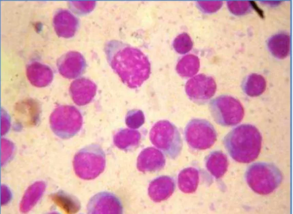
Fig. 2: Bone marrow aspirate–plasmablastic
cells. (leishman stain)
Fig. 3: Bone marrow biopsy-cart wheel
chromatin of plasma cells –diffuse
pattern.(H&E)
Fig. 4: Bone marrow biopsy with plasmablastic plasmacells (H&E)
J of Evolution of Med and Dent Sci/ eISSN- 2278-4802, pISSN- 2278-4748/ Vol. 3/ Issue 22/June 02, 2014 Page 6001 All the parameters taken in to consideration i.e. bone marrow plasma cell percentage, diffuse pattern of infiltration, plasmablastic morphology and grade 3 fibrosis showed significance [p<.05] with immediate prognosis. These results were also in concordance with study by Subramanian R, Basu D, Dutta TK.10
CONCLUSION: The correlation of biochemical and hematological parameters with immediate mortality showed direct link between raised blood urea, raised ESR, low hemoglobin, high percentage of plasma cells in marrow, plasmablastic morphology, diffuse pattern of marrow involvement, high grade [grade 3] fibrosis.
REFERENCES:
1. WHO classification of tumours of haematopoetic and lymphoid tissues 2008.
2. Grosbois B, Jego P, Attal M, et al. Familial multiple myeloma: report of fifteen families. Br J Haematol 1999; 105: 768–770.
3. Committee to Review Health Effects in Vietnam Veterans of Exposure to Herbicides. Division of Health Promotion and Disease Prevention. Institute of Medicine. Cancer in: Veterans and Agent Orange: Health Effects of Herbicides Used in Vietnam. Washington, D.C., National Academy Press, 1994, pp 433-590.
4. Schwartz GG. Multiple myeloma: clusters, clues and dioxins. Cancer Epidemiol Biomarkers Prev 1997; 6: 49–56.
5. Goedert JJ, Cote TR, Virgo P, et al. Spectrum of AIDS-associated malignant disorders. Lancet 1998; 351: 1833–1839.
6. Bartl R, Frisch B, Fateh-Moghadam A, Kettner G, Jaeger K, Sommerfeld W. Histologic classification and staging of multiple myeloma: A retrospective and prospective study of 674 cases. Am J Clin Pathol 1987; 87: 342-55.
7. Durie BG, Salmon SE. A clinical staging system for multiple myeloma. Correlation of measured myeloma cell mass with presenting clinical features, response to treatment, and survival. 1975; Cancer 36:842–854.
8. Sailer M, Vykoupil KF, Peest D, Coldewey R, Deicher H, Georgii A. Prognostic relevance of a histologic classification system applied in bone marrow biopsies from patients with multiple myeloma: A histopathological evaluation of biopsies from 153 untreated patients. Eur J Haematol 1995; 54: 137-46.
9. Thiele J, Kvasnicka HM, Facchetti F, Franco V, van der Walt J, Orazi A. European consensus on grading bone marrow fibrosis and assessment of cellularity. Haematologica 2005; 90: 1128-32. 10.Prognostic significance of bone marrow histology in multiple myeloma R Subramanian, D Basu,
J of Evolution of Med and Dent Sci/ eISSN- 2278-4802, pISSN- 2278-4748/ Vol. 3/ Issue 22/June 02, 2014 Page 6002
AUTHORS:
1. Kalaranjini K. V. 2. Vinukumar V. 3. Sankar S.
4. Sheela Vasudevan
PARTICULARS OF CONTRIBUTORS:
1. Assistant Professor, Department of Pathology, Sree Gokulum Medical College and Research Foundation.
2. Assistant Professor, Department of Pathology, Sree Gokulum Medical College and Research Foundation.
3. Professor, Department of Pathology, Priyadarsini Institute of Paramedical Sciences.
4. Professor and HOD, Department of Pathology, Sree Gokulum Medical College and Research Foundation.
NAME ADDRESS EMAIL ID OF THE CORRESPONDING AUTHOR:
Dr. Kalaranjini K. V, Assistant Professor, Department of Pathology,
Sree Gokulam Medical College and Research Foundation,
Venjaramoodu, Trivandrum, Kerala. Email: [email protected]