Erasmus Syndrome
A Rare Clinical Condition: Erasmus Syndrome
Nadir Görülen Klinik Bir Durum; Erasmus Sendromu
DOI: 10.4328/JCAM.4271 Received: 06.01.2016 Accepted: 25.01.2016 Printed: 01.07.2016 J Clin Anal Med 2016;7(4): 554-6 Corresponding Author: Yunus Ugan, Department of Internal Medicine, Division of Rheumatology, Suleyman Demirel University Faculty of Medicine, Isparta, Turkey. T.: +90 2462119220 F.: +90 2462370240 E-Mail: [email protected]
Özet
Sistemik Skleroz (SS), cildin ve iç organların fibrozisi ile giden, sebebi tam olarak aydınlatılamayan sistemik otoimmün bir hastalıktır. Hastalığın çevresel faktörler
-le ilişkisi olduğu bilinmektedir. Özellik-le silica tozlarıyla maruziyetin bir takım im
-mun reaksiyonların tetiklenmesiyle hastalığın patogenezinde rol aldığı düşünül
-mektedir. Silikozis ve SS birlikteliği Erasmus Sendromu (ES) olarak tanımlanmak
-tadır. Burada 6 yıldır silikozis nedeniyle takipli, kotlama işçisi olarak çalışan 30 ya
-şında sistemik sklerozlu bir olgu sunulmaktadır.
Anahtar Kelimeler
Silikozis; Sistemik Skleroz; Erasmus Sendromu
Abstract
Systemic sclerosis (SS) is a systemic autoimmune disease progressing with i
-brosis of the skin and internal organs, the cause of which cannot be precisely explained. The disease is known to be associated with environmental factors. In particular, exposure to silica powders is believed to have a part in the pathogen
-esis of the disease by the triggering of a number of immune reactions. Silicosis and SS association is deined as Erasmus Syndrome (ES). Here, we report on a 30-year-old patient working in denim sandblasting who developed SS while being
followed for 6 years due to silicosis.
Keywords
Silicosis; Systemic Sclerosis; Erasmus Syndrome
Yunus Ugan, Atalay Dogru, Mehmet Sahin, Sevket Ercan Tunc Department of Internal Medicine, Division of Rheumatology, Suleyman Demirel University Faculty of Medicine, Isparta, Turkey
Introduction
Systemic sclerosis (SS) is not only a disease progressing with
skin involvement but also a systemic autoimmune disease which afects internal organs such as the gastrointestinal system, kid
-ney, lung, and heart, and which is characterized by ibrosis [1].
The etiopathogenesis of the disease is not yet fully understood.
However, vinyl chloride, organic solvents, benzene, and silica powders have been shown to be associated with the disease [2]. Silicosis is an incurable disease characterized by an irreversible and progressive ibrotic reaction in the lung tissue due to silica crystals of respirable size. Silica is a mineral that occurs inten -sively in the earth’s crust. Exposure to silica powders is high in people workingin mines and quarries, ceramic manufacturing,
pottery making, and denim sandblasting works [3]. The associa
-tion between silicosis and SS is deined as Erasmus Syndrome (ES). Here, we report on a 30-year-old patient working in den
-im sandblasting who developed SS while being followed for 6
years due to silicosis.
Case Report
The patient, who was working in denim sandblasting for 10 years and had been followed with a diagnosis of silicosis for
6 years, applied to the rheumatology clinic with whitening and
bruising on the hands in response to cold exposure and pain
in the hand joints. The patient started to have pains that
in-creased especially at nights, accompanied by swellings in the
wrists and metacarpophalangeal joints. During the previous 3
weeks, a necrotic skin ulcer had occurred on the second inger of the let hand of the patient, who described recent hardening of
the skin. The patient had no relevant features in the family
his-tory. Physical examination found TA of 130/80 mmHg and body
temperature of 36.7°C. There were telangiectasias on the face and vertical lines on the lips. The mouth opening was narrowed.
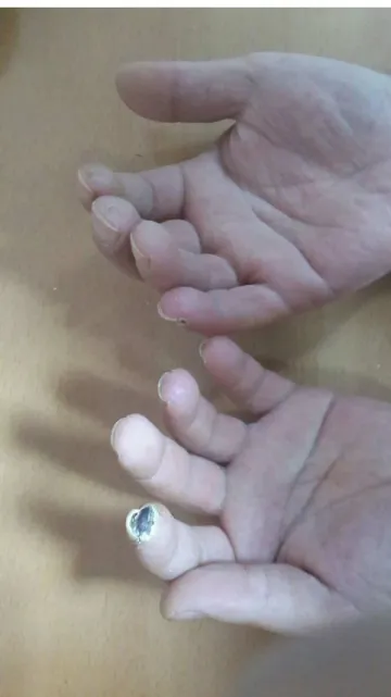
Bilateral ine crackles were found in breathing sounds. While hardening of the skin was detected on the back of the hand, a digital ulcer was detected on the second inger of the let hand
(Figure 1). In the thoracic tomography, while enlarged multiple lymph nodes measuring more than 40 mm in maximum size
were observed in the mediastinum and subcarinal space, septal thickening was observed in both lung bases (Figure 2). In the
transthoracic echocardiography, pulmonary artery pressure was
detected as 26 mmHg and EF was detected as 65%. In pulmo
-nary function test, FEV1, FVC, FEV1/FVC, and DLCO were 65%, 72%, 71%, and 79%, respectively. The sputum examination and culture for acid resistant bacillus (ARB) were negative and a tuberculin skin test (PPD) was measured as 7 mm. In the labo
-ratory examination the following values were detected: WBC: 8400/mm3, platelet: 365 × 103 per μL, Hb: 12.2 gr/dl, MCV: 81,
C-reactive protein: 78 mg/dl (N:0-3), erythrocyte sedimentation rate: 45 mm/h. Immunological investigations showed rheuma-toid factor (RF): 34 IU/l (N:0-15), anti cyclic citrullinated peptide
(anti-CCP): 5 (N:0-7), antinuclear antibody (ANA) homogeneous
and granular pattern 3+ with anti SCL 70. The patient was
di-agnosed with SS based on the available clinical and laboratory
results. The disease was considered as ES due to the
progres-sion of the silicosis. Lung involvement of SS was scanned by high resolution computed tomography but neither honeycomb nor ground glass opacity were detected. While methotrexate
15 mg/week was administered for the systemic disease of the
patient, calcium channel blocker, pentoxifylline, acetylsalicylic Figure 1. Digital ulcer on the 2nd inger of the let hand
Figure 2.Thoracic tomography; multiple lymph nodes in the mediastinum
Journal of Clinical and Analytical Medicine | 555
acid, and intravenous ilioprost treatment were administered for the digital ulcer. Recovering from the digital ulcer during follow-ups, the patient was discharged with recommendations.
Discussion
Erasmus Syndrome was irst reported by Erasmus in 1957 and its relationship between silicosis and SS was revealed. In addi
-tion, silicosis was also reported to be associated with pulmo
-nary tuberculosis, lung cancer, rheumatoid arthritis, and sys
-temic lupus erythematosus [4]. Although the pathogenesis of the relationship between silica powders and SS development has not been precisely explained yet, it is thought that silica particles are phagocytosed by macrophages, which ultimately leads to the release of lymphokine and chemokine by activating the ibroblasts. This also increases the synthesis of collagen and glycosaminoglycan and suppresses cellular immunity [5]. Also, Otsuki et al. reported that fasand caspase 8 antibodies
and the lymphocyte-mediated apoptosis play a key role in
tis-sue damage and immunity [6].
In SS cases developing secondary to silicosis, autoantibodies
such as anti-scl 70 and RF are generally detected as positive;
however, no clinical diference from the classical SS is observed [5]. Also in our patient, anti-scl 70 and RF were positive. The association of silica powders with tuberculosis has been known for many years. The incidence of tuberculosis in patients with
silicosis is higher than in people without silicosis. The
neces-sary examinations should be carried out when this diagnosis is suspected, because there is not always clear clinical evidence. In our patient, PPD was performed to rule out tuberculosis, and ARB and sputum culture were performed. However, there were no positive indings.
In SS cases developing with silicosis, decrease in difusion ca
-pacity in the lung is remarkable. Respiratory insuiciency is
generally seen in the form of restrictive pattern; dry cough and
exertional dyspnea constitute the clinical spectrum [7]. Our pa -tient’s pulmonary function test was in restrictive pattern and
there was a slight decrease in difusion capacity. While the risk
of secondary malignancy development increased with connec-tive tissue disease, the risk of lung canceris further increased
in patients with SS when in association with silica powders [8]. Therefore, attention should be paid to secondary malignancy
in patients with ES and the necessary periodic examinations
should be performed.
In silicosis-related SS cases, treatment does not vary much; it includes immunosuppressives such as corticosteroids,
cyclo-phosphamide, and azathioprine [3]. In patients with secondary
complications of SS such as Raynaud’s phenomenon, digital ulcer, gastrointestinal and cardiovascular system involvement, palliative treatments for these involvements are also used.
Thus, because digital ulcer was at the forefront in our patient,
treatment for this involvement was given.
Because silicosis is a refractory disease that occurs as a result
of exposure to silica, connective tissue diseases may develop
in these patients. Especially, attention should be paid to the possibility of rarely-seen ES and periodic follow-ups should be
performed with respect to potential lung cancer.
Competing interests
The authors declare that they have no competing interests.
References
1. Denton CP. Systemic sclerosis: from pathogenesis to targeted therapy. Clin Exp Rheumatol 2015;33(4):3-7.
2. Stern EP, Denton CP. The Pathogenesis of Systemic Sclerosis. Rheum Dis Clin North Am 2015;41(3):367-82.
3. Leung CC, Yu IT, Chen W. Silicosis. Lancet 2012;379(9830):2008-18. 4. Koeger AC, Lang T, Alcaix D, Milleron B, Rozenberg S et al. Silica - associ
-ated connective tissue disease. A study of 24 cases. Medicine (Baltimore)
1995;74(5):221-37.
5. Zaghi G, Koga F, Nisihara RM, Skare TL, Handar A et al. Autoantibodies in silico -sis patients and in silica-exposed individuals. Rheumatol Int 2010;30(8):1071-75.
6. Otsuki T, Maeda M, Murakami S, Hayashi H, Miura Y et al. Immunological efects of silica and asbestos. Cell MolImmunol 2007;4(4):261-8.
7. Martin RJ, Griin M, Moore E, Lochead JA, Edwards AC et al. Systemic sclerosis
(scleroderma ) in two iron ore mines. Occup Med 1999;49(3):161-9.
8. Chaouch N, Mjid M, Zarrouk M, Ammous I, Hantous S et al. Erasmus’ syndrome
with pseudo-tumour masses. Rev Mal Respir 2011;28(7):924-7.
How to cite this article:
Ugan Y, Dogru A, Sahin M, Tunc SE. A Rare Clinical Condition: Erasmus Syndrome. J Clin Anal Med 2016;7(4): 554-6.
| Journal of Clinical and Analytical Medicine 556