PL ISSN 0033-2674 www.psychiatriapolska.pl
Psychotic disorder in the course of Systemic Lupus
Erythematosus with subcortical calciications - case report
Michalina M a l e c1, Monika Rudzińska1, Dominika D u d e k2, Marcin S i w e k2, Marcin W n u k1, Andrzej S z c z u d l i k1
1 Department of Neurology, Jagiellonian University, Medical College, Krakow
Head: prof. dr hab. n. med. A. Szczudlik
2 Department of Affective Disorders Chair of Psychiatry, Jagiellonian University,
Medical College, Krakow Head: prof. dr. hab. n. med. D. Dudek
Summary
Systemic Lupus Erythematosus (SLE) is autoimmunological disease of connective tissue which is characterized with clinical symptoms of many systems and organs injury. There are often neuropsychiatric symptoms. Psychotic disorder is the least frequent syndrome. Neuropsychiatric symptoms are important because they deteriorate the quality of life and are poor prognostic factor.
Aim. The aim of the study is to present the patient with chronic, lasting for many years, skin lesions and laboratory tests results characteristic for SLE, who had psychotic disorder diagnosed as schizophrenia and in the next few years there were observed other neuropsy-chiatric symptoms including cognitive impairment and mood disorder.
Conclusions. Psychotic disorder is rare syndrome of neuropsychiatric SLE (NPSLE). It may primarily originate from SLE or be secondary either to the therapy or the complications
of the disease. It is not possible to deine if the psychosis is the primary schizophrenic process
or secondary to the autoimmune disease in presented patient. However the clinical picture pays
attention to the signiicance of careful diagnostic process, including neuroimaging. In head CT of presented patient there were revealed massive, bilateral, calciications of subcortical
structures which probably substantially enhanced neuropsychiatric symptoms.
Key words: Neuropsychiatric lupus erythematosus, schizophrenia, calciication of the basal
ganglia
Introduction
often are cognitive impairment or even dementia which is observed in 80% of patients, and depression in about 40% of patients [1]. The anxiety disorder and rarely in 3,5-8% of cases even the psychotic symptoms may occur [2]. In 1999 research committee
of American College of Rheumatology (ACR) deined 19 various syndromes of neu -rological and psychological symptoms because of the high prevalence of neuropsy-chiatric disorder, setting new special entity called Neuropsyneuropsy-chiatric Systemic Lupus Erythematosus (NPSLE). Neuropsychiatric symptoms are important clinical problem, because they worsen the quality of patient life and they are poor prognostic factors.
The aim of this study is to present the patient with long-lasting skin lesions and
the laboratory tests results speciic for SLE who had psychotic disorder and was treated
for schizophrenia with other neuropsychiatric symptoms such as cognitive impairment and mood disorder which occurred in later time.
Case report
The woman at the age of 47, married, mother of one child (27 years old nowa-days) was admitted to the Neurological Clinic of Medical College, Jagiellonian Uni-versity in Cracow because of progressing slowness of movement and gait disorder. Since the age of 12 she has suffered from extended skin lesions on the face and head (erythematous rash). The SLE was suspected, however the proper laboratory tests were not performed. At the age of 30 the skin lesions generalized, also appeared on
the trunk and legs. In the laboratory tests the titre of nonspeciic antinuclear antibodies
was increased (1:320), beside there was low level of C4 complement and tendency to thrombocytopenia. The SLE was diagnosed and the therapy with prednisone was performed (60 mg per day), and then continued for many years.
At the age of 39 in patient developed the persecutory delusions, delusions of refer-ence, unveiling delusions, delusions of stealing ideas and delusions with complex and bizarre substance (e.g. the soul of mother is altered, the mother has the head of frog)
accompanied by illusory and hallucinatory sensations. Under the inluence of mentioned
productive symptoms the patient was sometimes aggressive, with inadequate behavior. In the medical history documents there were observed formal thought disorder (per-severation, dissociation) and there was no insight into the disease. Due to the severity of the psychosis symptoms, the patient was hospitalized at the time in a psychiatric hospital. The schizophrenia was diagnosed and the therapy with antipsychotic drugs was performed, starting with haloperidol for 4 years, and then with aripiprazole and
lupenthixol.
Since the age of 41 the patient was observed by her husband with progressive
concentration disturbances, memory deicits and planning inability which deterio -rated daily functioning at last time. Since 4 years increases moving slowness, gait disturbances and involuntary twisting movements of left extremities have occurred, in particular distally.
dystonic movements of left extremities, slightly increased muscle tone (rigidity) of left
extremities, vivic and symmetrical deep tendon relexes of four limbs, Oppenheim
symptom in left lower limb, parkinsonian posture of anteversion, without left arm
swinging, abnormal postural relexes, MMSE- 24 points.
In psychiatric examination: the patient was properly orientated, in logical ver-bal contact, statements scarce, non-spontaneous. Pale affect, not adaptable, slightly depressed mood, periodical perseverations, incoherent thinking with disturbed train
of thought. The patient had dificulties with transferring the attention. There was seen
fatigability and psychomotor slowness. The patient was speaking about low functional
activity, dificulties in performing daily life tasks, apathy, loss of interests, anhedonia, abulia, impaired concentration and attention, dificulties in initiating the activity. There
were seen emptiness of thinking, purity of statements. She was talking about feeling of hopelessness, sadness, inability to think about future, resigned thoughts (actually without suicidal either thoughts or tendency). The patient was fully dependent from mother’s help, retired from social life, she was avoiding interpersonal contacts, non-spontaneous. She had fully preserved criticism for past delusions and hallucinations. Actually she did not have productive symptoms. The patient acknowledged that in the past she had delusions of unveiling and sending thoughts, persecutory delusions e.g. to the hospital personnel and delusions directed to herself. Criticism for the contem-porary symptoms was reduced. Working and recent memory was impaired while past memory was slightly reduced. Before the hospitalization Neurology Clinic she had suicidal thoughts however without evidence suicidal tendency.
In neuropsychological examination there were especially disturbed executive function, working memory, lower capacity of direct memory magazine for both visual
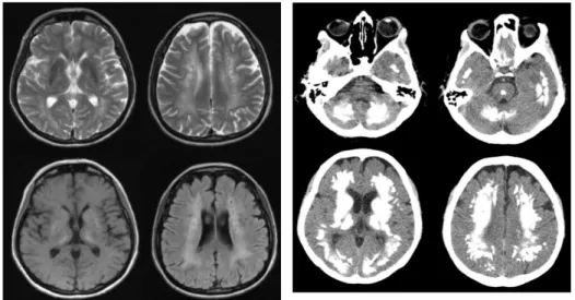
Fig.1. The MRI of patient’s head (July, 2011).
and auditory-verbal modality. In language processes there was signiicantly reduced verbal luency. There was observed bilateral, spatial dyspraxia. The disorder fulilled
the criterion for dementia by ICD-10 and DSM IV with dominant frontal and fronto-subcortical symptoms.
In the MRI of the head (Fig.1.) there were stated hiperintense areas bilaterally in T1 sequence and hypointense in T2 sequence in the region of basal ganglia. In T2 and FLAIR sequence there were also observed hiperintense areas subcortically in white matter.
Because the MRI was inconclusive the computed tomography (CT) was done
(Fig.2.). The images revealed massive, bilateral calciications in subcortical regions
such as basal ganglia, pons, cerebellum and centrum semiovale.
In the laboratory tests the levels of serum parathormone, ionised calcium, TSH and ceuloplasmin were within normal limits. There were detected antinuclear antibodies ANA (anti-Ro 52 kDa) with granular type of lightening in 1:640 titre and with cyto-plasmatic type of lightening in 1:640 titre. The level of C3c and C4 of the complement was within normal limits. There was tendency to thrombocytopenia (129 thousand/ µl). Moreover the genetic test for Huntington disease was negative.
Discussion
The presented case shows complex neurological and psychopathological phenom-enology in patient with the diagnosis of both SLE and schizophrenia. The important fact is that there is no possibility to resolve whether the psychotic symptoms are because of the primary schizophrenic process or they are similar to the schizophrenia syndrome and secondary to the pathophysiological process caused by SLE or the medications for autoimmune disease. First psychotic episodes occurred in relatively mature (39 years), professionally active, socially well adapted person, with stabilized family (husband, child). The psychosis was preceded by long-term SLE treatment. In the family there were no psychotic diseases. Mentioned above information argue for the conception
of the organic schizophrenic syndrome origin. On the other hand psychotic episodes
were recurrent and the exacerbations were independent from the SLE course and its treatment. The regression of psychosis was strictly dependent on the antipsychotic therapy and whether the patient was applying to the recommendations. Clinically acute psychotic episodes were very characteristic for paranoid schizophrenia, similarly long
lasting deicit symptoms after reduction of productive symptoms.
There is multifactor etiology of actual signiicant social deterioration. It is caused by
negative symptoms of psychosis (apathy, abulia, lack of willingness, non-spontaneity), depressive mood, cognitive impairment and motor disorder.
Nowadays there is no single test for strong diagnosis of NPSLE. The neuropsy-chiatric diagnosis in the course of SLE need the rheumathological examination, neu-roimaging, immunoserological tests, psychiatric examination and neuropsychological tests in terms of active phase of the disease. The diagnosis needs the exclusion of other
glucocortycosteroids, renal failure (uraemia), arterial emboli and primary psychiatric disease [2]. The last condition is inconclusive in presented case.
The psychotic symptoms are present the least frequently among the syndromes
of NPSLE. Usually they envelope in early phase of the disease, during the irst year.
Rarely they can be the only symptom of CNS disease [3]. It was evidenced that skin lesions coexists a little bit more often with psychotic disorder. In 14 years follow-up of patients with psychosis due to SLE it was observed that 60% of patients have the
remission during the irst year. Simultaneously the patients with fast remission are
characterized with deeper psychosis. Chronic psychotic disorder, lasting for longer than 10 years occurred in 20% of cases [4]. Typically the psychotic symptoms in NPSLE are disturbed form and substance of thinking, delusions, hallucinations, limited attention, tendency to get distracted, perceive disturbances, agitation and even aggression [5].
It is important to assess properly if the symptoms are caused by the therapy or by the failure of the CNS which is more often. It is proved that severe psychotic symptoms may be complication of steroid therapy only if daily dose is more than 40 mg of prednisone [6]. But in chronic therapy the depression likewise in short therapy
hypomania are more often than psychosis. Moreover the psychosis develops in irs 1-2 weeks of treatment [7]. In our patient irst symptoms occurred after 9 years of curing.
Until now there is only one case report of the patient with coexistence of
schizo-phrenia and SLE. But in that patient the psychotic symptoms were irst and for a long
time they were isolated. They occurred 14 years before other SLE symptoms, especially skin lesions, have occurred. After excluding lupus related to the antipsychotic drugs the authors suggested the participation of antibodies in pathogenesis of schizophrenia
[8]. There are several theories of the schizophrenia’s etiology such as the inluence of
genetic, civilizational, prenatal, social and economic factors, usage of psychoactive substances, psychological, emotional and biochemical elements. However in the last few years the relation between disturbed immunological processes and schizophrenia has been raised [9].
In the case of NPSLE the neuroimaging is recommended only in special conditions such as: the age over 60, after head injury, when the cognitive impairment progresses dynamically or when new neurological symptoms occurred. However the ICD-10 criteria require the brain disease exclusion for schizophrenia diagnosis. Because in patient’s head MRI the changes were inconclusive in the next step head CT was
done which revealed massive calciications of subcortical structures and cerebellum.
In the literature there are only 9 case reports of patients with SLE who had massive
calciications of cerebellum and subcortical structures including striatum and globus
pallidus [10-18]. In these case reports the dominant symptoms were cognitive impair-ment, depression and also neurological symptoms such as parkinsonism, involuntary movements, pyramidal signs, ataxia. Psychosis occurred in 2 cases. At any of two the psychotic symptoms were the only neuropsychiatric disorder. Both patients had recur-rent episodes of psychosis or needed chronic psychiatric care [15, 18].
complications of medicines [17]. In 30% of NPSLE some small calciications may be
seen in neuroimaging. Globus pallidus is the most common structures in which the
calciications occur, however they may be also seen in striatum, cerebellum and other brain structures such as thalamus and centrum semiovale. The reason of calciications
formation is not well known. There is hypothesis that primary vessels damage due to immunological processes may induce microinfarctions and secondary dystrophic
calciication, especially intensiied in basal ganglia, because of strong calcium concen
-tration in these regions [12]. In white matter the calciications probably are induced by exuded neurotoxic factors from vein vessels and local inlammation [13]. It is possible that calciication may coexist with demyelization caused by local encephalomyelitis
with immunological origin in the course of SLE [18].
Massive calciications are rarely seen in neuroimaging and because of that it is dificult to conirm its relation to the clinical symptoms. Calciications of basal gan -glia are relatively common phenomenon which exists in 0,3-1,5% CT scans made for another reason. It is connected with aging and usually asymptomatic. Typically the
calciications are small, symmetrical and occur only in globus pallidus [20]. In the case of symptomatic subcortical calciications the metabolic disorder should be excluded. The most common cause of calciication in globus pallidus, striatum and nucleus
dentatus in cerebellum are calcium metabolism disturbances (hypoparathyroidism, psudohypoparathyroidism) [20, 21]. However in our patient the levels of parathormone and calcium were within normal limits.
In differential diagnosis we were thinking about Fahr’s syndrome. It is a genetic disease, probably with autosomal dominant inheritance. There are many neuropsy-chiatric symptoms occurring at the age of 30-60 years in the disease [20, 22] with
existing subcortical and cerebellar calciications [23]. Neuropsychiatric symptoms
starting from mild cognitive and concentration impairment, through personality and behavior disorder, up to dementia and psychosis are the main early symptoms. Moreover at the beginning of the disease may occur: ataxia, fatigability, posture disturbances, slowness and/or slurred speech, muscle cramps and/or involuntary movements. Movement disorders such as parkinsonism (bradykinesia, rigidity, postural instability, mask face, rare blinking, dyzarthria) and less likely hyperkinetic (chorea, tremor, dystonia, athetosis, oro-facial dyskinezias) are the most frequent neurological symptoms connected with the disease [24]. Fahr’s disease is diagnosed
in case of fulilling speciic criteria which include positive family history and ex
-cluding other causes of calciications such as systemic diseases. Presented patient did not fulill the mentioned criteria.
In the diagnostic process we have paid attention to the side effects of neurolep-tics. Extrapyramidal symptoms such as parkinsonism with bradykinesia, rigidity, tremor or gait and posture disturbances are the main side effects of antipsychotic drugs. Moreover there may occur involuntary movements e.g. dystonic or facial, head and neck dyskinesias. Involuntary movements especially chorea may occur in SLE. They are relatively rare, however extrapyramidal signs as parkinsonism are
Conclusions
Psychotic disorder are rare syndrome of NPSLE. They can primarily be caused by SLE or secondary to the steroid therapy. In presented case it is not possible to clearly
deine if the psychosis is primary schizophrenic process independent to SLE or it is
secondary to the autoimmune disease. However presented clinical picture should pay
attention to the signiicance of the careful diagnostic process. Patients with SLE usu -ally are not performed neuroimaging. Meanwhile in presented patient head CT there
were revealed massive, bilateral, calciications of subcortical structures which probably
substantially enhanced neuropsychiatric symptoms.
References
1. Meszaros ZS, Perl A, Faraone SV. Psychiatric Symptoms in Systemic Lupus Erythematosus: A Systematic Review. Clin. Psychiatry 2012; 73(7): 993–1001.
2. Hanly JG. ACR classiication criteria for systemic lupus erythematosus: limitations and revisions
to neuropsychiatric variables. Lupus 2004; 13: 861–864.
3. Fernández Ga de las Heras V, Gorriti MA, García-Vicuńa R, Santos Ruiz JL. Psychosis leading
to the diagnosis of unrecognized systemic lupus erythematosus: a case report. Rheumatol. Int. 2007; 27:883–885
4. Pego-Reigosa J. M., D. Isenberg A. Psychosis due to systemic lupus erythematosus: characteristics
and long-term outcome of this rare manifestation of the disease. Rheumatology 2008; 47:1498–1502 5. Celińska-Löwenhoff M, Musiał J. Zaburzenia psychiczne w chorobach autoimmunologicznych
– problemy diagnostyczno-terapeutyczne. Psychiatr. Pol. 2012; 46(6): 1029–1042.
6. Warrington TP, Bostwick JM. Psychiatric Adverse Effects of Corticosteroids. Mayo Clin. Proc. 2006; 81: 1361–1367.
7. Bolanos SH, Khan DA, Hanczyc M, Bauer MS, Dhanani N, Brown ES. Assessment of mood
states in patients receiving long-term corticosteroid therapy and in controls with patient-rated and clinician-rated scales. Ann. Allergy Asthma Immunol. 2004; 92: 500–505.
8. Funauchi M, Yamagata T, Nozaki Y, Sugiyama M, Ikoma SY, Kinoshita K. A case of systemic
lupus erythematosus that manifested in the course of schizophrenia. Scand. J. Rheumatol. 2002; 31(6): 374–376.
9. Müller N, Myint AM, Schwarz MJ. Inlammation in schizophrenia. Adv. Protein Chem. Struct. Biol. 2012; 88: 49–68.
10. Nordstrom DM, West SG, Andersen PA. Basal ganglia calciications in central nervous system
lupus erythematosus. Arthritis Rheum. 1985; 28(12): 1412–1416.
11. García Raya P, Gil Aguado A, Simón Merlo MJ, Lavilla Uriol P, Vega Astudillo A, García Puig J. Massive cerebral calciication in systemic lupus erythematosus: report of unusual case. Lupus 1994; 3(2): 133–135.
12. Raymond AA, Zariah AA, Samad SA, Chin CN, Kong NC. Brain calciication in patients with
cerebral lupus. Lupus 1996; 5(2): 123–128.
13. Filloux V, Marotte H, Miossec P. Cerebral calciications in an erderly lupus patients. Ann. Rheum. Dis. 2003; 62(3): 283–284.
14. Cañas CA, Tobón GJ. Multiple brain calciications in a patient with systemic lupus
15. Andres RH, Schrot G, Remonda L. Extensive cerebral calciication in patient with systemic
lupus erythematosus. J. Neurol. Neurosurg. Psychiatry 2008; 79(4): 365.
16. Masuda Y, Uchiyama Y, Hashimoto S, Uchiyama S, Iwata M. Symmetrical progressive
intra-cranial calciication in a patient with SLE. Intern. Med. 2010; 49(4): 351.
17. Mathur A, Choudhary HR. Systemic lupus erythematosus causing cerebral infarction and basal
ganglion calciication: a case report. Neurol. India 2000; 48: 91–91.
18. Matsumo R, Shintaku M, Suzuki S, Kato T. Cerebral perivenous calciication in neuropsychiatric
lupus erythematosus: a case report. Neuroradiology 1998; 40: 583–586.
19. Kiroglu Y, Calli C, Karabulut N, Cagatay O. Intracranial calciication on CT. Diagn. Interv. Radiol. 2010; 16(4): 263–269.
20. Sobrido MJ, Hopfer S, Geschwind DH. Familial idiopathic basal ganglia calciication. W: Gene Reviews at GeneTests: Medical Genetics Information Resource (database online) University of Washington, Seattle. 1993-2012. http://www.genetests.org.
21. Baba Y, Broderick DF, Uitti RJ, Wszolek ZK. Heredofamilial brain calcinosis syndrome. Mayo Clin. Proc. 2005; 80(5): 641–651.
22. Manyam BV. What is and what is not ‘Fahr’s disease’. Parkinsonism Relat. Disord. 2005; 11(2): 73–80.
23. Wichowicz HM, Wilkowska A, Banecka-Majkutewicz Z, Kummer Ł, Konarzewska J, Raczak A. Mineralizacja jąder podstawy jako domniemana przyczyna złej tolerancji zuklopentyksolu
u chorej z wieloletnią nie leczoną schizofrenią paranoidalną. Psychiatr. Pol. 2013; 47(4): 599–605.
24. Manyam BV, Walters AS, Narla KR. Bilateral striopallidodentate calcinosis: clinical
charac-teristic of patients seen in a registry. Mov. Disord. 2001; 16(2): 258–264.