Recebido em 30.03.2001. / Received in March, 30thof 2001.
Aprovado pelo Conselho Consultivo e aceito para publicação em 02.4.2002. / Approved by the Consultive Council and accepted for publication in April, 2ndof 2002. * Trabalho realizado no Serviço de Dermatologia do Hospital das Clínicas de Franco da Rocha, Faculdade de Medicina de Jundiaí. / Work done al “Serviço de Dermatologia do Hospital das Clínicas de Franco da Rocha, Faculdade de Medicina de Jundiaí”.
1Médico Residente, Departamento de Dermatologia, Faculdade de Medicina de Jundiaí. / Resident Doctor, Department of Dermatology, “Faculdade de Medicina de Jundiaí”. 2Professora Adjunta; Doutora da Disciplina de Dermatologia /Assistant Professor, Doctor of Dermatology.
©2002by Anais Brasileiros de Dermatologia
Pitiríase Versicolor
*Pityriasis Versicolor
*Josenildo Rodrigues de Oliveira
1Viviane Tom Mazocco
1Denise Steiner
2Resumo:A expressão pitiríase versicolor define uma infecção fúngica superficial caracterizada por alter-ações na pigmentação cutânea. O distúrbio de pigmentação é devido à colonização do estrato córneo por um fungo dimórfico, lipofílico, encontrado na flora normal da pele, conhecido como Malassezia furfur. Trata-se de doença prevalente nos trópicos, mas também comum em climas temperados. Há vários trata-mentos disponíveis com taxas elevadas de cura, porém as recorrências são freqüentes.
Palavras-chave: Fungos; Malassezia; pitiríase.
Summary:Pityriasis versicolor is a superficial fungal infection characterized by changes in skin pig-ment due to colonization of the stratum corneum by a dimorphic lipophilic fungus of the normal flora of the skin, known as Malassezia furfur. This disease is most prevalent in the tropics, but is also com-mon in temperate climates. Treatment is available and cure rates are high, although recurrence is common.
Key words: Fungi; Malassezia; pityriasis.
Artigo de Revisão /
Review Article
INTRODUÇÃO
A Pitiríase versicolor (PV) é infecção fúngica super-ficial, caracterizada por mudanças de pigmentação da pele devido à colonização do estrato córneo por um fungo dimórfico, lipofílico, da flora normal da pele conhecido como Malassezia furfur.1,2,3A fase de levedura desse
orga-nismo apresenta duas formas morfologicamente distintas, uma ovóide, outra esférica, nas quais o fungo é denomina-do Pityrosporum ovalee Pityrosporum orbiculare,
respec-tivamente. A PVé também conhecida como tínea versico-lor, dermatomicose furfurácea e tínea flava. Embora possua distribuição universal, é mais comum nos trópicos. Pensava-se numa doença pós-puberal; entretanto, evidên-cias mostraram que a PV não é incomum em crianças.1
Atualmente, avanços terapêuticos estão sendo realizados no tratamento dessa infecção, como a substituição de anti-fúngicos tópicos por sistêmicos.4
INTRODUCTION
Pityriasis Versicolor (PV) is a superficial fungal infection, characterized by changes in skin pigment due to colonization of the stratum corneum by a dimorphic lipophilic fungus of the normal flora of the skin, known as Malassezia furfur.1,2,3
The organism’s yeast phase shows two morphologically distinct forms, one ovoid, the other spherical, in which the fungus is named
CONSIDERAÇÕES HISTÓRICAS
A PV foi reconhecida primeiramente como doença fúngica em 1846 por Eichstedt.5Em 1853, Robin descreveu
o fungo em escamas e nomeou-o Microsporum furfur.6
Malassez, em 1874, observou “esporos”7que foram então
denominados Pityrosporum ovale por Castellani e
Chalmers.8Em 1889, Baillon5usou o nome
Malassezia fur-furem seu texto. Em 1951, Gordon isolou outra levedura,
micromorfologicamente distinta do P. ovale, e
denominou-a Pityrosporum orbiculare.9
EPIDEMIOLOGIA
A PVé mais prevalente nos trópicos, com incidência de 40%,10 mas também é comum nas áreas temperadas.11
Ocorre em ambos os sexos e em todas as raças, e apresenta distribuição variável segundo a faixa etária, verificando-se a maioria dos casos em adultos jovens e pós-púberes,12e
sendo seus fatores predisponentes mudanças hormonais e/ou o aumento da secreção de sebo.13Culturas quantitativas
têm mostrado números bem maiores desse fungo em crian-ças abaixo de um ano de idade e nos pré-púberes.14A
sus-ceptibilidade em crianças é maior do que inicialmente se acreditava. Há um estudo confirmando cerca de 4,9% de casos em crianças entre cinco meses e 13 anos de idade.15,16
A distribuição da PV em crianças é diferente, pois a área mais acometida é a face.16 É importante lembrar que a PV
não é contagiosa e que hábitos de higiene precários não representam fator desencadeante dessa infecção.
ETIOLOGIA E PATOGÊNESE
A PVé causada pela Malassezia furfur, que pode
apre-sentar-se sob duas formas: oval – Pityrosporum ovale–,
fre-qüentemente no couro cabeludo, e cilíndrica – Pityrosporum orbiculare–, geralmente no tronco.17Esses fungos necessitam
da adição de substâncias lipídicas ao meio de cultura,18como
o óleo de oliva. Crescem melhor à temperatura média variável entre 32 e 37ºCem ambiente aeróbico. Filamentos vistos nor-malmente nas áreas da pele acometida pela infecção crescem quando as leveduras são incubadas em estrato córneo.19O
P. orbicularee o P. ovalesão similares na macromorfologia, mas
diferem na micromorfologia. Observações indicam que as duas formas são produzidas pelo mesmo organismo, podendo haver transformação de uma em outra;20algumas
similarida-des antigênicas também têm sido relatadas.21 Recentemente,
três formas distintas de Malassezia furfurforam descritas por
Cunnigham e col.22e designadas sorotipos A, Be C, que
dife-rem morfológica, fisiológica e sorologicamente, com antíge-nos de membrana celular distintos. Um estudo recente não descreveu nenhuma diferença entre a distribuição de sorotipos na pele lesada quando comparada à do controle.23,24
O organismo é encontrado na condição de flora nor-mal25,26em percentual que varia de 90 a 100% de indivíduos.
Parece ser oportunista, embora os fatores que aumentam a susceptibilidade ainda não tenham sido completamente defi-nidos. O simples supercrescimento não parece ser a causa
res-HISTORICAL CONSIDERATIONS
PV was first recognized as a fungal disease by Eichsedt in 1846.5
In 1853, Robin described the fungus in scales, naming it Microsporum furfur.6
In 1853, Malassez observed “spores”7
that were then named
Pityrosporum ovale by Castellani and Chalmers.8
In 1889, Baillon5
used the name Malassezia furfur in his text. In 1951, Gordon isolated other yeast, micromorpho-logically distinct from P. ovale, and named it
Pityrosporum orbiculare.9
EPIDEMIOLOGY
PV is most prevalent in the tropics with 40% inci-dence, 10 but it is also common in temperate areas.11
It occurs in both sexes and all races, and shows variable dis-tribution according to age range. Most cases of PV are in adults and post-pubescent children,12
its factors being a predisposition to hormonal changes and/or increase of sebum secretion.13
Quantitative cultures have shown much higher numbers of this fungus in children younger than one-year of age, and in pre-pubescents.14
Susceptibility in children is greater than what was initially believed. One study confirms about 4.9% of cases in children between five months and 13 years of age.15,16
Distribution of PV in children is different, yet the most affected area is the face.16
It is important to remember that PVis not contagious and that precarious hygiene habits do not represent a factor in the development of this infection.
ETIOLOGY AND PATHOGENESIS
PV is caused by Malassezia furfur, which may appear in two forms: oval – Pityrosporum ovale–, often in the scalp, and cylindrical – Pityrosporum orbiculare –gene-rally on the trunk.17
These fungi require the addition of lipi-dic substances in the middle of culture,18
like olive oil. They grow better on average in 32-37ºCenvironments. Filaments normally seen in areas of the skin affected by infection grow when the yeasts are incubated in stratum corneum.19P.
orbi-culareand P. ovaleare similar in macromorphology, but differ in micromorphology. Observations show that the two forms are produced by the same organism, with the trans-formation of one into another possibly taking place;20
some antigenic similarities have also been reported. Three dis-tinct forms of Malassezia furfur were recently described by Cunnigham et al.,22
and denominated serotypes A, Band C, which differ morphologically, physiologically and serologi-cally, with distinct cellular membrane antigens.23,24
A recent study did not describe any difference between the distribu-tion of serotypes in affected skin when compared to the con-trol group.23,24
The organism is found in normal flora conditions,
25,26
underlying cause,27,28
though some skin lesion cultures show much higher amounts of P. orbicularein comparison of non-lesional skin with the skin of healthy volunteers.17,25
PVoccurs when yeasts are converted by the micellar form due to certain predisposed factors, which may be clas-sified as endogenic or exogenic. The exogenous factors include heat and humidity, contributing to higher disease prevalence in the tropics and during the summer in tempe-rate climates. Other exogenous factor may be skin occlu-sion by clothing or cosmetics, resulting in an increase of carbon dioxide concentration 29 and leading to microflora and pH changes. The infection has been experimentally induced by occlusive clothing.30
On the other hand, endoge-nic factors31-33
are responsible due to disease prevalence in temperate climates, including seborrheic dermatitis, Cushing’s syndrome, treatment with immunosuppressor, malnutrition and (particularly flexural) hyperhydrosis. Hereditary factors appear to perform a certain role in the disease. Positive familial history was noted in various stu-dies, while conjugal cases are less commonly reported.32,33
Malassezia furfurhas been associated to folliculitis by Pityrosporum,34
reticular and confluent papilloma (Gougerot-Carteaud),19
seborrheic dermatitis, septicemic25
onychomycosis.36
In both the pathogenic and opportunist forms, the fungus resides inside of the stratum corneum and hair follicles, where it is fed by free fatty acids, sebum triglycerides and keratinized epidermis.
A possible factor in the development of PVis depres-sed cellular immunity. The lymphocytes of PV-carriers appear to produce a lower leucocyte migration factor of when stimulated with P. orbiculare stratum.37
In one study of a patient with PVand carrying the visceral leishmaniasis, a depressed cellular immunity disease, there was improve-ment in mycosis after treatimprove-ment of the leishmaniasis.
It has been suggested that the lipoperoxidation pro-cess produced by Pityrosporumcould be responsible for the clinical hypopigmented appearance of lesioned skin.39
Culture strata containing dicarboxilic acids, like azelaic acid, has shown strong inhibition in the in vitro dopa-tira-sinase reaction.40
Ultrastructural studies point to severe damage in the melanocytes, varying from melanossomas and altered mitochondria up to degeneration.41
These dicar-boxilic acids may be causing the cytotoxic effects.42
The damage to melanocytes explains why re-pigmentation may require periods varying from months to years. Another explanation is the fact that the PVscales prevent re-pigmen-tation. Soon after treatment, the area affected was still hypopigmented for a variable period of time.
The pathogenesis of hyperpigmentation in PVis not entirely understood. Two theories have been presented: (I) thickening of the keratine layer; and (II) presence of inten-se cellular inflamed infiltrate, which acts like a stimulus for melanocytes to produce more pigment, leading to an increa-se in the size of melanosomes and distribution changes in the epidermis.42
ponsável,27,28ainda que algumas culturas de lesão da pele
mos-trem números bem maiores de P. orbiculareem comparação
com a pele não lesional e a pele de voluntários saudáveis.17,25
A PVocorre quando as leveduras são convertidas para a forma micelar devido a certos fatores predisponentes, os quais podem ser classificados como fatores endógenos ou exó-genos. Os exógenos incluem calor e umidade, o que contribui para que a doença seja mais prevalente nos trópicos e no verão de climas temperados. Outro fator exógeno pode ser a oclusão da pele por roupas ou cosméticos, o que resulta no aumento da concentração de dióxido de carbono,29levando a alterações da
microflora e do pH. Experimentalmente a infecção tem sido induzida por roupas oclusivas.30Por outro lado, fatores
endó-genos31-33são responsáveis pela prevalência da doença em
cli-mas temperados, e neles incluem-se a dermatite seborréica, a síndrome de Cushing, o tratamento com imunossupressor, a desnutrição e a hiperidrose (particularmente flexural). Fatores hereditários parecem desempenhar algum papel na doença. História familiar positiva foi notada em vários estudos, enquanto casos conjugais são menos comumente relatados.32,33
A Malassezia furfurtem sido associada à foliculite
por Pityrosporum,34 papilomatose reticulada e confluente
(Gougerot-Carteaud),19 dermatite seborréica,35 septicemia25
onicomicose.36Tanto na forma patógena como oportunista o
fungo reside dentro do estrato córneo e de folículos pilosos, onde é nutrido por ácidos graxos livres, triglicerídeos do sebo e epiderme queratinizada.
Um possível fator no desenvolvimento da PVé a imuni-dade celular deprimida. Os linfócitos de pessoas portadoras de PV parecem produzir menor quantidade de fator de migração de leucócitos quando estimulados com estrato de P. orbicula-re.37Em um estudo de pacientes com PVe portadores de
leis-hmaniose visceral, doença com imunidade celular deprimida, houve melhora da micose após tratamento da leishmaniose.38
Tem sido sugerido que o processo de lipoperoxidação produzido pelo Pityrosporum poderia ser responsável pela
aparência clínica hipopigmentada da pele lesada.39 Estratos
de cultura contendo ácidos dicarboxílicos, como ácido aze-laico, têm mostrado forte inibição na reação dopa-tirosinase
in vitro.40Estudos ultra-estruturais apontaram grave dano aos
melanócitos, variando de melanossomas e mitocôndrias alte-rados até a degeneração.41Esses mesmos ácidos
dicarboxíli-cos podem estar causando os efeitos citotóxidicarboxíli-cos.42 O dano
aos melanócitos explica por que a repigmentação pode demandar período variável, de meses a anos. Outra explica-ção é o fato de as escamas da PVimpedirem a repigmenta-ção. Logo após tratamento, a área afetada permanece hipo-pigmentada por período de tempo variável.
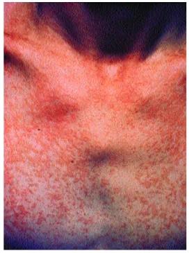
Figura 2: Lesões hiperpigmentadas lenticulares ora isoladas, ora confluentes no tronco.
Figure 2: Hyperpigmented lenticular lesions, either isolated or confluent on the trunk.
Figure 1: Hypopigmented lenticular lesions, either isolated or confluent on the trunk. Figura 1: Lesões
hipopigmentadas lenticulares ora isoladas, ora confluentes no tronco.
ACHADOS HISTOPA-TOLÓGICOS
A biópsia da pele é raramente necessária para confirmar o diagnóstico, podendo, entretanto, ser útil em alguns casos. A coloração pela hematoxilina-eosina
revela a presença de células globosas, células com formato de garrafa de boliche e pseudo-hifas curtas na camada cór-nea, além de leve hiperqueratose e acantose na epiderme, infiltrado linfocítico perivascular, e plasmócitos e histióci-tos na derme. A microscopia eletrônica revela degeneração de melanócitos, reação epidérmica manifestada por células de Langerhans mitóticas e reação celular alérgica ao fungo27
e inflamatória, generalizada na derme.
CARACTERÍSTICAS CLÍNICAS
Pacientes com PV geralmente apresentam múltiplas lesões no tronco, com regiões intercaladas de pele normal.43As
lesões podem também surgir no pescoço e extremidades supe-riores proximais. Sua distribuição normalmente é paralela à das glândulas sebáceas,26com ocorrência maior no tórax, dorso
e face. Entretanto, as lesões são encontradas em maior número no dorso. As que se localizam na face são mais comuns em crianças (incluindo recém-nascidos e lactentes) do que em adultos.44Um estudo revelou lesões na face de crianças
porta-doras de PV em aproximadamente 32% dos casos,15comumente vistas na
margem do couro cabeludo, como máculas acrômicas ou hipopigmenta-das, escamosas e de pequena dimen-são. Raramente, as lesões ficam limi-tadas aos membros inferiores, fossa poplítea, antebraço, axila, pênis/geni-tália,45ou em área de radioterapia.46A
distribuição também ocorre em áreas normalmente cobertas por roupas, enfatizando a teoria de que a oclusão das glândulas tenha um papel nessa doença. As lesões podem ser hipo ou hiperpigmentadas (figuras 1 e 2), eri-tematosas ou marrom-escuro;
justifi-HISTOPATHOLOGICAL FINDINGS
Skin biopsy is seldom necessary to confirm the diagnosis, though it may be useful in some cases. Staining due to hematoxili-ne-eosine reveals the presen-ce of globose presen-cells, bowling-pin-shaped presen-cells and short pseudo-hyphae in the corneal layer, in addition to mild hyperkeratosis and acanthosis in the epidermis, perivascu-lar lymphocytic infiltrate, and plasmocytes and histiocytes in the dermis. The electron microscope revealed melanocy-te degeneration, epidermic reaction manifesmelanocy-ted by mitotic Langerhans cells, and allergic and inflammatory cellular reaction to the fungus,27
generalized in the dermis.
CLINICAL CHARACTERISTICS
PVpatients generally show multiple lesions on the trunk, with intercalated regions of normal skin.43
The lesions may also erupt on the throat and proximal upper extremities. Its distribution is normally parallel to that of the sebaceous glands,26
with higher occurrence on the tho-rax, back and face. However, the lesions are found in a higher number on the back. Those located on the face are more common in children (including newborns and infants) than in adults.44
A study revealed face lesions in children of PV-carriers in approxi-mately 32% of cases,15
usually seen in the margins of the scalp, like achromic or hypopigmented, squa-mous and small macules. These lesions rarely remain limited to the lower limbs, were popliteal cavity, forearm, underarm, penis/genital,45
or in the area of radiotherapy.46
ery-thematous or dark-brown; justifying the versicolor name. In the surface of lesions, a fine scaling is found, as eviden-ced by the stretched skin, a birthmark named after Zileri. Although at times mild pruritus occurs, patients’ com-plaints are related to skin color changes more than to any other symptom.
PV as well as folliculities by Pityrosporum may occur in AIDS-patients. The disease then acquired an exten-ded appearance, but does not differ clinically from PV in non-HIVpatients.47,48
ULTRASTRUCTURAL ASPECTS
A study was performed in patients carrying PVwith the objective of establishing the ultrastructural changes of hyper- and hypopigmented skin, and compare them with the same patients’ non-affected skin. As such, four patients with confirmed diagnosis of PVwere selected and submitted to skin biopsies.
The hyperpigmented skin showed the stratum cor-neum to be leaner than in the hypopigmented skin, but in both the stratum corneum was thinner than in non-affected skin. A large number of tonofilaments were present in the granulous layer of the hyperpigmented skin.
In the hypopigmented skin, the melanosomes were dispersed and in a smaller amount than in the non-affected skin. In the hyperpigmented skin, a large number of mela-nosomes occupying the cytoplasm of some melanoctyes, while adjacent keratinocytes demonstrated few melanoso-mes in its cytoplasm, which suggests difficulty in transfer-ring the melanosome granules to the keratinosomes.
In the hypopigmented skin as in the hyperpigmented skin, the portion of the cell promptly affected is related to the cytoplasmatic organs, with a latent effect on the nucleus and nucleolus.49
DIAGNOSIS
The diagnosis of PV is usually performed only through clinical examination. Wood’s lamp is sometimes used to confirm the diagnosis and detect subclinical lesions. It presents yellow-gold fluorescence, probably due to excre-tion of (porphyrin) fungus metabolites, sensitive to ultravio-let radiation. The right mycological exam is performed on the lesion’s scraping, after clarification of the scales by 10% potassium, which allows the encounter with spores and pseudo-hyphae. The negative result excludes the diag-nosis. The characteristic distribution of PV on the trunk, throat and proximal extremities constitutes a relevant factor in the diagnosis.
DIAGNOSIS DIFFERENTIAL
Diagnosis differential includes other entities leading to cutaneous dispigmentation, namely vitiligo and alba pityriasis. None of them show furfuraceous scales or fluo-rescence under Wood’s lamp. Another differential diagnoses
cando a denominação versicolor. Na superfície das lesões encontra-se fina descamação, evidenciada pela distensão da pele, sinal que recebeu o nome de Zireli. Embora algumas vezes ocorra leve prurido, a queixa dos pacientes relaciona-se com a alteração da cor da pele mais do que com qualquer outro sintoma.
A PV, tanto quanto foliculite por Pityrosporum, pode
ocorrer em pacientes com Aids. A doença adquire então apresentação extensa, mas não difere clinicamente da PV
em pacientes não HIV.47,48
ASPECTOS ULTRA-ESTRUTURAIS
Foi realizado um estudo em pacientes portadores de
PVcom o objetivo de estabelecer as alterações ultra-estru-turais da pele hiper e hipopigmentada, e comparar com a pele não acometida do mesmo paciente. Desse modo quatro pacientes com diagnóstico confirmado de PVforam selecio-nados e submetidos a biópsias da pele.
A pele hiperpigmentada apresentava o estrato cór-neo mais delgado do que a pele hipopigmentada, mas em ambas o estrato córneo era mais fino do que a pele não aco-metida. Um grande número de tonofilamentos estava pre-sente na camada granulosa da pele hiperpigmentada.
Na pele hipopigmentada, os melanossomos estavam dispersos e em menor número do que na pele não acometida. Já na pele hiperpigmentada foi encontrado grande número de melanossomos ocupando o citoplasma de alguns melanócitos, enquanto queratinócitos adjacentes apresentavam poucos mela-nossomos em seu citoplasma, sugerindo dificuldade na transfe-rência dos grânulos de melanossomos para os queratinócitos.
Tanto na pele hipopigmentada como na hiperpig-mentada, a porção da célula prontamente afetada diz respei-to às organelas cirespei-toplasmáticas, com efeirespei-to latente no núcleo e nucléolo.49
DIAGNÓSTICO
O diagnóstico da PVé usualmente realizado apenas pelo exame clínico. A lâmpada de Wood é algumas vezes útil para confirmar o diagnóstico e detectar lesões subclíni-cas. Apresenta fluorescência amarelo-ouro, provavelmente devido à excreção de metabólitos (porfirinas) do fungo, sensíveis à radiação ultravioleta. O exame micológico dire-to é realizado do raspado da lesão, após clarificação das escamas pela potassa a 10%, permitindo o encontro de esporos e pseudo-hifas; o resultado negativo exclui o diag-nóstico. A distribuição característica da PVno tronco, pes-coço e extremidades proximais constitui fator relevante no diagnóstico.
DIAGNÓSTICO DIFERENCIAL
diagnósti-co diferencial que não deve ser esquecido é a hanseníase em sua forma indeterminada, principalmente se localizada na face, na qual a alteração de sensibilidade pode não estar pre-sente numa fase inicial.
Outras possibilidades incluem psoríase, dermatite seborréica (ambas podendo coexistir com PV), papilomato-se reticulada e confluente (Gougerot-Carteaud), eritrasma e dermatofitose. A luz de Wood é provavelmente uma das armas mais úteis na diferenciação da PVcom o eritrasma e a dermatofitose. O eritrasma adquire tonalidade vermelho-coral com a luz de Wood, e a dermatofitose não fluoresce.
TRATAMENTO
O tratamento da PVpode ser feito com grande núme-ro de agentes que estão divididos em dois grupos: tópicos e sistêmicos.
A – Agentes Tópicos
Nesse grupo o agente mais freqüentemente usado tem sido o sulfeto de selênio 2,5% a 5% na forma de xampu aplicado uma vez ao dia. O tratamento deve ser realizado durante período que varia de sete a 14 dias ou mais, algu-mas vezes indefinidamente.50
Todos os tópicos “azóis” parecem ter a mesma eficá-cia no tratamento da PV. Entretanto, nenhum deles foi tão completamente estudado como o cetoconazol creme.50
B – Agentes Sistêmicos
ßCetoconazol. A dose preconizada é de 200mg/dia por 10 dias. A taxa de cura é alta (de 90 a 100%). O risco de hepatotoxicidade existe e tem sido calculado em 1:500.000 pacientes que receberam cetoconazol oral por curto tempo (10 dias).
ß Fluconazol. A dose recomendada é de 150mg/semana por três semanas.
ßItraconazol. A dose preconizada é de 200mg/dia por sete dias. Trata-se de uma droga bem tolerada. Quando há indicação para uso prolongado, mais de sete dias, alguns efeitos gastrointestinais podem manifestar-se. Cefaléia, náusea e dor abdominal são efeitos colaterais possíveis em 7% dos casos.
ß Terbinafina. A terbinafina oral é efetiva contra muitas dermatofitoses, mas não no tratamento da PV. Talvez porque não atinja concentração suficiente na cama-da córnea. Já a terbinafina tópica mostrou-se eficaz no tra-tamento da infecção pela M. furfur.
Terapêutica tópica x terapêutica sistêmica
Do ponto de vista farmacoterapêutico, a PV sendo infecção superficial, deveria ser tratada com agentes tópi-cos. Entretanto, os pacientes defendem outro ponto de vista. Para eles o tratamento com agentes tópicos traz muitas des-vantagens, como o tempo necessário e a dificuldade na apli-cação da droga em grandes áreas afetadas, especialmente o
that must not be forgotten is leprosy in its indeterminate form, located mainly on the face, in which alteration to sen-sitivity may not be present in an initial phase.
Other possibilities include psoriasis, seborrheic der-matitis (both able to coexist with PV), recticular and con-fluent papillomatosis (Gougerot-Carteaud), erythrasma and dermatophitosis. Wood’s lamp examination is probably one of the most useful weapons in the differentiation of PV
with erythrasma and dermatophitosis. Erythrasma acquires a red-coral tonality with Wood’s lamp, and the dermatophi-tosis does not fluoresce.
TREATMENT
PVtreatment can be performed with a large amount of agents, subdivided into two groups: topical and systemic ones.
A –Topical Agents
In this group, the most frequently used agent has been selenium sulfide 2.5-5% in shampoo form applied once a day. Treatment must be realized over a period var-ying from seven to 14 days or more, and sometimes indefi-nitely.
All topical “azoles” seems to be equally effective in treating PV. Still, none of them have been as fully studied as ketoconazol cream.50
B – Systemic Agents
ßKetoconazol. The suggested dose is 200 mg daily for 10 days. The cure rate is high (from 90 to 100%). Risk of hepatotoxicity exists and has been calculated in 1:500,000 patients who received oral ketoconazol for a short duration (10 days).
ß Fluconazol. The recommended dose is 150 mg weekly for three weeks.
ßItraconazol. The suggested dose is 200 mg daily for seven days. This drug is well tolerated. When indicated for prolonged use, i.e. more than seven days, some gastroin-testinal effects may be manifested. Cephalea, nausea and abdominal pain are possible side effects in 7% of cases.
ßTerbinafine. Oral terbinafine is effective against many dermatofitoses, but not in PVtreatment, due perhaps to not reaching a high enough concentration in the cornea layer. Topical terbinafine proved to be effective in treating M. furfur infection.
Topical therapy vs. systemic therapy
reasons, patient adhesion is inadequate, which increases the rate of recurrence. Effectiveness of topical agents is lower, and rate of recurrence varies form 60 to 80%.50 As such, the systemic agents are those recommended in short-term treatment for many patients, in spite of the side effects they may bring.
Hepatic effects with antifungal therapy
Hepatotoxicity is a rare side-effect that may be asso-ciated with some oral antifugal medications. The report on hepatotoxicity with ketoconazol use seems to be most com-mon in women above the age of 50 who have been using the medication over long periods (from 51 to 60 days). Tests of hepatic function are found to be high, but normalization ensues by simply interrupting the drug.4
CONCLUSION
Pityriasis versicolor is a common superficial fungal infection caused by the Malassezia furfurfungus, incident mainly in the tropics. In the past it was thought to be a post-pubescent disease, however current evidence shows that versicolor pityriasis is not uncommon in school children. Diagnosis is easily performed by clinical exam.
Treatment shows changes in chronology, gradual substitution occurring of topical antifungal by systemic medication, for three reasons: (I) patient adhesion is inade-quate for the topical drugs due to the required time of use, difficult applicability in extensor areas and the unpleasant odor of certain agents; (II) effectiveness of topical agents is lower, and the rate of recurrence varies from 60 to 80%; (III) the emergence of systemic antifungal drugs at a lower rate of adverse events.
After treatment, the affected area remained hypopig-mented for a variable period of time. The mechanism of hypopigmentation has not been well established yet. But ultrastructural studies show severe damage to melanoctyes, which varies from melanosomes and altered mitochondria up to degeneration. q
tronco, e também o odor desagradável de certos agentes. Por essas razões, a adesão do paciente é inadequada, o que aumenta a taxa de recorrência. A eficácia dos agentes tópi-cos é menor, e a taxa de recorrência varia de 60 a 80%.50
Desse modo, os agentes sistêmicos são os recomendados no tratamento de curta duração em muitos pacientes, apesar dos efeitos colaterais que podem trazer.
Efeitos hepáticos com a terapia antifúngica
A hepatotoxicidade é efeito colateral raro, mas que pode estar associado a alguns antifúngicos orais. O relato de hepatotoxicidade com o uso de cetoconazol parece ser mais comum em mulheres com idade acima dos 50 anos e que fizeram uso da medicação por longo período (de 51 a 60 dias). Os testes de função hepática encontram-se elevados, mas há normalização com a simples interrupção da droga.4
CONCLUSÃO
A pitiríase versicolor é infecção fúngica superficial comum, causada pelo fungo Malassezia furfur, incidindo principalmente nos trópicos. Antigamente pensava-se numa doença pós-puberal, entretanto evidências atuais mostram que a pitiríase versicolor não é incomum em crianças. O diagnóstico é facilmente realizado pelo exame clínico.
O tratamento apresentou mudanças cronológicas, ocorrendo substituição gradativa de antifúngicos tópicos por sistêmicos, por três razões: (I) a adesão do paciente é inadequada às medicações tópicas pelo tempo de uso neces-sário, dificuldade de aplicação em áreas extensas e odor desagradável de certos agentes; (II) a eficácia dos agentes tópicos é menor, e a taxa de recorrência varia de 60 a 80%; (III) o surgimento de antifúngicos sistêmicos com menor índice de eventos adversos.
Após o tratamento, a área afetada pode permanecer hipopigmentada por período de tempo variável. O mecanis-mo da hipopigmentação ainda não está bem estabelecido, mas estudos ultra-estruturais mostram grave dano aos mela-nócitos, variando de melanossomos e mitocôndrias altera-dos até a degeneração. q
REFERÊNCIAS / REFERENCES
1. Michalowski R, Rodziewicz H. Pityriasis versicolor in chil-dren. Br J Dermatol 1963; 75: 397-400.
2. Adamski Z. Studies of a role played by lipophilic yeasts Malassezia furfur (Pityrosporum ovale, Pityrosporum orbiculare) in different dermatoses. Postepy Dermatol (Poznan) 1995; 12: 349-454.
3. Zaitz C – Dermatoses associadas às leveduras do gênero Malassezia. An Bras Dermatol 2000; 75 (2): 129-142.
4. Sunenshine PJ, Schwartz RA, Janniger CK. Review. Tinea ver-sicolor. Int J Dermatol 1998; 37: 648-655.
5. Bailon H. Traite de Botanique Medicale Cryptogamique. Paris: Octave Doin, 1889: 234.
6. Robin C. Historie Naturelle des Vegeaux Parasites. London: Balliere, 1853: 438.
7. Gordon MA. The lipophilic mycoflora of the skin. Mycologica 1951; 43: 524-534.
8. Castellani A, Chalmers AJ. Manual of Tropical Medicine, 2nd edn. New York: Wm. Wood & Co., 1913: 936-837.
9. Gordon MA. Lipophilic yeastlike organisms associated with tinea versicolor. J Invest Dermatol 1951; 17: 267-272.
10. Savin R. Diagnosis and treatment of tinea versicolor. J Fam Pract 1996; 43: 127-132.
11. Nowiscki R, Sadowska E. Mycotic infections in the Gdansk area. Przegl Dermatol 1993; 80: 245-250.
12. Akpata LE, Gugnani HC, Utsalo SJ. Pityriasis versicolor in school children in Cross River State of Nigeria. Mycoses 1990; 33: 549-551.
orbiculare in middle-aged and elderly individuals. Acta Derm Venereol 1988; 68: 537-540.
14. Bergbrant IM, Brobeg A. Pityrosporum ovale culture from the forehead of healthy children. Acta Derm Venereol 1994; 74: 260-261.
15. Terragni L, Lasagni A, Oriani A, Gelmetti C. Pityriasis versi-color in the pediatric age. Pediatr Dermatol 1991; 8: 9-12. 16. Miskeen AK, Kelkar SS, Shroff HJ. Pityriasis versicolor in children. Indian J Dermatol Venereol Leprol 1984; 50: 144-146. 17. Roberts SOB. Pityrosporum orbiculare: incidence and distri-bution on clinically normal skin. Br Dermatol 1969; 81: 264-269. 18. Faergemann J. Lipophilic yeasts in skin disease. Semin Dermatol 1985; 4: 173-184.
19. Faergemann J, Aly R, Maibach HI. Growth and filament pro-duction of Pityrosporum orbiculare and P. ovale on human stratum corneum in vitro. Acta Derm Venereol 1983; 63: 388-392. 20. Salkin IF, Gordon MA. Polymorphism of Malassezia furfur. Can J Microbiol 1977; 23: 471-475.
21. Faergemann J, Tjernlund U, Scheynius A, Sverker B. Antigenic similarities and differences in genus Pityrosporum. J Invest Dermatol 1982; 78: 28-31.
22. Cunningham AC, Leeming JP, Ingham E, Gowland G. Differentiation of three serovars of Malassezia furfur. J Appl Bacteriol 1990; 68: 439-446.
23. Ashbee HR, Ingham E, Holland KT, Cunliffe WJ. The carriage of Malassezia furfur serovars A, B, and C in patients with pityria-sis versicolor, seborrhoeic dermatitis and controls. Br J Dermatol 1993; 129: 533-540.
24. Ashbee HR, Ingham E, Holland KT, et al. The role of Malassezia furfur serovars A, B, and C in Pityriasis versicolor and seborrhoeic dermatitis. Br J Dermatol 1991; 125: 495.
25. Schmidt A. Malassezia furfur: a fungus belonging to the phys-iological skin flora and its relevance in skin disorders. Cutis 1997; 59: 21-24.
26. Leeming JP, Notman FH, Holland KT. The distribution and ecology of Malassezia furfur and cutaneous bacteria on human skin. J Appl Bacteriol 1989; 67: 47-52.
27. Breathnach AS, Nazzaro Porro M, Martin B. Ultrastructure of skin in pityriasis versicolor. Giornale Italiano di Dermatologia-Minerva Dermatologica 1975; 110:457-469.
28. Scheynius A, Faergemann J, Forsum U, Sjoberg O. Phenotypic characterization in situ of inflammatory cells in pityriasis (tinea) versicolor. Acta Derm Venereol 1984; 64: 473-479.
29. King RD, Cunico RL, Maibach HI, et al. The effect of occlu-sion on carbon dioxide emisocclu-sion from human skin. Acta Derm Venereol 1978; 58: 135-138.
30. Faergemann J, Bernander S. Tinea versicolor and Pityrosporum orbiculare. A mycological investigation. Sabouraudia 1979; 17: 171-179.
31. Congly H. Pityriasis versicolor in a 3-month-old boy. Can Med Assoc J 1984; 130: 844-845.
32. Roberts SOB. Pityriasis versicolor: a clinical and mycological investigation. Br J Dermatol 1969; 81: 315-326.
33. Burke RC. Tinea versicolor: susceptibility factors and
experimen-tal infections in human beings. J Invest Dermatol 1961; 36: 389-402. 34. Elewski BE, Hazen PG. The superficial mycoses and the der-matophytes. J Am Dermatol 1989; 21: 655-673.
35. Heng MCY, Henderson CL, Barker DC, Haberfelde G. Correlation of Pityrosporum ovale density with clinical severity of seborrheic dermatitis as assessed by a simplified technique. J Am Acad Dermatol 1990; 23: 82-86.
36. Crozier WJ, Wise KA. Onychomycosis due to Pityrosporum. Australas J Dermatol 1993; 34: 109-112.
37. Sohnle PG, Collins-Lech C. Cell-mediated immunity to Pityros-porum orbiculare in tinea versicolor. J Clin Invest 1978; 62: 45-53. 38. Hashim FA, Elhassan AM. Tinea versicolor and visceral leish-maniasis. Int J Dermatol 1994; 33: 258-259.
39. De Luca C, Picardo M, Breathnach A, et al. Lipoperoxidase activity of Pityrosporum: characterization of by-products and pos-sible role in pityriasis versicolor. Exper Dermatol 1996; 5: 49-56. 40. Nazzaro-Porro M. Identification of tyrosinase inhibitors in cul-tures of Pityrosporum. J Invest Dermatol 1978; 71: 205-208. 41. Soyer HP, Cerroni L. The significance of histopathology in the diagnosis of dermatomycoses. Acta Dermatovenerologica Alpina Pannonica et Adriatica 1992; 1: 84-87.
42. Galadari I, El-Komy M, Mousa A, et al. Tinea versicolor: his-tologic and ultrastructural investigation of pigmentary changes. Int J Dermatol 1992; 31: 253-256.
43. Schwartz RA, Fox MD. Office Dermatology. Kansas City, Missouri: American Academy of family Physicians Publications, 1992.
44. Terragni L, Lasagni A, Oriani A. Pityriasis versicolor of the face. Mycoses 1991; 34: 345-347.
45. Daneshvar SA, Hashimoto K. An unusual presentation of tinea versicolor in an immunosuppressed patient. J Am Acad Dermatol 1987; 17: 304-305.
46. Conill C, Azón-Masoliver A, Verger E, et al. Pityriasis versi-color confined to the radiation therapy field. Acta Oncol 1990; 29: 949-950.
47. Aly R, Berger T. Common superficial fungal infections in patients with AIDS. Clin Infect Dis 1996; 22: S128-S132. 48. Elmets CA. Management of common superficial fungal infec-tions in patients with AIDS. J Am Acad Dermatol 1994; 31: S60-S63.
49. Karaoui R, Bou-Resli M, Al-Zaid NS, Mousa A. Tinea versi-color: Ultrastructural studies on hypopigmented and hyperpig-mented skin. Dermatologica 1981; 132: 69-85.
50. Savin R. Diagnosis and treatment of tinea versicolor. J Fam Pract 1996; 43: 127-132.
ENDEREÇO PARA CORRESPONDÊNCIA: / MAILINGADDRESS: Profa. Dra. Denise Steiner
Rua Engº Edgar Egídio de Souza, 420 Pacaembú São Paulo SP 01233-020