Recebido em 20.09.2000. / Received in September, 20thof 2000.
Aprovado pelo Conselho Consultivo e aceito para publicação em 10.03.2003. / Approved by the Consultive Council and accepted for publication in March, 10thof 2003. * Trabalho realizado na Universidade Federal de Juiz de Fora. / Work done at "Universidade Federal de Juiz de Fora".
1Acadêmico de medicina, bolsista do CNPq. Universidade Federal de Juiz de Fora. / Medical student, grant from CNPq, Federal University of Juiz de Forma (UFJV). 2Pós-Graduandos em Dermatologia, Hospital Universitário da Universidade Federal de Juiz de Fora. / Postgraduate students of Dermatology, Teaching Hospital-UFJF.
3Professor auxiliar, Serviço de Dermatologia - Hospital Universitário da Universidade Federal de Juiz de Fora. / Auxiliary Professor, Dermatology Service, Teaching Hospital-UFJF. 4Professor de Micologia da UFJF. / Professor of Mycology at UFJF
5Professora de Dermatologia da UFJF. / Professor of Dermatology at UFJF.
©2003 by Anais Brasileiros de Dermatologia
Pitiríase Versicolor e Síndrome da Imunodeficiência
Adquirida (SIDA)
*Pityriasis Versicolor and AIDS
*Marlon Soares Roza
1David Dornellas
2Márcio Tavares Rodrigues
4Patrícia Viana Vieira
2Marco Andrey Cipriani Frade
3Maria Teresa Feital de Carvalho
5Re sum o : FUNDAMENTOS- A p itiríase versicolor é u ma in fecção crôn ica recorren te da camada córn ea
cau sada p ela Ma la ssezia fu rfu r, geralmen te assin tomática, p oden do ser cau sa de fu n gemia. A sín -drome da imu n odeficiên cia adqu irida caracteriza-se p or u ma imp ortan te dep ressão celu lar, o qu e p rop icia a ocorrên cia de in fecções op ortu n istas.
OBJETIVOS- Dados a imu n odep ressão cau sada p elo HIVe o risco da p resen ça de M. fu rfu r em su a forma p atogên ica n o imu n ocomp rometido, realizou -se este trabalho comp arativo bu scan do as reais
diferen ças da p itiríase versicolor n o imu n ocomp eten te e n o imu n odep rimido p elo HIV.
PACIENTESe MÉTODOS- Foram avaliados, n o p eríodo de ju lho de 1998 a ju n ho de 1999, 50 p acien tes
HIVp ositivos do Hosp ital Un iversitário da UFJF. Como gru p o con trole, foram examin ados de forma
aleatória 50 p acien tes soron egativos p ara HIV. Para cada p acien te foi p reen chido u m p rotocolo con
s-tan do de iden tificação e exames clín icos e laboratoriais.
RESULTADOS - A p itiríase versicolor foi diagn osticada clin icamen te n o gru p o HIVp ositivo em sete (14%) p acien tes, resu ltado coin ciden te n o gru p o con trole. Qu an to ao p assado p ara a p itiríase
ver-sicolor, 12 (24%) p acien tes já haviam ap resen tado tal doen ça n o gru p o HIVp ositivo, e igu al in cidên
-cia ocorreu n o gru p o con trole.
CONCLUSÕES- A p itiríase versicolor n ão se mostrou mais freqü en te e n em mais agressiva n a p op u
la-ção imu n odep rimida p elo HIVem relação ao gru p o con trole, en qu an to a dermatite seborréica ap
re-sen tou -se mais freqü en te n o gru p o in fectado p elo HIV.
Palavras-chave: Malassezia; p itiríase; Sín drome da Imu n odeficiên cia Adqu irida; tin ha versicolor.
Su m m a r y: BACKGROUND- Pityria sis versicolor (PV) is a recu rren t, chron ic in fection of the stratu m corn eu m d u e to Malassezia fu rfu r tha t is gen erally asym ptom a tic an d has the poten tial of lead in g to fu n gem ia. Acqu ired Im m u n od eficien cy Syn d rom e (AIDS) is cha racterized by a severe cellu lar d epression , w hich fa cilita tes the occu rren ce of opportu n ist in fection s.
OBJECTIVES- Given the im m u n od epression cau sed by HIVan d risk this en tails for the presen ce of the pa thogen ic form of M. fu rfu r, this w ork sou ght the real d ifferen ces in PVa ffectin g im m u n ocom pe-ten t hosts com pa red to im m u n ocom prom ised HIV-patien ts.
PATIENTS ANDMETHODS- From Ju ly 1998 to Ju n e 1999, 50 HIV-positive ou tpatien ts from the Teachin g Hospital w ere stu d ied . The con trol grou p com prised of 50 HIV-n egative patien ts. The protocol for ea ch patien t in clu d ed id en tifica tion , clin ical an d laboratory ex a m in ation .
RESULTS- PVw as clin ica lly d iagn osed in the HIV-positive grou p in 7 (14%) patien ts, w ith the sam e n u m ber in the con trol grou p. As for prior history, 12 (24%) patien ts in the HIV-positive grou p repor-ted havin g previou sly presen repor-ted PV, the sam e in cid en ce w as fou n d in the con trol grou p.
CONCLUSIONS- PVw a s n either m ore frequ en t n or m ore severe in the HIV-im m u n ocom prom ised grou p com pared to the con trol grou p, w hereas seborrheic d erm a titis w as m ore frequ en t in the form er. Key w ord s: Ma la ssezia ; pityriasis; Acqu ired Im m u n od eficien cy Syn d rom e; tin ea versicolor.
INTRODUÇÃO
A pitiríase versicolor (PV) é a infecção pitirospórica
mais comum da pele.1É uma infecção crônica e recorrente
da camada córnea causada pela Malassezia furfur, geral-mente assintomática, podendo ser causa de fungemia adqui-rida por introdução de cateter, tanto em adultos como, espe-cialmente, em neonatos que recebem alimentação parente-ral rica em lipídios.2,3
Clinicamente a pitiríase versicolor caracteriza-se por máculas ou placas numulares ou gutatas, amareladas, acin-zentadas ou hipocrômicas, comprometendo principalmente ombros e a parte superior do tronco de adultos jovens que
perspiram acentuadamente.4
Na pitiríase versicolor, o Pityrosporum ovale muda de sua forma leveduriforme (blastosporo) para a forma miceliana devido a fatores predisponentes, como alta tem-peratura e umidade, ou fatores endógenos, como oleosidade cutânea, hereditariedade, tratamentos ou distúrbios
imuno-depressivos.5
Para Lacas tudo faz crer que os gêneros Malassezia
e Pityrosporum sejam sinônimos.6
A síndrome da imunodeficiência adquirida (SIDA) caracteriza-se por uma importante depressão celular, o que propicia a ocorrência de infecções oportunistas. As mani-festações tegumentares da enfermidade são extremamente freqüentes, atingindo mais de 90% dos doentes em alguma
fase de sua evolução.7
Mathes & Douglas afirmaram que 80% dos
pacien-tes com SIDA apresentavam dermatite seborréica.8
Em pacientes com dermatite seborréica, grande
quantidade de P. ovale foi encontrada por Mc Ginley et al.9
Shuster10e Faergemann5acreditam que P. ovale seja a
prin-cipal causa da dermatite seborréica, que, sendo comum em
pacientes HIV positivos, lhes provoca provavelmente
aumento de susceptibilidade à infecção por essa levedu-ra.11,12,13Assim, espera-se maior número de casos de
pitiría-se versicolor na SIDA. Contrariamente a essa suposição, podemos notar que em diversos trabalhos publicados sobre lesões cutâneas em SIDA, essa micose superficial é pouco
citada ou mesmo não o é, o que leva a concluir que a PVnão
parece ser mais freqüente nesses pacientes.8,14,15
Manifestações sistêmicas graves por M. furfur são
cita-das em outras doenças capazes de causar imunodepressão.16,17
Dados a imunodepressão causada pelo HIVe o risco
da presença de M. furfur em sua forma patogênica no imu-nocomprometido, realizou-se este trabalho comparativo buscando as reais diferenças da pitiríase versicolor no
imu-nocompetente e no imunodeprimido pelo HIV.
Po pulação e studada
Foram avaliados, no período de julho de 1998 a
junho de 1999, 50 pacientes HIVpositivos, na faixa etária
de 20 a 50 anos, dos ambulatórios de Dermatologia e Doenças Infectoparasitárias do Hospital Universitário da UFJF.
INTRODUCTION
Pityriasis versicolor (PV) is the most common pity-rosporic infection of the skin.1It is a chronic and recurrent infection of the stratum corneum caused by Malassezia
fur-fur. Usually asymptomatic, it can be the cause of fungemia
acquired by catheter introduction, in adults and especially in neonates that receive parental feeding rich in lipids.2,3
Clinically pityriasis versicolor is characterized by num-mular or guttate stains or plaques, these being yellowish, gra-yish or hypochromic, mainly involving the shoulders and the superior part of the trunk of young adults that perspire heavily.4 In pityriasis versicolor, the Pityrosporum ovale changes from its yeast form (blastospore) to a mycelian form due to predisposing factors such as high temperatures and humidity, or to endogenous factors such as cutaneous oiliness, hereditary predisposition, or to immunodepressant treatments or disturbances.5
According to Lacas all indications lead one to believe that the genera Malassezia and Pityrosporum are synonymous.6 Acquired immunodeficiency syndrome (AIDS) is cha-racterized by an important cellular depression that facilita-tes the occurrence of opportunistic infections. The tegumen-tary manifestations of the illness are extremely frequent, affecting more than 90% of the patients in some phase of its clinical development.7
Mathes & Douglas affirmed that 80% of the patients with AIDSpresented seborrheic dermatitis (SD).8
In patients with seborrheic dermatitis, McGinley et
al. found a significant amount of P. ovale.9 Shuster10and
Faergemann5 consider that P. ovale is the main cause of seborrheic dermatitis, which, since it is common in HIV -positive patients, probably provokes increased susceptibi-lity to infection by this yeast.11,12,13 Thus, a larger number of cases of pityriasis versicolor could be expected in patients with AIDS.But contrary to this supposition, it is noticeable that in several works published on cutaneous lesions in AIDS, superficial mycosis is infrequently mentioned or even not at all, which leads to the conclusion that PV does not apparently occur more frequently in these patients.8,14,15
Serious systemic manifestations of M. furfur are mentioned in other diseases capable of causing immunode-pression.16,17
Given the immunodepression caused by HIVand the risk of the presence of M. furfur in its pathogenic form in the immunocompromised, this work was undertaken to com-pare any real differences to be found between pityriasis ver-sicolor in immunocompetent patients and in those immuno-depressed by HIV.
Stud y p op ula tion
As a control group, 50 patients of the same age group that were HIV-negative were selected at random and exami-ned. These were from the sector of voluntary blood donors of the Hemotherapy Service of Juiz de Fora (Hemominas).
METHODS
For each patient a form was filled out, the informa-tion consisting of name, sex, age, color, profession, medical history and the result of the ELISAexam for HIV.
The form used for the HIVpositive patients or those suffering from AIDS also included: serum grouping accor-ding to the CDC revision of 1993, epidemiology, date of diagnosis of HIV, CD4cell count and viral load.
Questions were asked regarding any history of pity-riasis versicolor or seborrheic dermatitis and about the use of systemic or topical fungicides.
In the dermatological exam it was considered impor-tant to look for the signs of Zireli and Besnier.
All the patients that presented lesions suggestive of PV, lesions that were hyper or hypochromic, erythematose or dark-brown that showed on distention of the skin (Zireli's sign) a fine desquamation, were submitted to direct mycological exam using transparent adhesive tape technique. This was applied on the affected areas of the skin and later onto a glass slide to be examined by optical microscopy.
The data were analyzed with aid of the software EpInfo version 6.04 of the STATCALC module. Statistical association was analyzed by means of odds-ratio and its significance in the chi-square test, using the 95% confiden-ce limits. This was done separately in the presenconfiden-ce of PV and seborrheic dermatitis.
RESULTS
In the HIV-positive patients the age ranged from 24 to 62 years. There was a male sex bias (29 male patients against 21 female patient) and most were Caucasians (Table 1).
Como grupo controle, foram examinados de forma aleatória, na mesma faixa etária, 50 pacientes soronegativos
para HIV, do setor de doação voluntária do Serviço de
Hemoterapia de Juiz de Fora (Hemominas).
MÉTODOS
Para cada paciente foi preenchido um protocolo em que constavam nome, sexo, idade, cor, profissão, prontuário
e resultado do exame Elisa para o HIV.
Em se tratando dos pacientes HIVpositivos ou
aidé-ticos fizeram parte do protocolo: sorogrupo segundo CDC
revisão de 1993, epidemiologia, tempo de diagnóstico do
HIV, contagem de células CD4e carga viral.
Interrogou-se sobre o passado de pitiríase versicolor e dermatite seborréica, e sobre o uso de antifúngico sistêmi-co ou tópisistêmi-co.
No exame dermatológico deu-se importância para os sinais de Zireli e Besnier.
Todos os pacientes que apresentavam lesões
sugesti-va de PV, lesões hiper ou hipocrômicas, eritematosas ou
marrom-escuras que evidenciavam, pela distensão da pele (sinal de Zireli), uma fina descamação foram submetidos ao exame micológico direto pela técnica da fita adesiva trans-parente, que foi aplicada sobre as áreas lesadas da pele e posteriormente sobre uma lâmina de vidro, sendo examina-das por microscopia óptica.
Os dados foram analisados com auxílio do
progra-ma EpInfo versão 6.04 do módulo STATCALC.
Analisaram-se a associação estatística por meio da razão dos produtos cruzados (odds-ratio) e sua significância pelo teste de qui-quadrado e intervalo de confiança de 95%, separadamente
para a presença das doenças PVe DS.
RESULTADOS
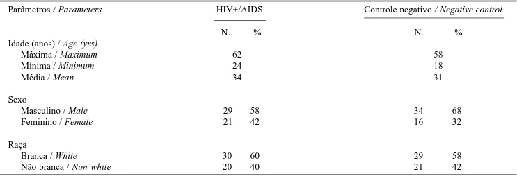
Nos pacientes HIVpositivos a idade variou de 24 a
62 anos. Houve predomínio do sexo masculino (29 pacien-tes) sobre o feminino (21 pacienpacien-tes), e a maioria foi da raça branca (Tabela 1).
Parâmetros / Parameters HIV+/AIDS Controle negativo / Negative control N. % N. %
Idade (anos) / Age (yrs)
Máxima / Maximum 62 58
Mínima / Minimum 24 18
Média / Mean 34 31
Sexo
Masculino / Male 29 58 34 68
Feminino / Female 21 42 16 32
Raça
Branca / White 30 60 29 58
Não branca / Non-white 20 40 21 42
Tabela 1: Relação idade, sexo e raça en tre gru p o HIV+ e gru p o con trole n egativo.
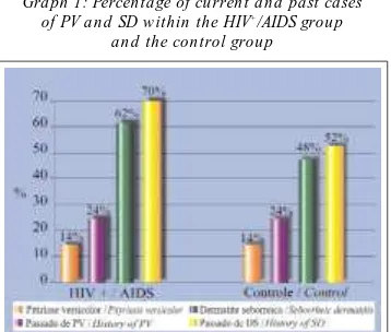
PVwas diagnosed clini-cally in seven (14%) patients of the group that was HIV positi-ve, coincident with the result in the control group. In the HIV -positive group all the patients presented only involvement of the dorsal area of the thorax, while in the control group five patients also presented macu-lo-squamous lesions in the dorsal area, and two also on the face and on the superior members. Reference to pre-vious incidence of PV was also coincident, reported in both groups by 12 (24%) patients (Graph 1).
In all the patients with PVthe signs of Zireli and Besnier were positive. In all of these under optical microscopy it was observed that the scales collected with transparent adhesive tape revealed the presence of ovoid cells and mycelian filaments.
Seborrheic dermatitis was present at the time of the exam in 31 (62%) patients of the HIV-positive group, while in the control group it occurred in 24 (48%) indivi-duals (Graph 1).
In the HIV-positive group, 35 (70%) patients refer-red to seborrheic dermatitis in the past, against 26 (52%) in the control group (Graph 1).
As for the affected area, of the 31 patients in the HIV-positive group, seborrheic dermatitis was isolated in the scalp in 19 (61.3%), associated with involvement of the face in 10 (32.3%), associated with the dorsal area of the thorax in only one (3.2%), isolated involvement of the dorsal area of the thorax in one (3.2%). Also in one (3.2%) patient there was a disseminated picture.
In the control group seborrheic dermatitis was pre-sented by 24 (48%) individuals involving the scalp separa-tely in 23 (95.8%) and associated with the face in one (4.2%).
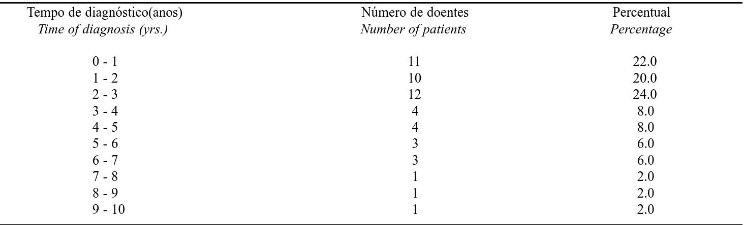
In the patients with HIVthe time period from the diagnosis of the disease up to the clinical consultation for PV varied from zero to 10 years, most of them (33 patients) had acquired HIV less than three years pre-viously (Table 2).
Sporting activities that cause increased perspiration were practiced by 13 (26%) of the HIV-positive patients, compared to 28 (56%) in the control group. Although
appa-A pitiríase versicolor
(PV) foi diagnosticada
clini-camente no grupo HIV
posi-tivo em sete (14%) pacien-tes, resultado coincidente com o do grupo controle. No
grupo HIVpositivo todos os
pacientes apresentaram ape-nas comprometimento da região dorsal do tórax,
enquanto no grupo controle cinco pacientes mostravam lesões máculo-escamosas na região dorsal, e dois também
na face e nos membros superiores. Referência à PVno
pas-sado também foi coincidente, sendo relatada nos dois gru-pos em 12 (24%) pacientes (Gráfico 1).
Em todos os pacientes portadores de PVos sinais de
Zireli e Besnier foram positivos. Em todos observou-se, à microscoscopia óptica das escamas colhidas com fita adesi-va transparente, presença de células ovóides e filamentos micelianos.
A dermatite seborréica (DS) estava presente no
momento do exame em 31 (62%) pacientes do grupo HIV
positivo, enquanto no grupo controle ocorreu em 24 (48%) indivíduos (Gráfico 1).
No grupo HIVpositivo 35 (70%) pacientes referiram
dermatite seborréica no passado, enquanto no grupo contro-le esse relato foi feito por 26 (52%) indivíduos (Gráfico 1).
Quanto ao local de comprometimento, nos 31
pacientes do grupo HIV positivo, foi observada dermatite
seborréica no couro cabeludo isoladamente em 19 (61,3%), associada ao comprometimento da face em 10 (32,3%), associada à região dorsal do tórax em um (3,2%), compro-metimento isolado da região dorsal do tórax em um (3,2%) e compromentimento disseminado também em um (3,2) paciente.
No grupo controle a dermatite seborréica acometeu 24 (48%) indivíduos compromentendo o couro cabeludo isola-damente em 23 (95,8%) e associado à face em um (4,2 %).
O tempo de diagnóstico de HIVdos pacientes até a
consulta clínica para pesquisa de PV variou de zero a 10
anos, estando a maioria (33 pacientes) com menos de três anos de evolução (Tabela 2).
Entre os HIVpositivos 13 (26%) e no grupo
contro-le 28 (56%) pacientes praticavam alguma atividade esporti-va que leesporti-vaesporti-va a aumento da perspiração, sem que isso
Gráfico 1: Porcen tagem de casos atu ais e p assados
de PV e DS n o gru p o HIV+
/SIDA e gru p o con trole
Gra ph 1: Percen ta ge of cu rren t an d past cases of PV a n d SD w ithin the HIV+
rently this was unrelated to the presence of PV.
As to the means of infection, there was a prevalen-ce of sexual transmission over the other probable paths. Since a single patient might report more than one possible source of infection, the number of patients exceeds the population of the group (Table 3).
In the laboratory exams, 41 (82%) patients were evaluated in relation to the CD4 cell count (Table 4).
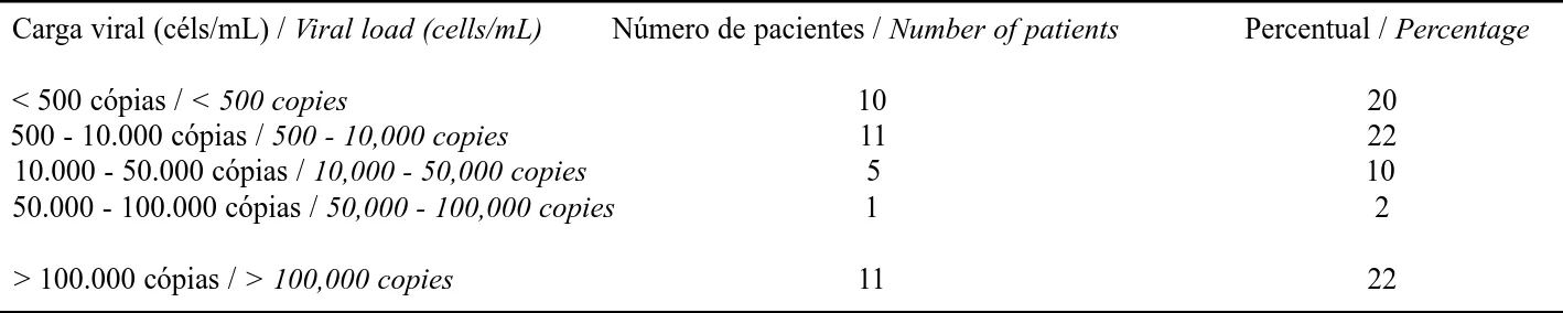
As for viral load of the HIV-positive/AIDSpatients, 38 (76%) were evaluated (Table 5).
The HIV-positive patients were classified in accor-dance with the revision of 1993 proposed by CDC, into: category A, 17 patients; B, 11 patients; and C, 22 patients (Table 6).
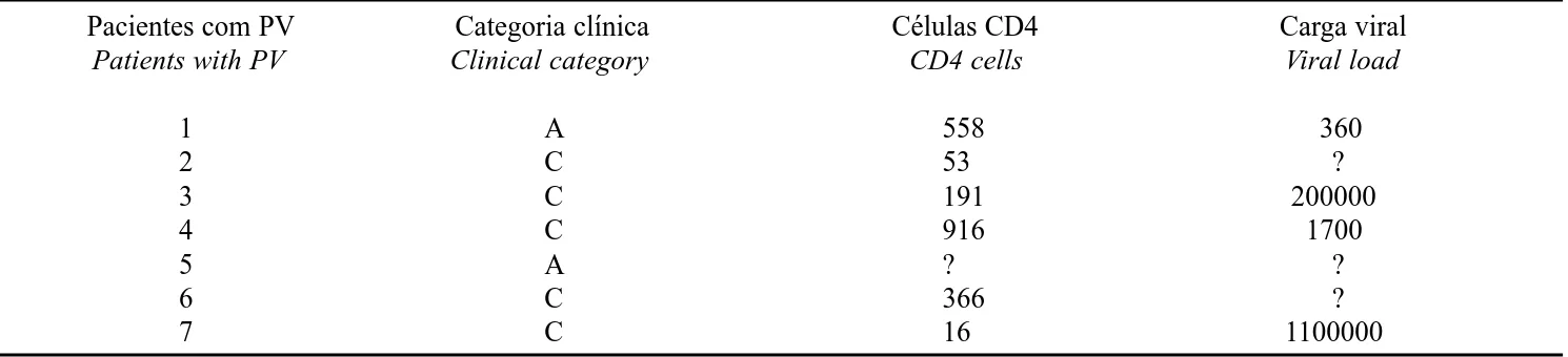
There was no relationship between pityriasis versicolor and clinical category, CD4cell count or viral load (Table 7).
DISCUSSION
Analysis of the age of the patients in the HIV -positi-ve/AIDSgroup gave a mean of 34 years, which is similar to that found by Friar et al.,18in a work done in the same institution. It also correlates with the age group most affected by HIVin the world population, which is from 20 to 40 years.18,19
The control group presented much the same characte-ristics, with a mean age of 31 years.
In Brazil, since the beginning of the epidemic, AIDShas shown a male sex bias, however that pattern is changing with the increase of cases among women. The male/female ratio was
pudesse ser relacionado com presença de PV.
Houve predomínio da via sexual sobre as outras possíveis vias de infecção. Devido ao fato de um mesmo paciente poder apresentar mais de uma via provável de infecção, o número de pacientes excede a população do grupo (Tabela 3).
Laboratorialmente foram avaliados 41 (82%) em
relação à contagem de células CD4(Tabela 4).
Quanto à carga viral do paciente HIV positivo/SIDA, foram avaliados 38 (76%) dos pacientes (Tabela 5).
Os pacientes HIVpositivos foram distribuídos
con-forme a revisão de 1993 proposta pelo CDC,
observando-se, na categoria A, 17 pacientes, na B, 11 pacientes e na C, 22 pacientes (Tabela 6).
Não houve relação da pitiríase vesicolor com
catego-ria clínica, contagem de célula CD4e carga viral (Tabela 7).
DISCUSSÃO
Analisando a idade dos pacientes, o grupo HIV
posi-tivo/SIDA apresentou a média de idade de 34 anos,
seme-lhante à encontrada por Frade et al.,18em trabalho
realiza-do na mesma instituição, e também em conformidade com
a faixa etária mais atingida pelo HIVna população mundial,
que é de 20 a 40 anos.18,19
O grupo controle apresentou as mesmas caracterís-ticas, com a média de idade de 31 anos.
No Brasil, desde o início da epidemia, a SIDA repercute mais no sexo masculino, porém esse padrão
vem-Tempo de diagnóstico(anos) Número de doentes Percentual
Time of diagnosis (yrs.) Number of patients Percentage
0 - 1 11 22.0
1 - 2 10 20.0
2 - 3 12 24.0
3 - 4 4 8.0
4 - 5 4 8.0
5 - 6 3 6.0
6 - 7 3 6.0
7 - 8 1 2.0
8 - 9 1 2.0
9 - 10 1 2.0
Tabela 2: In tervalo de temp o de diagn óstico p ara o HIVn o gru p o HIV+ /SIDA.
Ta ble 2: Tim e in terva l for d iagn osis of HIVin the HIV+ /AIDSgrou p.
Via de infecção / Means of infection Número de pacientes / Number of patients Percentual / Percentage
Sexual / Sexual 44 88
Drogas injetáveis / Intravenous drug abuse 7 14 Transfusão sangüínea / Blood transfusion 0 0
Acidental / Accidental 0 0
Desconhecida / Unknown 2 4
Tabela 3: Avaliação ep idemiológica qu an to à via de in fecção p ara HIVn os p acien tes do gru p o HIV+ /SIDA.
se alterando, com o aumento de casos em mulheres. A rela-ção masculino/feminino, que em 1985 era de 25/1, passou
em 1996 para 3/1 e em 1997 para 2/1.19,20
Na amostra aqui apresentada houve preponderância do sexo masculino (58%) em relação ao feminino (42%) no
grupo HIV positivo. Essa informação torna-se relevante,
uma vez que no estudo de Frade et al.,18realizado de
agos-to/95 a agosto/96 na mesma instituição, a participação das mulheres foi de apenas 17,39%. Tal fato indica um
aumen-to da infecção pelo HIV na população feminina conforme
dados da literatura atual.
No grupo controle a população masculina corres-pondeu a 68%, enquanto a feminina a 32%.
Segundo alguns autores, a pitiríase versicolor
aco-mete jovens com acentuada perspiração.4 Tendo tentado
relacionar a freqüência de PV com atividade física que
aumentasse a sudorese, os autores perceberam que em ambos os grupos tal afirmação não procedeu. Apesar de a atividade esportiva ter sido mais freqüente no grupo
contro-le (56%) em relação ao grupo HIVpositivo (26%), a
pitiría-se versicolor não aprepitiría-sentou prevalência estatística em nenhum grupo.
Muitas doenças dermatológicas têm sido associadas
à HIVinfecção ou SIDA.18,21Elas atingem mais de 90% dos
pacientes em alguma fase de sua evolução.7
Vários pesquisadores observaram que a dermatite seborréica é uma das dermatoses mais freqüentes em
pacientes com SIDA.8 Tal entidade aparece em cerca de
50% dos casos, em algum momento do espectro da infecção
pelo HIV. Acredita-se que a dermatite seborréica dos
pacientes HIVpositivos seja uma condição peculiar,
asso-ciada à reatividade anormal ao Pityrosporum ovale.22
A Malassezia furfur é também citada como tendo
alta incidência em pacientes com dermatite seborréica.8,9
Seria de esperar que a pitiríase versicolor tivesse sua
preva-25/1 in 1985, then passed to 3/1 in 1996, and in 1997 had rea-ched 2/1.19,20
In the sample population presented here there was a male sex bias (58% against 42%) in the HIV-positive group. This information becomes relevant, considering that in the study by Frade et al.18 carried out from August/95 to August/96 again in the same institution, the proportion of women was only 17.39%. This fact reflects the increase of HIV infection in the female population, corroborating the data of the current literature.
In the control group the male population correspon-ded to 68%, while the female population was 32%.
According to some authors, pityriasis versicolor affects young people that have accentuated perspiration.4 attempts to relate the frequency of PVwith physical activity that increased sudoresis, in this research showed that in both groups this hypothesis was not true. Although sporting activity was more frequent in the control group (56%) in relation to the HIV-positive group (26%), pityriasis versico-lor did not present a statistical prevalence in either group.
Many dermatological diseases have been associated with HIVinfection or AIDS.18,21They affect more than 90% of patients in some phase of the clinical course.7
Several researchers have observed that seborrheic dermatitis is one of the most frequent dermatoses in patients with AIDS.8It appears in about 50% of the cases at some point in the spectrum of the HIVinfection. It is believed that seborrheic dermatitis in HIV-positive patients is an excep-tional condition, associated with an abnormally low resis-tance to Pityrosporum ovale.22
Malassezia furfur is also mentioned as having a high
incidence in patients with seborrheic dermatitis.8,9 It is to be expected that pityriasis versicolor has a high prevalence in immunodepressed HIV-patients due to the fact that they present an elevated frequency of SD.
Contagem de céls. CD4 / CD4 cell count Número de pacientes / Number of patients Percentual / Percentage Menor que 200 céls/mL / Less than 200 cells/mL 17 41.46
200 - 500 céls/mL / 200 - 500 cells/mL 14 34.15 Maior que 550 céls/mL / Greater than 550 cells/mL 10 24.40
Tabela 4: Con tagem de célu las CD4de 41p acien tes do gru p o HIV+ /AIDS.
Table 4: CD4cell cou n t of 41 patien ts in the HIV+ /AIDSgrou p.
Carga viral (céls/mL) / Viral load (cells/mL) Número de pacientes / Number of patients Percentual / Percentage < 500 cópias / < 500 copies 10 20
500 - 10.000 cópias / 500 - 10,000 copies 11 22 10.000 - 50.000 cópias / 10,000 - 50,000 copies 5 10 50.000 - 100.000 cópias / 50,000 - 100,000 copies 1 2 > 100.000 cópias / > 100,000 copies 11 22
Tabela 5: Avaliação da carga viral de 38 p acien tes do gru p o HIV+ /SIDA.
In this sample of 50 HIV-positive or AIDSpatients, seborrheic dermatitis was present in 62%, the most affected areas being the scalp and the face. In the control group the frequency of seborrheic dermatitis was accentuated also, found in 48% of the cases and affecting primarily the scalp. The observed differences were not statistically significant (OR=1.77, 95% CI=0.74 to 4.24, p=0.159).
Pityriasis versicolor was diagnosed in 14% of the HIV-positive/AIDS patients, a prevalence exactly equal to that of the immunocompetent population of the control group (p=1.000).
In spite of the small sample of seven patients with pityriasis versicolor in both groups, it was notable that pity-riasis versicolor in the HIV-positive group did not present more aggressive characteristics than in the group control, in that two (4%) patients presented the disseminated form of pityriasis versicolor.
It is important to underscore that the use of systemic antimycotic agents during the previous year was not repor-ted by the HIV-positive patients.
A greater frequency of pityriasis versicolor was not observed in the patients with severe seborrheic dermatitis.
In relation to the medical history of seborrheic der-matitis, it was found to be frequent in both groups, but with a predominance in the HIVpositive group.
It was verified that the lowering of cellular immunity was not an important factor in the spread of M. furfur in the HIV-positive patients that were studied.
It was demonstrated that the main path in the spread of AIDS continues to be mainly through sexual contact (88%), this is in agreement with data of the literature.
The direct mycological exam of the patients that pre-sented pityriasis versicolor was positive, demonstrating the
lência elevada em pacientes imunodeprimidos pelo HIV
devido à alta freqüência de DSneles encontrada.
Nesta amostra de 50 pacientes HIVpositivos ou
por-tadores de SIDA a dermatite seborréica esteve presente em 62%, sendo o couro cabeludo e a face as regiões mais com-prometidas. No grupo controle a freqüência de dermatite seborréica foi também acentuada, encontrada em 48% dos casos, comprometendo predominantemente o couro cabelu-do. As diferenças observadas não foram estatisticamente significativas (OR=1,77, IC de 95% = 0,74 a 4,24, p=0,159). A pitiríase versicolor foi diagnosticada em 14% dos
pacientes HIV positivos/SIDA, prevalência exatamente
igual à da população imunocompetente do grupo controle (p=1,000).
Apesar da pequena amostragem de sete pacientes com pitiríase versicolor, em ambos os grupos, ficou
eviden-ciado que a pitiríase versicolor no grupo HIVpositivo não
apresentou características mais agressivas do que no grupo controle, em que foram encontrados dois (4%) pacientes com pitiríase versicolor em sua forma disseminada.
É importante ressaltar que não foi citado o uso de
antimicóticos sistêmicos no último ano pelos pacientes HIV
positivos.
Não foi observada maior freqüência de pitiríase ver-sicolor nos pacientes com dermatite seborréica grave.
Em relação à história pregressa de dermatite sebor-réica, esta se mostrou freqüente nos dois grupos,
predomi-nando no grupo HIVpositivo.
Verificou-se que a baixa de imunidade celular não foi fator importante na disseminação de M. furfur nos
pacientes HIVpositivos estudados.
Demonstrou-se que a principal via de disseminação da SIDA continua sendo em alta escala a via sexual (88%),
Categoria clínica / Clinical category Número de pacientes / Number of patients Percentual / Percentage
A 17 34
B 11 22
C 22 44
Tabela 6: Classificação clín ica dos p acien tes HIV+ (CDC-revisão de 1993).
Table 6: Clin ica l cla ssification of HIV+ patien ts (CDC-revision , 1993).
Pacientes com PV Categoria clínica Células CD4 Carga viral
Patients with PV Clinical category CD4 cells Viral load
1 A 558 360
2 C 53 ?
3 C 191 200000
4 C 916 1700
5 A ? ?
6 C 366 ?
7 C 16 1100000
Tabela 7: Distribu ição dos p acien tes HIV+ /SIDAcom p itiríase versicolor de acordo com categoria clín ica, CD4e
Carga viral / Ta ble 7: Distribu tion of HIV+ /AIDSpatien ts w ith pityria sis versicolor, a ccord in g to clin ica l
o que está de acordo com dados da literatura.
O exame micológico direto dos pacientes que apre-sentaram pitiríase versicolor foram positivos, comprovando a presença da Malassezia furfur.
Os valores de CD4 e carga viral (Tabela 7) não
influenciaram a presença e intensidade da manifestação da micose, o mesmo tendo sido observado em relação à
classi-ficação clínica do HIV(Tabela 6).
São escassos os dados imunológicos da pitiríase versi-color na literatura. Deficiências de anticorpos não específicos
e fatores de complementos têm sido associados com a doença.3
Níveis elevados de anticorpos específicos têm sido vistos em
população doente e também em população controle normal.1,8
Sohnle e Collins-Lech demonstraram que M. furfur induz à formação de anticorpos IgA, IgG e IgM, e que estes ativam os fatores de complemento por meio das vias
alter-nativa e clássica.2,7Nos pacientes crônicos de pitiríase
ver-sicolor, um defeito na produção de linfocinas tem sido demonstrado, mas seu papel, bem como o de outros fatores
imunológicos, no processo patológico é desconhecido.3,8
Vê-se, tanto pelos dados da literatura acima como pelos achados nesta pesquisa, que a ocorrência de pitiríase versicolor está relacionada a uma alteração de resposta imune humoral e a fatores de complemento, não parecendo sofrer influência da resposta imune celular.
Foram encontrados 66% de pacientes HIVpositivos
com diagnóstico de infecção pelo HIV nos últimos três
anos, sendo 10 anos o maior tempo de evolução da doença. Não foi observada relação entre o tempo de evolução da
infecção pelo HIV e a pitiríase versicolor.
Todos os dados encontrados discordam do estudo de
Faergemann,5que cita maior predisposição para a pitiríase
versicolor em decorrência de fatores como transpiração, pele oleosa e imunodepressão.
CONCLUSÕES
A dermatite seborréica apresentou-se mais freqüente
no grupo infectado pelo HIV, embora não tenha atingido a
significância estatística na amostragem, enquanto a pitiría-se versicolor não foi mais freqüente e nem mais agressiva
na população imunodeprimida pelo HIV em relação ao
grupo controle. !
presence of Malassezia furfur.
The values for CD4count and viral load (Table 7) did not reflect the presence or intensity of the manifestation of mycosis, the same having been observed in relation to the clinical classification of HIV(Table 6).
There is little immunological data in the literature regarding pityriasis versicolor. Deficiencies of nonspecific antibodies and complementary factors have been associa-ted with the disease.3High levels of specific antibodies have been observed in both patients and normal control popula-tions.1,8
Sohnle and Collins-Lech have reported that M.
fur-fur induces the formation of IgA, IgG and IgM antibodies,
and that these activate complementary factors through alternative and classical means.2,7In patients with chronic pityriasis versicolor, a defect in lymphokine production has been demonstrated, but its role, as well as that of other immunological factors in the pathological process is unk-nown.3,8
It can be seen, as much in the data of the literature above as by the findings in this research, that the occurren-ce of pityriasis versicolor is related to an alteration of humoral immune response and to complementary factors. It does not seem to be influenced by cellular immune res-ponse.
In 66% of HIV-positive patients, the diagnosis of HIV infection had occurred during the previous three years. The longest disease duration was 10 years. No relationship was observed between the duration of the clinical course of the HIVinfection and pityriasis versicolor.
All the data found contradicted with the study by Faergemann5 that reported a greater predisposition for pityriasis versicolor due to factors such as perspiration, oily skin and immunodepression.
CONCLUSIONS
Seborrheic dermatitis was most frequent in the group infected by HIV, although it did not reach statistical significance in the sample. Pityriasis versicolor was neither more frequent nor more aggressive in the population that was immunocompromised due to HIVwhen compared with
the control group. !
AGRADECIMENTO
O trabalho foi realizado com ajuda finan-ceira do CNPq.
ACKNOLEDGEMENT
REFERÊNCIAS / REFERENCES
1. Roberts SOB: Pityriasis versicolor: A clinical and mycological investigation. Br J Dermatol 81:315, 1969 MUID 4239493. 2. Zaitz C, Campbell I, Marques AS et al. Compêndio de Micologia Médica. Micoses superficiais propriamente ditas. 1ª ed. São Paulo: Ed. Medsi, 1998; 6:65.
3. Fitzpatrick, Thomas B, Eisen, Arthur Z, Wolff, Klaus, Freedberg Irwin M, Austen, K. Frank. Dermatology in General Medicine: Pityriasis Versicolor. McGraw - Hill, Inc. 1999; 2462 - 2465. 4. Arnold HL, Odom RB, James WD. Andrews'disases of the skin: clinical dermatology.Diseases due to fungi and yeasts. 8ª edição. Philadelphia: Ed. W.B. Sauders Company,1990; 15: 347. 5. Faergemann MDJ. Antibodies to Pityrosporum orbiculare in Patients with Tinea Versicolor and Controls of Various Ages. J Invest Dermatol. 1983; 80:133-135.
6. Lacaz CS, Porto E, Martins J E C Micologia Médica 8ª edição. São Paulo: Editora Ed. Sarvier Editora de Livros Médicos Ltda, 1991; 158.
7. Sampaio SAP, Rivitti E. Dermatologia 1ª Edição. São Paulo: Ed. Artes médicas Ltda, 1998; 65:737.
8. Mathes BM, Douglas MC. Seborhoeic dermatitis im patients with acquired immunodeficiency syndrome. J Am Soc Dermatol 1985;13:947-951.
9. McGinley KJ, Leyden JJ, Marples BN, Kigliman AM. Quantitive microbiology of the scalp in non-danfruff, dandruff and sebohoeic dermatitis. J Invest Dermatol. 1975; 64: 401.
10. Shuster S. The aetiology of dandruff and the mode of action of therapeutic agents. Br J Dermatol 1984; 111:235-42.
11. Champion RH, Burton JL, Ebling FJG. Textbook of Dermatology. Eczema, Lichenification, Prurigo and Erythroderma. 5th ed. Oxford: Blackwell Scientific Publications, 1992; 14: 546.
12. Berger RS. Cutaneous manifestations of early human immunod-eficiency virus exposure. J Am Acad Dermatol 1988; 19:298-303. 13. Soeprono FF, Schinella RA, Cockerell CJ et al. Seborrhoeic-like dermatitis of AIDS. J Am Acad Dermatol 1986;14:242-8. 14. Champion RH, Burton JL, Ebling FJG. Textbook of
Dermatology. Mycology. 5th ed. Oxford: Blackwell Scientific Publications, 1992; 27:1176.
15. Rosatelli J B, Machado A A, Roselino A M F Dermatoses among Brazilian HIV-positive patients: correlation with the evo-lutionary phases of AIDS. Int J Dermatol 1997; 38, 729 - 734 16. Yohn JJ, Lucas J, Camisa C. Malassezia folliculitis in immunocompromised patients. Cutis 1985; 35(6):536-8.
17. Redline RW, Dahms BB. Malassezia pulmonary vasculitis in na infant on long-term intralipid therapy. The New England J Med 1981; 305(23):1395-8.
18. Frade MAC, Carvalho MTF, Valverde RV, Roland RK. Prurido e AIDS. An bras Dermatol 1998;73(4):299-305.
19. Schechter M, Marangoni DV. Doenças Infecciosas: conduta diagnóstica e terapêutica. 1ª edição. Ed. Guanabara Koogman 1994; 20:423.
20. Veronesi R, Focaccia R, Lomar AV. Retroviroses Humanas HIV/AIDS: etiologia, patogenia e patologia clínica: tratamento e prevenção.Epidemiologia. 1ª edição. São Paulo. Ed. Atheneu, 2000; 4:33.
21. Ministério da Saúde. Coordenação Nacional de DST e AIDS. Boletim Epidemiológico - AIDS ano XI nº 1- Semana Epidemio-lógica 49/97 a 08/98, 1998.
22. Shapiro RS. Samorodin C, Hood AF. Pruritus as a presenting sign of acquired immunodeficiency syndrome. J Am Acad Dermatol 1987; 16:1115-7.
ENDEREÇO PARA CORRESPONDÊNCIA: / MAILINGADDRESS: Maria Teresa Feital d e Ca rva lho
Serviço de Derm atologia - Hospital Universitário - UFJF Rua Catulo Breviglieri, s/n. - Bairro Santa Catarina Ju iz d e Fora MG 36036-110