84 Haddad R, Lima CET, Boasquevisque CH, Haddad GS, Ferreira TD
J Bras Pneumol. 2006;32(1):84-7
Pneumothorax and tension pneumopericardium
following cardiothoracic surgery*
Pneumotorax e pneumopericárdio hipertensivo em cirurgia cardiotorácica
RUI HADDAD1, CARLOS EDUARDO TEIXEIRA LIMA2, CARLOS HENRIQUE BOASQUEVISQUE3,
GUILHERME SARAIVA HADDAD4, TADEU DINIZ FERREIRA2
RESUMO
São apresentados dois casos de pacientes com pneumotorax e pneumopericárdio hipertensivo, em pós-operatório de cirurgia cardiotorácica. Ambos tiveram abertura do pericárdio como um dos tempos cirúrgicos da operação inicial e apresentaram sintomas de tamponamento pericárdico como complicação. O tratamento foi uma drenagem pleural nos dois casos, que evoluíram para resolução do processo.
Descritores: Pneumopericário/etiologia; Pneumotórax/etiologia; Procedimentos cirúrgicos torácicos/efeitos adversos;
Complicações pós-operatórias
* Trabalho realizado no Departamento de Cirurgia Torácica da Faculdade de Medicina, Universidade Federal do Rio de Janeiro - UFRJ - Rio de Janeiro (RJ) Brasil.
1. Doutor em Cirurgia Torácica da Faculdade de Medicina, Universidade Federal do Rio de Janeiro - UFRJ - Rio de Janeiro (RJ) Brasil.
2. Mestre em Medicina pela Faculdade de Medicina, Universidade Federal do Rio de Janeiro - UFRJ - Rio de Janeiro (RJ) Brasil.
3. Chefe da Divisão de Cirurgia Torácica do IDT Faculdade de Medicina, Universidade Federal do Rio de Janeiro -UFRJ - Rio de Janeiro (RJ), Brasil.
4. Acadêmico do Curso de Medicina da Universidade Estácio de Sá -UNESA - Rio de Janeiro (RJ), Brasil.
Endereço para correspondência: Rui Haddad Av. Aquarela do Brasil 333 - Bloco 1 - Apto. 2401 - São Conrado - CEP: 22610-010 - Rio de Janeiro, RJ, Brasil
Recebido para publicação, em 27/2/05. Aprovado, após revisão, em 2/5/05.
Relato de Caso
ABSTRACT
Herein, we report two cases of pneumothorax and tension pneumopericardium after cardiothoracic surgery. Both patients underwent pericardiotomy during the primary operation and developed pericardial tamponade as a complication. The treatment was tube thoracostomy, and both patients recovered completely.
Keywords: Pneumopericardium/etiology; Pneumothorax/etiology; Thoracic surgical procedures/adverse effects;
Postoperative complications
INTRODUCTION
Pneumopericardium is defined as a collection of air or gas in the pericardial sac. Tension pneumopericardium is a rare complication, few cases of which have been published in the literature. It is defined as a pneumopericardium large enough to cause diastolic restriction and characteristic symptoms. Causes of pneumopericardium include chest trauma, mechanical ventilation (particularly in children), propagation of diseases in contiguous
organs, cardiothoracic surgery, infection and pericardiocentesis.(1) Tension pneumopericardium is
an acute and life-threatening condition that must be promptly recognized and treated in order to obtain a favorable outcome.
Case 1
J Bras Pneumol. 2006;32(1):84-7
Pneumothorax and tension pneumopericardium following cardiothoracic surgery 85
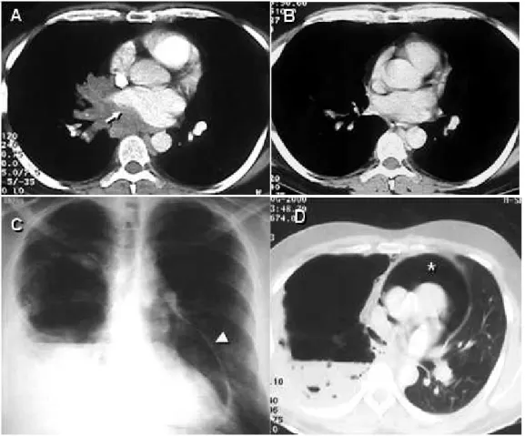
and his chest X-rays showed hilar adenopathy in the right lower lobe. A computed tomography (CT) scan revealed a central mass invading the inferior pulmonary vein and compressing the left atrium (Figure 1A). An endobronchial lesion was seen in the right lower lobe bronchus, and a biopsy was performed. The results were consistent with neuroendocrine large cell carcinoma. The patient was submitted to three cycles of cisplatin-based chemotherapy and radiotherapy. Subsequent CT scans (Figure 1B) and bronchoscopic examinations revealed a highly positive response to the treatment. He was submitted to radical right pneumonectomy including a portion of the border of the left atrium (first resection margin positive at the level of the inferior pulmonary vein) and the perihilar pericardium. The pericardial defect was grafted with bovine pericardium and a 2-cm
orifice was left in the suture line to allow fluid or air to freely exit the pericardial sac. The patient was discharged on the fifth postoperative day after a short and uneventful postoperative period. Ten days later, he returned to the hospital presenting postural hypotension and constant substernal pain. A chest X-ray showed pneumopericardium (Figure 1C) confirmed on a CT scan, which also revealed multiple air pockets and an air-fluid level in the pleural space, suggesting a bronchopleural fistula (Figure 1D). He was admitted to the hospital, a (36FR) chest tube was inserted into the right pleural space, and a bronchoscopy showed a tiny (1.0-mm) bronchial stump fistula. There were no signs or symptoms of infection, and the pleural fluid was clear, with fewer than 300 cells, 80% of which were eosinophils. The patient did not present intraoperative or postoperative air leakage through
86 Haddad R, Lima CET, Boasquevisque CH, Haddad GS, Ferreira TD
J Bras Pneumol. 2006;32(1):84-7
the chest tube. Antibiotics were given, and the evolution was excellent. The chest tube was withdrawn on the tenth postoperative day. The symptomatic pneumopericardium disappeared immediately after tube thoracostomy, and no empyema or other complications developed.
Case 2
A 64-year-old male patient with chronic obstructive pulmonary disease was submitted for coronary artery bypass graft (CABG) with left internal mammary artery-left anterior descending anastomosis. In the intensive care unit, at two hours after extubation, he presented a spontaneous left-sided pneumothorax that was controlled through insertion of a 14FR pigtail catheter, after which the patient improved. Two days later, however, he presented respiratory discomfort and a second pneumothorax was seen despite the fact that the pigtail catheter remained in the pleural cavity and was unobstructed. A second pigtail catheter was inserted in an anterior position. The problem was resolved, and the catheters were removed. On the following day, the patient presented hypotension, tachycardia and dyspnea. For technical reasons, it was not possible to perform an echocardiogram. A CT scan showed an anterior left pneumothorax and a large pneumopericardium (Figure 2). A 32FR chest tube was inserted into the left pleural cavity, and the problem was completely resolved. The patient also underwent talc pleurodesis. The same simple solution (tube thoracostomy) was applied in this second case.
DISCUSSION
Tension pneumopericardium in the absence of chest trauma or mechanical ventilation is an uncommon entity. It usually involves communication between the pericardium and the tracheobronchial tree or digestive tract. When there is no communication between the pericardial sac and the pleural cavity, the treatment of choice is pericardial drainage. When such communication exists, chest tube insertion is preferred. In patients undergoing intrapericardial lung resection, the main etiologic factor for tension pneumopericardium is bronchopleural fistula, whereas it can be related to pneumothorax or barotrauma following cardiac surgery. The common causes of pneumopericardium are penetrating chest trauma, mechanical ventilation (barotrauma), propagation of diseases in contiguous organs, cardiothoracic surgery, invasive procedures (pericardiocentesis), infection, and abnormal communication between the airways and the pericardium.(1) In the two cases presented herein,
the patients underwent different procedures, but both had abnormal communication between the airways and the pericardial sac related to postoperative complications. Both patients presented pneumothorax, the first caused by a small bronchopleural fistula and the second by an alveolopleural fistula as a result of post-CABG barotrauma. In fact, Capizzi et al.(2) found a
correlation between pneumothorax and tension pneumopericardium in approximately 87.5% of the cases evaluated. It is important to point out that pneumopericardium is more prevalent in nontrauma settings in which greater ventilatory support is necessary, such as in acute respiratory distress syndrome or neonatal respiratory support.(3) It is also
important differentiate between pneumopericardium and pneumomediastinum, especially when the pneumopericardium is small. A very interesting indicator, the continuous left hemidiaphragm sign, has been reported in a case of pneumopericardium(4)
and can inform in the diagnosis. Benedík et al.(5)
reported a case of pneumopericardial tamponade after coronary artery bypass surgery. The authors found that the condition was related to bullae rupture into the pericardial sac (adhesions prevented concurrent pneumothorax) and treated it using emergency thoracotomy. Kim et al.(6) published a
case of pneumopericardium and pneumothorax caused by the rupture of a cancerous lung tumor
J Bras Pneumol. 2006;32(1):84-7
Pneumothorax and tension pneumopericardium following cardiothoracic surgery 87
into the pericardial sac. Brandenhoff et al.(7) published
two cases of pneumopericardium occurring after lung resection, one of which was a case of tension pneumopericardium. Both patients had been on prolonged ventilatory support and presented air leakage prior to the pneumopericardium. The authors treated both cases with thoracotomy and pericardiotomy. Stuklis et al.(8) published a case of tension
pneumopericardium after middle lobectomy that was treated using the same simple technique employed in our two cases, tube thoracostomy.
CONCLUSION
Acute hemodynamic deterioration in patients with pneumothorax or requiring mechanical ventilation should prompt further investigation, and cardiac tamponade should be actively ruled out. In cases of tension pneumopericardium in which there is no evidence of communication between the pericardial sac and the pleura, emergency p e r i c a r d i o c e n t e s i s o r d r a i n a g e s h o u l d b e performed. However, if there is such evidence, tube thoracostomy is indicated.
REFERENCES
1. Boyce SH, Corfield AR, McGuffie AC, Stevenson J, Rawlings D. Spontaneous tension pneumopericardium. Eur J Emerg Med. 2004; 11(3):181-4.
2. C a p i z z i P J , M a r t i n M , B a n n o n M P. Te ns i o n pneumopericardium following blunt injury. J Trauma. 1995; 39(4):775-80. Review.
3. Clouse WD, Dent DL, Stewart RM, Gray GA. Tension pneumopericardium from blunt chest trauma. Contemp Surg. 2003; 59(6):271-5.
4. Brander L, Ramsay D, Dreier D, Peter M, Graeni R. Continuous left hemidiaphragm sign revisited: a case of spontaneous pneumopericardium and literature review. Heart [serial on the Internet]. 2002; [cited 2005 Sep 17]: 88(4): [about 5p.]. Available from. http:// www.heartjnl.com/cgi/content/full/88/e5
5. Benedik J, Uchytil B, Cernosek J. Pneumopericardial tamponade after coronary artery bypass operation. Eur J Cardiothorac Surg. 2002;21(3):585-6. Review. 6. Kim YI, Goo JM, Im JG. Concurrent pneumopericardium
and pneumothorax complicating lung cancer: a case report. Korean J Radiol. 2000;1(2):118-20.
7. Brandenhoff P, Hoier-Madsen K, Struve-Christensen E. Pneumopericardium after pneumonectomy and lobectomy. Thorax. 1986:41(1):55-7.