290
1. Pediatrics Cardiac Surgery Service - Hospital de Base, State Medical School of São José do Rio Preto - FAMERP
Correspondence: Ulisses Alexandre Croti
Hospital de Base – FAMERP – Avenida Brigadeiro Faria Lima, 5544. CEP 15090-000 – São José do Rio Preto – São Paulo
Phone (Fax): 17 – 3201-5025 / 3222-6450 / 9772-6560. E-mail address: [email protected]
Ulisses Alexandre CROTI1, Domingo Marcolino BRAILE1, Lílian BEANI1, Maria Cristina Passos FLEURY1 Rev Bras Cir Cardiovasc 2008; 23(2): 290-291
CLINICAL-SURGICAL CORRELATION
RBCCV 44205-990
Monocúspide de homoenxerto decelularizado no tratamento do truncus arteriosus com a técnica de
Barbero Marcial
The use of decellularized homograft monocuspid
in the treatment of truncus arterious by Barbero
Marcial technique
Article received on Abril 20th, 2008 Article accepted on May 21st, 2008 CLINICAL DATA
A twenty-one-day-old male infant, 3.1kg, from Penápolis, Brazil. Since birth, very tearful and with slight respiratory distress that developed into tachydyspnea. In the Service of origin, the patient was diagnosed with cyanogenic congenital heart disease with pulmonary hyperflow and was immediately sent for surgical treatment. During physical examination, he presented in poor condition: afebrile, tachydyspneic, cyanotic + / 4 + and icteric + / 4 +. Hyperdynamic precordium, regular heart rhythm, systolic murmur +++/6+ in median and low left sternal margin; and hyperphonetic second heart sound. Normal pulmonary auscultation. Flaccid abdomen, liver at 3 cm from the costal edge, no palpable spleen and presence of fluid-air sounds. Extremities with poor perfusion, without edema, palpable and symmetrical pulses in all limbs, peripheral saturation of 87%. Even in the preoperative, an episode of convulsive crisis that was investigated and which excluded the presence of arteriovenous malformation.
ELECTROCARDIOGRAM
Sinus rhythm, frequency 150 beat/min, SÂP +90º, SÂQRS + 90°, PR interval 0.2 s. Mild standard block in the right branch.
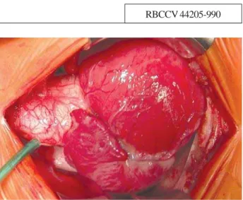
Fig. 1 – Tourniquet for ligation of the right pulmonary artery immediately after opening the pericardium flap.Important maneuver for better surgical outcome
RADIOGRAM
291
CROTI, UA ET AL - The use of decellularized homograft monocuspid in the treatment of truncus arterious by Barbero Marcial technique
Rev Bras Cir Cardiovasc 2008; 23(2): 290-291
ECHOCARDIOGRAM
Situs solitus with levocardia. Normal venoatrial and atrioventricular connections. Abnormal ventriculoarterial connection with single common outflow tract, leading to a diagnosis of truncus arteriosus Type I. The pulmonary trunk originated from the truncal vessel at the left lateral face at 4cm to the valvar plane. Truncal valve with stenosis and slight insufficiency, with peak systolic gradient (through Doppler) of 21mmHg by the truncal valve. Presence of interatrial communication (oval fossa) of 2.3 mm, wide interventricular communication and slight mitral valve insufficiency.
DIAGNOSIS
The clinical presentation of irritability and manifestations of heart failure is typical in children with truncus arteriosus. The echocardiogram clearly showed the single origin of the systemic, pulmonary and coronary arteries, confirming the diagnosis and excluding severe deffects that may be associated with the disease, such as interruption of the aortic arch. In the differential diagnosis, we must consider the transposition of the large arteries with interventricular communication, hypoplastic left heart syndrome, Tetralogy of Fallot with agenesis of the pulmonary valve and complex heart diseases without pulmonary stenosis.
OPERATION
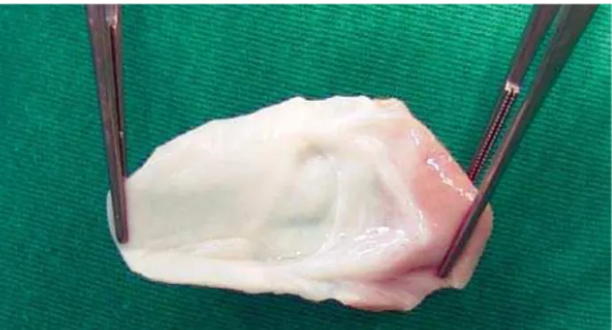
Median transsternal thoracotomy, pericardium opening, ligation of the right pulmonary artery for restriction of the flow in order to avoid myocardial ischemia (situation that may be crucial in the prognosis [Figure 1]). Installation of the conventional cardiopulmonary bypass (CPB) with the use of cannulas in cavae and aorta. Occlusion of the pulmonary branches, deep hypothermia at 25°C, aortic clamping the most distal possible level of the truncal valve; opening of the right atrium and left atrium aspiration by the interatrial communication. Blood anterograde cardioplegia, intermittent and hypothermic at 4ºC. Longitudinal incision on the anterio-posterior wall of the truncal vessel toward the left pulmonary artery inferior up to the Valsalva sinus. Through this opening, the systemic components, origins of the pulmonary arteries and coronary ostia were observed. With a bovine pericardium patch of the appropriate size, the neoaorta was separated from the pulmonary trunk. With a vertical incision in the right ventricle wall and by means of this surgical approach, the interventricular communication was closed with another bovine pericardium patch. The lateral left flap of the truncal component was sutured to the right ventricle wall, creating the posterior wall of the neopulmonary with autologous tissue [1]. The anterior face was rebuilt with a decellularized homograft monocuspid patch from a pulmonary homograft 22 (Figures 2 and 3). The time of infusion was 114 minutes, plus 75 minutes of myocardial ischemia. After the CPB, there was
bleeding caused by a coagulation disorder, and was needed a new CPB assistance to heat and correct metabolic alterations over 30 minutes.In the immediate postoperative period, even with alterations in the coagulation cascade, the patient developed retained clot syndrome and needed exploratory thoracotomy. In the intensive care unit, because the patient was a newborn with severe heart disease and had undergone CPB, he underwent peritoneal dialysis to remove liquids and components of the acute inflammatory response. The patient developed pulmonary infection which was treated with appropriate antibioticotherapy, and he was discharged from the hospital on the 16th postoperative day with good clinical conditions with the use of digoxin and aldactone. After 3 months of postoperative, the child is asymptomatic, gaining weight appropriately, with a diet of only breast milk. The controlled echocardiogram confirmed the excellent surgical outcome.
REFERENCE
1. Barbero-Marcial M, Riso A, Atik E, Jatene A. A technique for correction of truncus arteriosus types I and II without extracardiac conduits. J Thorac Cardiovasc Surg. 1990;99(2):364-9.
Fig. 2 – Decellularized homograft monocuspid prepared for implantation in the anterior wall