Non-Conventional Models Correlates with the Yeast
In
Vitro
Susceptibility Profile
Liliana Scorzoni1,2, Maria Pilar de Lucas3, Ana Cecilia Mesa-Arango1,4, Ana Marisa Fusco-Almeida2, Encarnacio´n Lozano3, Manuel Cuenca-Estrella1, Maria Jose Mendes-Giannini2*, Oscar Zaragoza1*
1Mycology Reference Laboratory, National Centre for Microbiology, Instituto de Salud Carlos III, Madrid, Spain,2Laborato´rio de Micologia Clı´nica, Faculdade de Cieˆncias Farmaceˆuticas, Universidade Estadual Paulista de Sa˜o Paulo, Araraquara, Brazil,3Department of Cellular Biology, National Centre for Microbiology, Instituto de Salud Carlos III, Madrid, Spain,4Group of Investigative Dermatology, University of Antioquia, Medellı´n, Colombia
Abstract
The incidence of opportunistic fungal infections has increased in recent decades due to the growing proportion of immunocompromised patients in our society.Candida krusei has been described as a causative agent of disseminated fungal infections in susceptible patients. Although its prevalence remains low among yeast infections (2–5%), its intrinsic resistance to fluconazole makes this yeast important from epidemiologic aspects. Non mammalian organisms are feasible models to study fungal virulence and drug efficacy. In this work we have used the lepidopteranGalleria mellonellaand the nematodeCaenorhabditis elegansas models to assess antifungal efficacy during infection byC. krusei. This yeast killedG. mellonellaat 25, 30 and 37uC and reduced haemocytic density. Infected larvae melanized in a dose-dependent manner. Fluconazole did not protect againstC. kruseiinfection, in contrast to amphotericin B, voriconazole or caspofungin. However, the doses of these antifungals required to obtain larvae protection were always higher duringC. kruseiinfection than during C. albicansinfection. Similar results were found in the model hostC. elegans. Our work demonstrates that non mammalian models are useful tools to investigatein vivoantifungal efficacy and virulence ofC. krusei.
Citation:Scorzoni L, de Lucas MP, Mesa-Arango AC, Fusco-Almeida AM, Lozano E, et al. (2013) Antifungal Efficacy duringCandida kruseiInfection in Non-Conventional Models Correlates with the YeastIn VitroSusceptibility Profile. PLoS ONE 8(3): e60047. doi:10.1371/journal.pone.0060047
Editor:Gustavo Henrique Goldman, Universidade de Sao Paulo, Brazil
ReceivedOctober 4, 2012;AcceptedFebruary 20, 2013;PublishedMarch 28, 2013
Copyright:ß2013 Scorzoni et al. This is an open-access article distributed under the terms of the Creative Commons Attribution License, which permits unrestricted use, distribution, and reproduction in any medium, provided the original author and source are credited.
Funding:O.Z. is funded by grant SAF2011-25140 from the Spanish Ministry for Economics and Competitiveness. L.S. is funded by a fellowship from the Agencia Espan˜ola para la Cooperacio´n Internacional y Desarrollo. C. elegans strain AU37 was provided by the CGC (Caenorhabditis Genetics Center), which is funded by National Institutes of Health Office of Research Infrastructure Programs (P40 OD010440). The funders had no role in study design, data collection and analysis, decision to publish, or preparation of the manuscript
Competing Interests:The authors have the following interests: In the past 5 years, M.C.E. has received grant support from Astellas Pharma, bioMerieux, Gilead Sciences, Merck Sharp and Dohme, Pfizer, Schering Plough and Soria Melguizo SA. He has been an advisor/consultant to Gilead Sciences, Merck Sharp and Dohme, Pfizer, and Schering Plough. He has been paid for talks on behalf of Gilead Sciences, Merck Sharp and Dohme, Pfizer, and Schering Plough. There are no patents, products in development or marketed products to declare. This does not alter the authors’ adherence to all the PLOS ONE policies on sharing data and materials.
* E-mail: giannini@fcfar.unesp.br ( JMG); ozaragoza@isciii.es (OZ)
Introduction
Fungal infections have emerged worldwide due to a growing population of immunosuppressed patients, including patients with cancer, AIDS, solid-organ and hematopoietic stem cell transplant recipients, premature neonates, and patients recovering from major surgery [1–5]. These infections have significant morbidity and mortality rates and are difficult to prevent, diagnose and treat [6–8].
Candida spp are commensal yeasts responsible for different clinical manifestations, from mucocutaneous overgrowth to blood stream infections [1,9–12].Candida albicansis still the major cause of invasive fungal disease. However, a growing number of infections produced by non-albicans Candidaspp has been reported in the last years [1,13–15]. Among them, there are some species that are intrinsically resistant or have reduced susceptibility to antifungals. The massive use of antifungals in prophylaxis, such as fluconazole, has facilitated the selection of pathogenic fungi resistant to these agents [16–19].
Candida krusei is an opportunistic pathogen which presents intrinsic resistance to fluconazole. The infection is associated with the prophylactic or therapeutic use of this antifungal agent [20– 23]. Two mechanisms of azole resistance inC. krusei have been described: overexpression of drug efflux pumps [24] and diminished sensitivity of the target enzyme, the cytochrome P450 sterol 14-demethylase (encoded by the CYP51 gene) [25]. Diseases caused byC. krusei have high associated mortality (30– 60%) [26,27]. Despite the intrinsic resistance to fluconazole, C. kruseiis usually susceptible to voriconazolein vitro, which correlates with the binding of this drug to the target enzyme [21].
Antifungal resistance in vitro does not always correlate with clinical resistance. The best correlation betweenin vitroand clinical efficacy is found in HIV-positive patients with oropharyngeal candidiasis [22,28,29]. In contrast, althoughC. parapsilosisshows reduced in vitrosusceptibility to echinocandins, these antifungals have been shown to be effective in the treatment of invasive candidiasis caused by this species [30].
The use of invertebrate hosts has recently emerged and facilitated the study of fungal pathogenesis. Among these non-mammalian hosts, amoebae (Acanthamoeba castellanii, Dictyostellium discoideum), nematodes (Caenorhabditis elegans) and insects (Drosophila melanogaster,Galleria mellonella) have been successfully used to study the virulence of some fungi [31–35]. Moreover, some aspects of the innate response are conserved between these hosts and mammals [36].Galleria mellonellais a lepidopteran (Pyralidae) that provides important advantages as host model. The larvae can be incubated in a range of temperature between 25 to 37uC, so it is possible to simulate the natural fungal habitat and the infection conditions in mammals. In addition, as in mammalian models, it is possible to introduce by injection exact doses of pathogens to the larvae, which poses a technical improvement over other non-conventional hosts. Galleria mellonella has six types of phagocytic cells that play an important role in the defense system [37,38]. The density of these cells in the haemolymph is not constant, and changes during infection can be easily measured and used as a parameter of the response of the larvae after exposure to pathogens [39]. The viability of the larvae can be easily recorded by the lack of movement and the massive melanization induced by G. mellonella in response to infection [40–42]. Another organism that is used as model host is the soil nematodeCaenorhabditis elegans, which feeds on microorganisms, but is susceptible to bacterial and fungal pathogens [33,43–45].Caenorhabditis eleganshas been used to study virulence, filamentation and antifungal efficacy of antifungal drugs [44,46].
In this study, we initially aimed to characterize the interaction between G. mellonellaand C. krusei with two purposes: 1) To get insights about virulence traits of this pathogenic yeast, and 2) to investigate if antifungal efficacy in vivo correlates with the susceptibility profile shown byC. krusei in vitro. Furthermore, we have complemented our studies with C. elegans, and observed similar behaviors, indicating that non-conventional models can be used to investigateC. kruseivirulence and antifungal efficacy.
Materials and Methods
Strains and media
Candida albicans SC5314 [47], C. krusei ATCC 6258 and two clinical isolates (CL8053 and CL80317) from the Yeast Collection of the Mycology Reference Laboratory of the Spanish National Centre for Microbiology and Cryptococcus neoformans varietygrubii (H99 strain, ATCC 20882) were used in this study. The yeasts were grown overnight in liquid Sabouraud medium (Difco, BD, USA) at 30uC with shaking. Escherichia coli OP50 strain was obtained from the Caenorhabditis Genetics Center (University of Minnesota) and was maintained on LB agar plates at 37uC.
Antifungal susceptibility testing (AFST)
Minimum inhibitory concentration (MIC) values were deter-mined using the EUCAST protocol [48,49]. For AFST, 10 different clinical isolates ofC. albicansand 10 clinical isolates ofC. krusei were obtained from the yeast collection of the Mycology Reference Laboratory of the Spanish National Centre for Microbiology. Data were expressed as geometric mean, mode, range (minimum-maximum) and MIC frequency distribution.
Insect larvae manipulation and incubation conditions
Galleria mellonella larvae (0.3–0.5 g, R.J. Mous Livebait, The Netherlands) were placed in Petri dishes and incubated at 37uC in the dark the night before the experiments. Larvae with color alterations (i.e. dark spots or with apparent melanization) were excluded. Antifungals and yeast suspensions were injected in the
haemocele through the last left pro-leg of the larvae using a 10mL
Hamilton syringe (Hamiltion, USA). The pro-leg had been previously cleaned with 70% ethanol. A total of 10mL were
injected in each larva. Larvae death was monitored by visual inspection of the color (brown-dark brown) and by lack of movement after touching them with forceps. For each condition, a total of 20 larvae were used, and each experiment was repeated at least twice. After infection, larvae were incubated at 25, 30 or 37uC.
Survival assay
Yeasts were grown overnight in liquid Sabouraud, washed with PBS, and suspended in the same buffer. Cell density was estimated with an Automatic Cell Counter TC10 (Bio Rad). For survival assays, larvae were inoculated with 107, 56106and 2.56106cells/ larva ofC. kruseiand 106, 5
6105and 105cells/larva ofC. albicans. The inocula were prepared in PBS plus 20 mg/L of ampicillin to prevent bacterial contamination. The infected larvae were incubated at 25uC, 30 or 37uC, and the death was daily monitored during 7 days.
Growth curve at different temperatures
Yeast strains were grown overnight and diluted in fresh Sabouraud liquid medium at 103 cells/mL. Two hundred microliters of this suspension were placed in 96-well microdilution plates, and incubated at 25, 30 or 37uC in a Labsystems IEMS Reader MF spectrophotometer. Optical density (OD) was determined at 530 nm every hour during 72 hrs.
In vivophagocytosis assay
Yeast cells were stained with 10mg/mL Calcofluor white
(Sigma, St. Louis, MO) for 30 min at 37uC. Then, these cells were injected intoG. mellonellalarvae (107cells/larva, 5 per group), and phagocytosis was analyzed after 3 hrs of incubation at 25 and 37uC. Haemolymph was collected in 1.5 mL tubes and diluted 1:1 in IPS buffer (Insect Physiological Saline: 150 mM sodium chloride, 5 mM potassium chloride, 10 mM Tris-HCl pH 6.9, 10 mM EDTA and 30 mM sodium citrate) to avoid coagulation and melanization of the haemolymph. Haemocytes were placed on a slide and phagocytosis was visually quantified using a Leica DMI 3000B microscope. One hundred haemocytes from each larva were counted in each case, and the percentage of haemocytes containing yeasts was calculated and plotted.Cryptococcus neoformans H99 strain was used as control. Phagocytosis was also analyzed in larvae infected in the same way, but treated with 64 mg/kg fluconazole or 4 mg/kg amphotericin B.
Determination of haemocyte density
Groups of fiveG. mellonellawere infected with 107yeast cells/ larvae and incubated at 37uC for 3 hrs. The haemolymph of each larva was collected in 1.5 mL tubes and diluted 1:10 in IPS buffer. The cells were counted using a haemocytometer.
Measurement of in vivo filament formation
Galleria mellonellawas infected with 107cells/larva ofC. albicans and C. krusei strains. The larvae were incubated at 37uC for 24 hours. Larvae were macerated in 100mm nylon cell strainers
(Falcon, BD, USA) with 1 mL of IPS. The liquid was then collected, centrifuged and suspended in 1 mL of the same buffer. Samples were stained with Calcofluor white (Sigma, St. Louis, MO), as described above, and yeast morphology was observed with a Leica DMI 3000B fluorescence microscope.
Melanization quantification
Larvae were infected with PBS, 56105, 106and 56106cells/ larva of C. krusei. Then, the haemolymph of each larva was collected after 3 and 24 hrs in 1.5 mL tubes and diluted 1:10 with IPS buffer. The samples were placed in 96 well microdilution plates. To quantify melanin levels, we took advantage of existing protocols that quantify laccase activity by detecting the final product of the reaction by measuring the OD in the visible range (400–500) [50,51]. In our conditions, we observed that 405 nm was an optimal OD to quantify larval melanin and to correlate the results with the visualization of the dark compound. So the OD at 405 nm was measured using a Labsystems IEMS Reader MF spectophotometer. Melanization of larvae infected withC. albicans (56105cells/larva) and C. krusei (56106cells/larva) and treated with 64 mg/kg fluconazole and 4 mg/kg amphotericin B was also evaluated.
Treatment with antifungal drugs
Infected larvae were treated with amphotericin B (1, 2 or 4 mg/ kg, Sigma Aldrich Quimica, Madrid, Spain), fluconazole (128, 64, 32, 12, or 4 mg/kg, Pfizer SA, Madrid, spain), voriconazole (7.5 or 10 mg/kg, Pfizer SA, Madrid, Spain) or caspofungin (1, 2 or 4 mg/kg Merck & Com, Inc, NJ, USA). In some experiments, a combination of fluconazole and amphotericin B was also used. Antifungals were applied immediately after the infection. Groups of 10 larvae were treated with the antifungals alone to test the toxicity.
Fungal burden determination
Infected larvae were selected at different times of infection, washed with 70% ethanol and cut into small pieces with a scalpel. Two mL of PBS-ampicillin were added and the mix was homogenized gently with a vortex and glass beads for 10 seconds. The mix was finally suspended in 9 mL of PBS-ampicillin. Different dilutions were made for each sample and 50mL from
these dilutions were placed on Sabouraud-cloramphenicol agar plates (Oxoid). The plates were incubated at 37uC for 48 h, and the number of colony forming units (CFUs) was determined.
Histology
Three larvae from different groups (uninfected, infected and/or treated with antifungals) were collected on different days of the infection. The larvae were preserved in 70% ethanol and longitudinal incisions were made with a scalpel in the dorsal part. The samples were fixed with 10% buffered formaline for 24 hrs. Then, the samples were dehydrated with increasing concentrations of ethanol, rinsed with xylol, and embedded in paraffin. Tissue sections (5 microns) were stained with periodic acid Schiff (PAS) solution and observed with a Leica DMI3000 microscope.
Caenorhabditis elegansstrain and infection conditions The followingC. elegansmutant strain, obtained from CGC, was used in all experiments: AU37 (glp-4(bn2) I; sek-1(km4) X). This strain was grown on agar plates seeded with E. coli OP50 and incubated at 15uC according to standard procedures [52]. This strain is usually chosen for virulence and antifungal efficacy assays because glp-4 mutants are sterile at 25uC. This allows to easily following up the survival of the individual animals from the beginning to the end of the experiment and avoids mixing with their progeny [33,44]. Thesek-1gene encodes a mitogen-activated protein kinase which is important for the defense of C. elegans against microbial infections [33,44]. Therefore worms defective for sek-1are more susceptible to infection and die earlier than
wild-type C. elegans animals. Candida strains were cultivated in liquid Sabouraud medium (Difco, BD, USA) at 35uC with shaking. One hundredmL from this culture were inoculated on solid BHI media (Difco) containing kanamycin (90mg/mL) and ampicillin (200mg/
mL) and incubated at 30uC for 24 hours. Synchronized worms in the L4 stage were added to the center of the agar plates inoculated with the yeast strains lawns and incubated for three hours at 25uC. In parallel, L4 worms were placed on agar plates containing lawns of E. coliOP50 strain. After the three hours incubation, worms were washed with M9 and transferred to 12-well plates with 1 mL 60% M9 buffer [45], 40% BHI, 10mg/mL cholesterol in ethanol,
200mg/mL ampicilin and 90mg/mL kanamycin. Around 20–30
worms were placed in each well. For antifungal efficacy, amphotericin B (1 and 2mg/mL), fluconazole (12mg/mL),
voriconazole (0.25, 7.5 and 10mg/mL), caspofungin (2, 4 and
6mg/mL), or a combination of amphotericin B (1mg/mL) plus
fluconazole (12mg/mL) were added to the media. Plates were incubated at 25uC and individual worm survival was monitored daily. Nematodes were considered dead when they did not respond to touching. A minimum of two independent experiments was carried out for each treatment. Images were captured with a video camera (JVC KY-F550) attached to a dissecting microscope (Leica MZ7.5).
Statistics
Graphs and Statistics analyzes were performed with Graph Pad Prisma 5 (La Jolla CA, USA). Survival curves were analyzed by Log-rank (Mantel-Cox) Test and phagocytosis assays, haemocyte density, melanization quantification and fungal burden were analyzed using t-Test.
Results
Candida kruseikilledG. mellonellain a dose dependant manner
We first investigated ifC. kruseikilledG. mellonella. Our results showed that G. mellonella is susceptible to C. krusei infection (Figure 1A). The death rate of the larvae depended on the yeast dose injected. Most reproducible results were found when larvae were infected with 56106C. kruseicells. However,C. kruseiwas less virulent than other fungi, such as C. albicans, which killed G. mellonellaat lower doses (56105, Figure 1B). To confirm that the death was not a consequence of a shock due to big amounts of yeast injected in the larvae, we inoculated a group of larvae with yeast inactivated by incubation in 4% paraformaldehyde. As shown in Figure 1C, inactivated yeast did not killG. mellonella, confirming that larvae death was dependent on living yeast.
compared to 30uC (two-fold reduction in generation time). In contrast, C. krusei grew similarly at both temperatures (0.85 fold decrease in generation time when the cells were grown at 37uC compared to 30uC). We found similar results at 25uC (data not shown), supporting that C. krusei growth is not affected by the incubation temperature. The final OD reached at the stationary phase at different temperatures was different with both species. Candida albicansreached higher OD at 37uC, which differed from the situation found in C. krusei, where the final OD at the stationary phase was almost identical at 30 and 37uC. Latency period was longer at 30uC, but the same trend was observed in both species (Figures 1E and 1F).
Yeast inoculation caused early melanization of the larvae
Galleria mellonellalarvae appeared melanized after a few minutes ofC. kruseiinjection (Figure 2A). To quantify this phenomenon, we collected the haemolymph and measured its optical density at 405 nm. When larvae were infected with 56106 C. krusei cells, there was a significant accumulation of melanin in the haemo-lymph (4.3 times compared to the non-infected larvae), and this melanization increased over time (5 times at 24 hrs, Figure 2B). Clinical isolates showed a similar behavior (Figures 2C and D). We evaluated ifC. kruseiinduced melanization ofG. mellonellaat lower temperatures, and we found that this phenomenon also occurred at 25uC (data not shown).
Phagocytosis and effect ofC. kruseion haemocyte density
We examined if different C. krusei strains had any effect on haemocyte density. As shown in Figure 3A,C. kruseiproduced a decrease in haemocyte density in a similar manner toC. albicans.
We then investigated ifC. krusei cells were phagocytosed byG. mellonella haemocytes. We compared the phagocytosis of this pathogen to the one measured withC. albicansand C. neoformans. The phagocytosis for all Candida strains (albicans and krusei) was significantly lower to the phagocytosis observed withC. neoformans (Figure 3B). The same result was found when phagocytosis was assessed at 25uC (data not shown).
Candida krusei can filament in vitro, so we investigated if this change also took place during infection in G. mellonella. We included C. albicans in these experiments as control, since it has been reported that this yeast can form hyphae in this model host [53]. As expected,C. albicansefficiently produced filaments in the larvae.Candida kruseialso produced filaments, and inG. mellonella crude extracts they were frequently found in clumps of fat body of dark color, which we believe that are composed mainly of insect melanin. This fact may explain the fast melanization ofG. mellonella when infected withC. krusei.
Antifungal efficacy duringC. kruseiinfection inG. mellonella
One of the main features forC. kruseiis itsin vitrosusceptibility profile. As shown in Figure 4, C. krusei is less susceptible to amphotericin B, voriconazole and caspofungin than C. albicans, and intrinsically resistant to fluconazole. So we studied if this phenotype correlated with a lack of response to the antifungal during infection inG. mellonella. For this purpose, we infectedG. mellonellawithC. kruseiorC. albicans, and treated the larvae with different antifungals (fluconazole, voriconazole, amphotericin B and caspofungin). In the case of larvae infected with C. krusei, treatment with fluconazole, even at very high doses (32 or 64 mg/ kg) did not increase the survival (Figures 5A and B). At higher
Figure 1. Comparison of the virulence ofC. kruseiandC. albicansinG. mellonella.(A) Survival curve ofG. mellonellainfected with different inocula ofC. kruseiATCC 6258
N
PBS;&107cells/larva;m56106cells/larva;.2.56106cells/larva incubated at 37uC (B). Survival curve ofG. mellonella
infected with different inocula ofC. albicansSC5314
N
PBS;&106cells/larva;m56105cells/larva;.105cells/larvae (C) Survival ofG. mellonella
infected with inactivated yeast. Control dead cells
N
PBS;&C. kruseiATCC 6258 56106cells/larva;mC. kruseiATCC 6258 56106cells/larva (dead);. C. albicansSC5314 106cells/larva;¤
C. albicansSC5314 106cells/larva (dead) (D); Effect of the incubation temperature on the virulence ofC. albicansandC. krusei.
N
PBS;mC.kruseiATCC 6258 (37uC);.C.kruseiATCC 6258 (30uC);¤
C. albicansSC5314 (37uC);&C. albicansSC5314 (30uC); Growth curves ofC. albicans(E) andC. krusei(F) at different temperatures.#37uC;m30uC.doi:10.1371/journal.pone.0060047.g001
concentrations (128 mg/kg), there was a decrease in the survival, which is explained by the toxicity of the antifungal at this high concentration, which induced 25% of death after 7 days of treatment (data not shown). When the same experiments were performed with C. albicans, treatment with all the fluconazole concentrations produced significant survival (Figures 5C and 5D). Concerning other azoles, C. krusei is considered susceptible to
voriconazole, although it presents higher MIC values to this antifungal thanC. albicans(see Figure 4). So we studied the efficacy of voriconazole during infection in G. mellonella. We found that both voriconazole concentrations tested (7.5 and 10 mg/kg) protected larvae fromC. albicansinfection (Figure 6A). In contrast, larvae infected with C. krusei were only protected with higher
Figure 2. Melanization ofG. mellonellainfected withC. krusei.(A)Visual appearance ofG. mellonellalarvae infected with differentC. krusei
doses. (B, C and D) Optical Density (OD) of the haemolymph ofG. mellonellainfected withC. kruseiATCC 6258 (B), clinical isolate CL8053 (C) and CL80317 (D) with 56105, 106, 56106cells/larva. The different size inoculum reveals dose-response melanization (* p,0.05). All the experiments in this figure were performed at 37uC.
doi:10.1371/journal.pone.0060047.g002
Figure 3. Interaction betweenC. kruseiand haemocytes.(A) Changes in haemocyte density duringC. kruseiinfection. The haemolymph of infected larvae withC. neoformans, C. albicansSC5314,C. kruseiATCC 6258, CL8053 and CL80317 clinical isolates and PBS was collected and the concentration of haemocytes was estimated using a haemocytometer (B). Phagocytosis percentage ofC. neoformans,C. albicansSC5314,C. krusei
voriconazole concentrations (Figure 6B). Lower doses did not have any effect on survival.
Amphotericin B (4 mg/kg) prolonged survival of larvae infected with C. albicans at all the concentrations tested (Figure 6A). In contrast, amphotericin B only protected larvae infected with C. krusei at the highest dose (4 mg/kg), which produced a 60% survival at the fourth day (Figure 6B). In a similar way, caspofungin was effective during C. albicans infection at all the doses tested (Figure 6C), while it only protected larvae inoculated withC. krusei at the highest dose (4 mg/kg) (Figure 6D). We also used an antifungal combination with fluconazole (12 or 4 mg/kg) and amphotericin B at a sub-therapeutic dose in G. mellonella (1 mg/kg), but we found no synergic effect between the antifungals (data not shown).
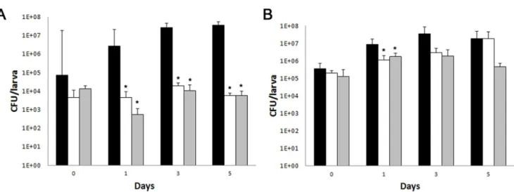
Fungal burden determination and histopathology The fungal burden was determined by recovering the yeast cells from the larvae infected withC. albicans orC. krusei and treated with fluconazole (12 mg/kg) or amphotericin B (4 mg/kg). The
number of CFUs increased in larvae infected with both pathogens with the time of infection (Figure 7). Treatment of larvae infected withC. albicanswith fluconazole or amphotericin B decreased the number of CFUs by 1000-fold (Figure 7A). In larvae infected with C. krusei, amphotericin B reduced the fungal burden by 10-fold. Curiously, fluconazole also reduced the initial fungal burden, although it did not have an effect after longer times (5 days, Figure 7B).
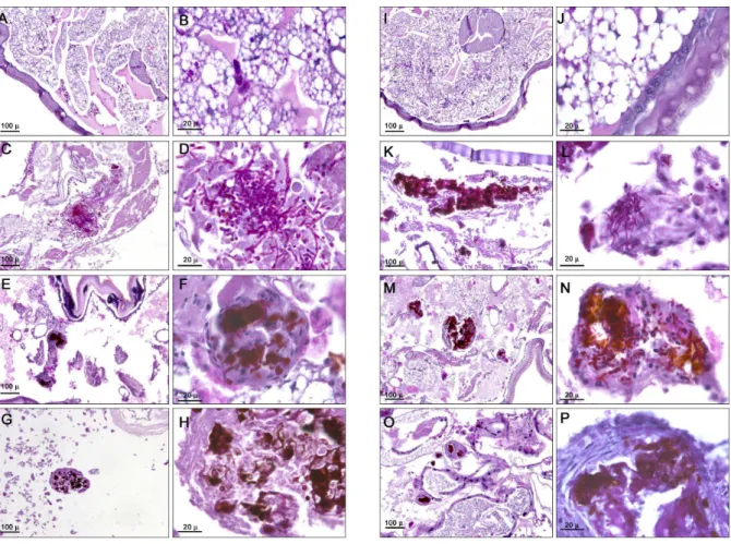
To complement these studies, we performed histopathology of infected and treated larvae. Candida albicans (Figure 8C and 8D) and C. krusei(Figure 8K and 8L) were found both in yeast and filament forms. The antifungal treatment with fluconazole (12 mg/kg) in larvae infected withC. albicansorC. kruseidecreased the number of yeasts. Moreover, the fungi were mainly found in defined structures surrounded byG. mellonella cells (Figure 8E, F, M, N). Amphotericin B (4 mg/kg) had the same effect as fluconazole, although fewer yeast cells were found with this treatment (Figure 8G, H, O, P). The antifungals did not have a different effect on larvae infected with C. albicans or C. krusei.
Figure 4. Antifungal susceptibility profile ofC. kruseiandC. albicans.A) Distribution of MIC values (n = 10) to amphotericin B, caspofungin, fluconazole and voriconazole ofC. albicans(white bars) andC. krusei(black bars). B) Description of antifungal susceptibility ofC. albicansandC. krusei
to different antifungals. N = 10. The geometric mean (GM), mode, minimum (Min) and maximum (Max) are shown. doi:10.1371/journal.pone.0060047.g004
Treatment with the antifungals alone did not have any effect on the histopathology of the larvae (result not shown).
Effects of amphotericin B and fluconazole on the physiology ofG. mellonella duringC. albicans andC. kruseiinfection
Antifungals have immunomodulatory properties in mammals and inG. mellonella[54–56]. We studied the effect of Amphotericin B (4 mg/kg) and fluconazole (12 and 64 mg/kg) on haemocyte density, melanization and phagocytosis during G. mellonella infection by C. krusei and C. albicans. None of the antifungal treatments influenced the haemocyte density ofC. kruseiinfected larvae. However, fluconazole (64 mg/kg) reduced the haemocyte density in larvae infected withC. albicansby two fold (p = 0.017, Figure 9A).
None of the antifungals had a significant effect on the melanization of larvae infected with C. krusei. In contrast, antifungal treatment of larvae infected with C. albicans reduced melanization after 24 hours of infection. Fluconazole (64 mg/kg) and amphotericin B (4 mg/kg) reduced the melanization of these larvae by 1.8 (p = 0.0139) and 1.5 fold, respectively (p = 0.003, Figure 9B). No differences were observed in melanization or phagocytosis after 3 hours of infection withC. albicansorC. krusei. Antifungal drugs alone did not cause any effect inG. mellonellaon the parameters analyzed.
Virulence and antifungal efficacy inC. elegansmodel The nematodeC. elegansis another non mammalian model that has been used as a host to study microbial virulence in this study. We also used this model to evaluate the in vivo protection of antifungals during C. krusei infection such as amphotericin B, fluconazole, voriconazole, caspofungin, and a combination of amphotericin B plus fluconazole.Candida albicansandC. kruseiboth killedC. elegansworms. In both Candida strains, worm death was
associated with filamentation of the yeast in the worms (Figure 10A). When we investigated the protection of the different antifungal treatments, we found that all the antifungals protected during C. albicans infection at all the concentrations tested (Figure 10B). In contrast, in nematodes infected with C. krusei, the behavior of the antifungals was different: amphotericin B only protected at concentrations$2mg/mL and fluconazole was not protective at any of the concentrations used (Figure 10C). Caspofungin showed similar protection as the one observed when the worms were infected with C. albicans (Figure 10C). The antifungal combination of fluconazole (12mg/mL) and
ampho-tericin B (1mg/mL) did not show any synergistic effect in this model (result not shown). We also studied how voriconazole protected the worms from infection. As shown in Figure 10D, all the concentrations used (0.25, 7.5 and 10 mg/L) protected larvae from infection byC. albicans. However, only the higher doses (7.5 and 10 mg/L) showed efficacy duringC. kruseiinfection, while the lowest dose (0.25 mg/L) was not protective.
Discussion
The use of invertebrate hosts to study the virulence of microbial pathogens presents advantages over conventional mammals. Amoebae, nematodes and insect hosts are good models to study virulence and to elucidate host–pathogen interaction. Ethical issues, cost and faster results are other benefits of these models [41,42,57]. During evolution, non vertebrate animals have developed immunity against microbial pathogens [42], and for this reason, there are functional and structural similarities between the innate immune system of mammals and insects. So, these models can be used to study immune responses [57].
In this work, we have used two different hosts,G. mellonellaand C. elegans, to investigate virulence of C. krusei and antifungal efficacy. Compared to other non-conventional models,G. mellonella allows the use of precise pathogen doses by injection, low cost and
Figure 5. Efficacy of fluconazole duringG. mellonellainfection withC. kruseiorC. albicans.Effect of fluconazole during infection of larvae with 56106cells ofC. krusei(ATCC 6258) per larvae (A and B) and 56105cells ofC. albicanscells (SC5314) per larva (C and D) inG. mellonella.
Figure 6. Efficacy of voriconazole, amphotericin B or caspofungin duringC. kruseiandC. albicansinfection inG. mellonella. A and B) Voriconazole treatment efficacy (7 and 10 mg/kg) in G. mellonellainfected withC. albicans SC5314 (A) orC. krusei ATCC 6258 (B).C and D) Amphotericin B treatment efficacy (1, 2, 4 mg/kg) in G. mellonellainfected withC. albicans SC5314 (C) orC. kruseiATCC 6258 (D).E and F) Caspofungin treatment efficacy (1, 2, 4 mg/kg) inG. mellonellainfected withC. albicansSC5314 (E) orCandida kruseiATCC 6258 (F). In all the cases, the larvae were infected with 56105C. albicanscells/larva and 56106C. kruseicells/larva.
doi:10.1371/journal.pone.0060047.g006
Figure 7. Effect of antifungal treatment on fungal burden inG. mellonellainfected withC. albicansorC. krusei.Galleria mellonellalarvae were infected withC. kruseiATCC 6258 (A, 56106cells/larva) orC. albicansSC5314 (B, 56105cells/larva) and CFUs recovered fromG. mellonella. Black
bars, no treatment, white bars, fluconazole (12 mg/kg), grey bars, amphotericin B (4 mg/kg). doi:10.1371/journal.pone.0060047.g007
evaluation of survival at different temperatures. The virulence of five C. albicans strains with mutations in genes related to filamentation was evaluated in G. mellonella and it was
demon-strated that this model is useful as a filamentation assay [53]. In the case ofC. neoformans, the virulence of different isolates, morpho-genesis and antifungal treatments in G. mellonella showed good
Figure 8. Histopathology ofG. mellonella infected withC. kruseiandC. albicansand treated with different antifungals. Galleria mellonellawas infected with 56105cells/larva ofC. albicansSC5314 (C–H), or with 56106cells/larva ofC. kruseiATCC 6258 (K–P). After 96 hours of
infection, larvae were processed for histopathology as described in Material and Methods. (A, B, I, J), uninfected controls; (C, D, K and L), untreated controls; (E, F, M and N), larvae treated with fluconazole (12 mg/kg); (G, H, O and P), larvae treated with amphotericin B (4 mg/kg). (A, C, E, G, I, K, M, O), low magnification; (B, D, F, H, J, L, N and P), high magnification.
doi:10.1371/journal.pone.0060047.g008
Figure 9. Effect of antifungal treatment of haemocyte density and melanization ofG. mellonellainfected withC. kruseiorC. albicans.
(A) Hemocytic density ofG. mellonellainfected withC. albicansSC5314 orC. kruseiATCC 6258 treated with amphotericin B (4 mg/kg) or fluconazole (64 mg/kg). (B) Optical Density (OD) of the haemolymph ofG. mellonellainfected withC. albicansorC. kruseitreated with amphotericin B (4 mg/kg) or with fluconazole (64 mg/kg). Black bars, no treatment; grey bars, fluconazole; white bars, amphotericin B. * p,0.05.
correlation with mammalian system [57,58]. Previous work demonstrated that C. elegans is susceptible to different Candida species. For that reason, this host has been used to look for new compounds with antifungal activity [44,46]. Besides, the available C. elegansmutant animals defective in signaling pathways involved in the immune system allows the study of the molecular mechanisms of host-pathogen interaction [59]. However, there are also some cases in which there is no correlation between virulence in mammals andG. mellonella[60], so further studies are required to validate the use of these models.
For these reasons,C. krusei offers a good model to validate the use of invertebrate models. This yeast shows reduced virulence in mammalian systems and fungal burden is significantly lower in animals infected withC. kruseithan in animals infected with other fungal pathogens, such as C. albicans [61], so it is possible to compare its virulence with other highly virulent yeasts. Moreover, C. krusei is intrinsically resistant to fluconazole, so it offers an excellent model to correlate antifungal efficacyin vitroandin vivo.
Previous work showed that G. mellonella infected with 26106 cells/larva ofC. kruseikilled 20% of the larvae after 72 hrs [62]. In our work, we have reproduced the model and observed that larvae killing can be faster by increasing the pathogen concentration. However,C. krusei was less virulent thanC. albicans because the amount of yeast required to cause 100% death on the fourth day was 10 times higher. This is also in agreement with the reduced virulence of C. krusei in mammalian models [63,64], but also indicates thatG. mellonellaoffers a simple method to study virulence traits ofC. krusei. This finding is of particular interest, since not every microorganism (i.e.,Pneumocystis murina) can cause disease in G. mellonella[65].
The possibility to incubateG. mellonellaat different temperatures is one of the best advantages of this model because it permits to study virulence at both environment and mammalian tempera-tures. The virulence of some pathogenic microorganisms (such as Cryptococcus neoformans,Fusariumspp andAcinetobacter baumannii) inG. mellonellais affected by the incubation temperature of the larvae
Figure 10. Virulence ofC. kruseiandC. albicansinC. elegansand antifungal efficacy.Caenorhabditis eleganswas infected as described in material and methods withC. krusei(ATCC 6258),C. albicans(SC5314) andE. coliOP50. (A) Visual appearance of infected worms (506magnification).
(B) Antifungal efficacy inC. elegansinfected withC. albicans.
¤
OP50,N
C. albicans,&C. albicansand treated with 2mg/mL amphotericin B(p,0.0001),mFluconazole 12mg/mL (p,0.0001);.Caspofungin 2mg/mL (p,0.0001). (C) Antifungal efficacy duringC. kruseiinfection
¤
OP50;N
C. krusei;&C. kruseitreated with amphotericin B 2mg/mL; (p,0.0001);mFluconazole 12mg/mL (p = 0.1207);.Caspofungin 2mg/mL (p,0.0001). (D)Effect of voriconazole on survival of C. elegans worms infected with C. albicans (
N
, C. albicans, m, voriconazole 0.25 mg/L (p,0.0001); &,voriconazole, 7.5 mg/L (p,0.0001);.voriconazole 10 mg/L (p,0.0001)). (E), Efficacy of voriconazole during infection ofC. elegansbyC. krusei(
N
C. krusei;mvoriconazole 0.25 mg/L (p = 0.1217);.voriconazole 7.5 mg/L (9,0.0001);&voriconazole 10 mg/L (p,0.0001)).doi:10.1371/journal.pone.0060047.g010
after inoculation [66,67]. In contrast, no statistical difference in the virulence ofC. kruseiwas observed between the two temperatures. This correlates with the growth rate of C. krusei at both temperatures. In contrast toC. albicans, C. kruseigrowth was less affected by the temperature. Interestingly, G. mellonella seems to better tolerate environmental temperature than physiological temperature, and it would be expected that immunity is impaired at high temperature. However, our data indicate that in the case of fungi with reduced virulence, immunity at high temperature can control infection, and virulence of the fungus is more dependent on virulence traits of the yeast. Candida krusei and C. albicans produced filaments inG. mellonella, although the morphology was different. Candida krusei tended to form cell aggregates with melanization, characteristic of encapsulation. This result indicates thatG. mellonelladifferentially recognizes pathogenic yeasts, which can be related to the different virulence exhibited by these two Candidaspecies.
Decrease in the amount of haemocytes has been associated with increased susceptibility to fungal infections [39]. Candida krusei induced a reduction in the proportion of haemocytes in a similar way as C. albicans. This result suggests a mechanism of phagocytosis avoidance by which Candida species induce killing of G. mellonella, but does not explain the difference in virulence shown by the different Candida spp. This reduction might be a consequence of haemocyte explosion after filamentation of these yeasts after internalization. Interestingly, Cryptococcus neoformans does not cause a reduction in the number of hemocytes in the first two hours post-infection [58,66], which might correlate with the fact that this fungus is an intracellular pathogen and can survive in phagocytic cells without affecting their viability [68,69]. In addition, haemocytes are recruited at infection sites to form clumps or nodules [38,70], so it is also possible that a proportion of the haemocytes migrate from the haemolymph to the infection sites. In agreement with our findings, it has been described thatC. albicansinduced a reduction in the concentration of haemocytes. In contrast, larvae infected with S. cerevisiae showed higher survival and haemocytic concentration [39]. Moreover, the compound [Ag2 (mal) (phen3)] increased the survival of larvae infected with C. albicans, and also increased the haemocytic concentration [71]. Phagocytosis ofC. kruseiandC. albicanswas also lower compared to other fungi, such asC. neoformans. There are several mechanisms that could account for this phenomenon. Candida spp might be poorly phagocytosed due to impaired pathogen recognition by insect haemocytes. In addition to the reduction of haemocyte density and haemocyte explosion after filament formation discussed above could also explain the reduced phagocytosis observed. The future development ofin vitromodels to study yeast-haemocyte interaction will be of great help to fully characterize these phenomena.
Melanization is a humoral response of the insect that is catalyzed by the enzyme phenoloxidase, and the reaction occurs through the formation of capsules that surround foreign particles [72]. We observed a fast melanization process after infection with C. krusei. The degree of melanization depended on the inoculum concentration, but not on the viability of the cells, indicating that melanization is an unspecific process that depends on the presence of foreign particles.
One of the main findings of our work is the correlation between the in vivo efficacy of antifungals during C. albicansand C. krusei infection and their in vitro susceptibility profiles. Fluconazole did not have any protective effect duringC. kruseiinfection in bothG. mellonella and C. elegans models. Similar findings have been obtained with protection in immunosuppressed mice [63,73], which validates the use of non-mammalian models to study
antifungal efficacy. Due to the simplicity of these models, we believe that these hosts offer suitable and reliable systems to evaluate antifungal efficacyin vivo. In this sense,C. eleganshas been successfully used to perform high-throughput assays to evaluate fungal susceptibility to different types of compounds [46,74–76]. However, more information with resistant strains is required to fully validate their use. We also noticed differences in the protection betweenC. albicansandC. krusei in vivoafter treatment with voriconazole, amphotericin B and caspofungin. During C. krusei infection, the caspofungin and amphotericin B concentra-tions required for protection were always higher than duringC. albicansinfection. AlthoughC. kruseiis considered susceptible to the three drugs, it has reduced susceptibility to caspofungin and amphotericin B compared toC. albicans[20,77,78]. So our data are again in agreement with the different susceptibility profile shown by these species in vitro. While several articles suggest molecular mechanisms for the resistance to fluconazole exhibited byC. krusei, it is not known why this species is less susceptible to amphotericin B and caspofungin than C. albicans. The survival experiments correlated with the fungal burden observed in the larvae. Reduction of the fungal burden was very significant during C. albicanswith all the antifungals. In contrast, in larvae infected withC. krusei, fluconazole had no effect on CFUs and the effect of amphotericin B was less pronounced than in larvae inoculated withC. albicans. These data indicate that larvae response is less dynamic during C. krusei infection, which poses a limitation to perform pharmacodynamic studies in this infection model. Similar findings have been found in mammalian models. In immunosup-pressed mice, fluconazole does not protect during C. krusei infection and amphotericin B had a partial effect, while anidulafungin treatment resulted in almost full survival of the animals [79]. In agreement, in another study, fluconazole had a very moderate effect in reducing fungal burden in neutropenic mice compared to other azoles, such as isavuconazole [64]. The use of antifungal combinations has not been sufficiently explored to study the pharmacodynamics response duringC. kruseiinfection, and we believe that non mammalian models might offer a simple and easy procedure to evaluate this important issue.
Caenorhabditis elegans is also useful to test antifungal efficacy against several pathogenic fungi, including Candida spp and Fusariumspp [44,80]. We found very similar results withC. elegans, and these results are comparable with the ones found in G. mellonella. This finding is important in the context of our work, because we have been able to reproduce very similar results using two different and independent host models. Despite the differences in the immune responses between nematodes and insects,C. krusei andC. albicanswere virulent in both hosts. These results strongly support the use of these hosts as screening models. Interestingly, we could not find significant differences in the virulence of these species inC. elegans, in contrast to the results found inG. mellonella, whereC. albicanswas more virulent thanC. krusei. We believe that this difference between the behavior of the different yeast species in G. melonella and C. elegans is the temperature at which the virulence experiments are performed, which is significantly lower inC. elegans.
some virulence phenotypes are reproduced in invertebrate models. For this reason, we believe that more studies to validate the full use of these hosts are required in the future.
Acknowledgments
We thank Raquel Pe´rez Tava´rez for the histology experiments.
Author Contributions
Conceived and designed the experiments: LS MPL EL MCE MJMG OZ. Performed the experiments: LS MPL ACMA EL. Analyzed the data: LS MPL ACMA EL OZ. Contributed reagents/materials/analysis tools: EL MCE OZ. Wrote the paper: LS MPL ACMA AMFA EL MCE MJMG OZ.
References
1. Pfaller MA, Diekema DJ (2007) Epidemiology of invasive candidiasis: a persistent public health problem. Clin Microbiol Rev 20: 133–163. 2. Almirante B, Rodriguez D, Park BJ, Cuenca-Estrella M, Planes AM, et al. (2005)
Epidemiology and predictors of mortality in cases of Candida bloodstream infection: results from population-based surveillance, barcelona, Spain, from 2002 to 2003. J Clin Microbiol 43: 1829–1835.
3. Chen S, Slavin M, Nguyen Q, Marriott D, Playford EG, et al. (2006) Active surveillance for candidemia, Australia. Emerg Infect Dis 12: 1508–1516. 4. Labelle AJ, Micek ST, Roubinian N, Kollef MH (2008) Treatment-related risk
factors for hospital mortality inCandidabloodstream infections. Crit Care Med 36: 2967–2972.
5. Pfaller MA, Diekema DJ (2010) Epidemiology of invasive mycoses in North America. Crit Rev Microbiol 36: 1–53.
6. Colombo AL, Tobo´n A, Restrepo A, Queiroz-Telles F, Nucci M (2011) Epidemiology of endemic systemic fungal infections in Latin America. Med Mycol 49: 785–798.
7. Warnock DW (2006) Fungal diseases: an evolving public health challenge. Med Mycol 44: 697–705.
8. Shorr AF, Gupta V, Sun X, Johannes RS, Spalding J, et al. (2009) Burden of early-onset candidemia: analysis of culture-positive bloodstream infections from a large U.S. database. Crit Care Med 37: 2519–2526; quiz 2535.
9. Eggimann P, Garbino J, Pittet D (2003) Epidemiology of Candida species infections in critically ill non-immunosuppressed patients. Lancet Infect Dis 3: 685–702.
10. Colombo AL, Nucci M, Park BJ, Nouer SA, Arthington-Skaggs B, et al. (2006) Epidemiology of candidemia in Brazil: a nationwide sentinel surveillance of candidemia in eleven medical centers. J Clin Microbiol 44: 2816–2823. 11. Shao PL, Huang LM, Hsueh PR (2007) Recent advances and challenges in the
treatment of invasive fungal infections. Int J Antimicrob Agents 30: 487–495. 12. Pema´n J, Salavert M (2012) General epidemiology of invasive fungal disease.
Enferm Infecc Microbiol Clin 30: 90–98.
13. Arendrup MC (2010) Epidemiology of invasive candidiasis. Curr Opin Crit Care 16: 445–452.
14. Peman J, Canton E, Quindos G, Eraso E, Alcoba J, et al. (2012) Epidemiology, species distribution and in vitro antifungal susceptibility of fungaemia in a Spanish multicentre prospective survey. J Antimicrob Chemother 67: 1181– 1187.
15. Trick WE, Fridkin SK, Edwards JR, Hajjeh RA, Gaynes RP (2002) Secular trend of hospital-acquired candidemia among intensive care unit patients in the United States during 1989–1999. Clin Infect Dis 35: 627–630.
16. Pfaller MA, Diekema DJ, Gibbs DL, Newell VA, Nagy E, et al. (2008)Candida krusei, a multidrug-resistant opportunistic fungal pathogen: geographic and temporal trends from the ARTEMIS DISK Antifungal Surveillance Program, 2001 to 2005. J Clin Microbiol 46: 515–521.
17. Horn DL, Neofytos D, Anaissie EJ, Fishman JA, Steinbach WJ, et al. (2009) Epidemiology and outcomes of candidemia in 2019 patients: data from the prospective antifungal therapy alliance registry. Clin Infect Dis 48: 1695–1703. 18. Miceli MH, Dı´az JA, Lee SA (2011) Emerging opportunistic yeast infections.
Lancet Infect Dis 11: 142–151.
19. Rodrı´guez-Tudela JL, Arendrup MC, Cuenca-Estrella M, Donnelly JP, Lass-Flo¨rl C (2010) EUCAST breakpoints for antifungals. Drug News Perspect 23: 93–97.
20. Abbas J, Bodey GP, Hanna HA, Mardani M, Girgawy E, et al. (2000)Candida kruseifungemia. An escalating serious infection in immunocompromised patients. Arch Intern Med 160: 2659–2664.
21. Fukuoka T, Johnston DA, Winslow CA, de Groot MJ, Burt C, et al. (2003) Genetic basis for differential activities of fluconazole and voriconazole against
Candida krusei. Antimicrob Agents Chemother 47: 1213–1219.
22. Pappas PG, Rex JH, Sobel JD, Filler SG, Dismukes WE, et al. (2004) Guidelines for treatment of candidiasis. Clin Infect Dis 38: 161–189.
23. Spanakis EK, Aperis G, Mylonakis E (2006) New agents for the treatment of fungal infections: clinical efficacy and gaps in coverage. Clin Infect Dis 43: 1060– 1068.
24. Venkateswarlu K, Denning DW, Manning NJ, Kelly SL (1996) Reduced accumulation of drug inCandida krusei accounts for itraconazole resistance. Antimicrob Agents Chemother 40: 2443–2446.
25. Orozco AS, Higginbotham LM, Hitchcock CA, Parkinson T, Falconer D, et al. (1998) Mechanism of fluconazole resistance inCandida krusei. Antimicrob Agents Chemother 42: 2645–2649.
26. Pema´n J, Canto´n E, Orero A, Viudes A, Frasquet J, et al. (2002) Epidemiology of candidemia in Spain - Multicenter study. Rev Iberoam Micol 19: 30–35.
27. Mun˜oz P, Sa´nchez-Somolinos M, Alcala´ L, Rodrı´guez-Cre´ixems M, Pela´ez T, et al. (2005) Candida krusei fungaemia: antifungal susceptibility and clinical presentation of an uncommon entity during 15 years in a single general hospital. J Antimicrob Chemother 55: 188–193.
28. Rogers TR (2006) Antifungal drug resistance: limited data, dramatic impact? Int J Antimicrob Agents 27 Suppl 1: 7–11.
29. Rodriguez-Tudela JL, Alcazar-Fuoli L, Cuesta I, Alastruey-Izquierdo A, Monzon A, et al. (2008) Clinical relevance of resistance to antifungals. Int J Antimicrob Agents 32 Suppl 2: S111–113.
30. Colombo AL, Ngai AL, Bourque M, Bradshaw SK, Strohmaier KM, et al. (2010) Caspofungin use in patients with invasive candidiasis caused by common non-albicansCandidaspecies: review of the caspofungin database. Antimicrob Agents Chemother 54: 1864–1871.
31. Steenbergen JN, Shuman HA, Casadevall A (2001) Cryptococcus neoformans
interactions with amoebae suggest an explanation for its virulence and intracellular pathogenic strategy in macrophages. Proc Natl Acad Sci U S A 98: 15245–15250.
32. Steenbergen JN, Nosanchuk JD, Malliaris SD, Casadevall A (2003)Cryptococcus neoformansvirulence is enhanced after growth in the genetically malleable host
Dictyostelium discoideum. Infect Immun 71: 4862–4872.
33. Mylonakis E, Ausubel FM, Perfect JR, Heitman J, Calderwood SB (2002) Killing ofCaenorhabditis elegans byCryptococcus neoformansas a model of yeast pathogenesis. Proc Natl Acad Sci U S A 99: 15675–15680.
34. Glavis-Bloom J, Muhammed M, Mylonakis E (2012) Of model hosts and man: usingCaenorhabditis elegans,Drosophila melanogaster andGalleria mellonellaas model hosts for infectious disease research. Adv Exp Med Biol 710: 11–17. 35. Alarco AM, Marcil A, Chen J, Suter B, Thomas D, et al. (2004)
Immune-deficientDrosophila melanogaster: a model for the innate immune response to human fungal pathogens. J Immunol 172: 5622–5628.
36. Fallon AM, Sun D (2001) Exploration of mosquito immunity using cells in culture. Insect Biochem Mol Biol 31: 263–278.
37. Boman HG, Hultmark D (1987) Cell-free immunity in insects. Annu Rev Microbiol 41: 103–126.
38. Kavanagh K, Reeves EP (2004) Exploiting the potential of insects for in vivo pathogenicity testing of microbial pathogens. FEMS Microbiol Rev 28: 101– 112.
39. Bergin D, Brennan M, Kavanagh K (2003) Fluctuations in haemocyte density and microbial load may be used as indicators of fungal pathogenicity in larvae of
Galleria mellonella. Microbes Infect 5: 1389–1395.
40. Fuchs BB, O’Brien E, Khoury JB, Mylonakis E (2010) Methods for usingGalleria mellonellaas a model host to study fungal pathogenesis. Virulence 1: 475–482. 41. Desalermos A, Fuchs BB, Mylonakis E (2012) Selecting an invertebrate model
host for the study of fungal pathogenesis. PLoS Pathog 8: e1002451. 42. Fuchs BB, Mylonakis E (2006) Using non-mammalian hosts to study fungal
virulence and host defense. Curr Opin Microbiol 9: 346–351.
43. Mahajan-Miklos S, Tan MW, Rahme LG, Ausubel FM (1999) Molecular mechanisms of bacterial virulence elucidated using a Pseudomonas aeruginosa-Caenorhabditis eleganspathogenesis model. Cell 96: 47–56.
44. Breger J, Fuchs BB, Aperis G, Moy TI, Ausubel FM, et al. (2007) Antifungal chemical compounds identified using aC. eleganspathogenicity assay. PLoS Pathog 3: e18.
45. Tampakakis E, Okoli I, Mylonakis E (2008) AC. elegans-based, whole animal, in vivo screen for the identification of antifungal compounds. Nat Protoc 3: 1925– 1931.
46. Okoli I, Coleman JJ, Tampakakis E, Tempakakis E, An WF, et al. (2009) Identification of antifungal compounds active againstCandida albicansusing an improved high-throughputCaenorhabditis elegansassay. PLoS One 4: e7025. 47. Gillum AM, Tsay EY, Kirsch DR (1984) Isolation of theCandida albicansgene for
orotidine-59-phosphate decarboxylase by complementation of S. cerevisiae ura3 and E. coli pyrF mutations. Mol Gen Genet 198: 179–182.
48. Rodriguez-Tudela JL, Arendrup M, Barchiesi F, Bille J, Chryssanthou E, et al. (2008) EUCAST definitive document EDef 7.1: method for the determination of broth dilution MICs of antifungal agents for fermentative yeasts. Clin Microbiol Infect 14: 398–405.
49. Arendrup MC, Cuenca-Estrella M, Lass-Florl C, Hope W (2012) EUCAST technical note on the EUCAST definitive document EDef 7.2: method for the determination of broth dilution minimum inhibitory concentrations of antifungal agents for yeasts EDef 7.2 (EUCAST-AFST). Clin Microbiol Infect 18: E246– 247.
50. Alvarado-Ramirez E, Torres-Rodriguez JM, Sellart M, Vidotto V (2008) Laccase activity inCryptococcus gattiistrains isolated from goats. Rev Iberoam Micol 25: 150–153.
51. Williamson PR (1994) Biochemical and molecular characterization of the diphenol oxidase ofCryptococcus neoformans: identification as a laccase. J Bacteriol 176: 656–664.
52. Sulston J, Hodgkin J (1988) The NematodeCaenorhabditis elegans. Cold Spring Harbor, New York: Cold Spring Harbor Laboratory Press.
53. Fuchs BB, Eby J, Nobile CJ, El Khoury JB, Mitchell AP, et al. (2010) Role of filamentation inGalleria mellonellakilling byCandida albicans. Microbes Infect 12: 488–496.
54. Ben-Ami R, Lewis RE, Kontoyiannis DP (2008) Immunocompromised hosts: immunopharmacology of modern antifungals. Clin Infect Dis 47: 226–235. 55. Kelly J, Kavanagh K (2011) Caspofungin primes the immune response of the
larvae ofGalleria mellonellaand induces a non-specific antimicrobial response. J Med Microbiol 60: 189–196.
56. Mesa-Arango AC, Scorzoni L, Zaragoza O (2012) It only takes one to do many jobs: Amphotericin B as antifungal and immunomodulatory drug. Frontiers in Microbiology 3: 286.
57. Mylonakis E, Casadevall A, Ausubel FM (2007) Exploiting amoeboid and non-vertebrate animal model systems to study the virulence of human pathogenic fungi. PLoS Pathog 3: e101.
58. Garcı´a-Rodas R, Casadevall A, Rodrı´guez-Tudela JL, Cuenca-Estrella M, Zaragoza O (2011) Cryptococcus neoformanscapsular enlargement and cellular gigantism duringGalleria mellonellainfection. PLoS One 6: e24485.
59. Pukkila-Worley R, Ausubel FM, Mylonakis E (2011)Candida albicansinfection of
Caenorhabditis elegans induces antifungal immune defenses. PLoS Pathog 7: e1002074.
60. Jackson JC, Higgins LA, Lin X (2009) Conidiation color mutants ofAspergillus fumigatusare highly pathogenic to the heterologous insect hostGalleria mellonella. PLoS One 4: e4224.
61. Arendrup M, Horn T, Frimodt-Moller N (2002) In vivo pathogenicity of eight medically relevantCandidaspecies in an animal model. Infection 30: 286–291. 62. Cotter G, Doyle S, Kavanagh K (2000) Development of an insect model for the
in vivo pathogenicity testing of yeasts. FEMS Immunol Med Microbiol 27: 163– 169.
63. Karyotakis NC, Anaissie EJ, Hachem R, Dignani MC, Samonis G (1993) Comparison of the efficacy of polyenes and triazoles against hematogenous
Candida kruseiinfection in neutropenic mice. J Infect Dis 168: 1311–1313. 64. Majithiya J, Sharp A, Parmar A, Denning DW, Warn PA (2009) Efficacy of
isavuconazole, voriconazole and fluconazole in temporarily neutropenic murine models of disseminated Candida tropicalis and Candida krusei. J Antimicrob Chemother 63: 161–166.
65. Fuchs BB, Bishop LR, Kovacs JA, Mylonakis E (2011)Galleria mellonellaare resistant toPneumocystis murinainfection. Mycopathologia 171: 273–277. 66. Mylonakis E, Moreno R, El Khoury JB, Idnurm A, Heitman J, et al. (2005)
Galleria mellonellaas a model system to studyCryptococcus neoformanspathogenesis. Infect Immun 73: 3842–3850.
67. Coleman JJ, Muhammed M, Kasperkovitz PV, Vyas JM, Mylonakis E (2011)
Fusariumpathogenesis investigated usingGalleria mellonellaas a heterologous host. Fungal Biol 115: 1279–1289.
68. Feldmesser M, Tucker S, Casadevall A (2001) Intracellular parasitism of macrophages byCryptococcus neoformans. Trends Microbiol 9: 273–278. 69. Garcia-Rodas R, Zaragoza O (2012) Catch me if you can: phagocytosis and
killing avoidance byCryptococcus neoformans. FEMS Immunol Med Microbiol 64: 147–161.
70. Mesa-Arango AC, Forastiero A, Bernal-Martinez L, Cuenca-Estrella M, Mellado E, et al. (2012) The non-mammalian hostGalleria mellonellacan be used to study the virulence of the fungal pathogen Candida tropicalisand the efficacy of antifungal drugs during infection by this pathogenic yeast. Med Mycol.
71. Rowan R, Moran C, McCann M, Kavanagh K (2009) Use ofGalleria mellonella
larvae to evaluate the in vivo anti-fungal activity of [Ag2(mal)(phen)3]. Biometals 22: 461–467.
72. Bidla G, Hauling T, Dushay MS, Theopold U (2009) Activation of insect phenoloxidase after injury: endogenous versus foreign elicitors. J Innate Immun 1: 301–308.
73. Anaissie EJ, Karyotakis NC, Hachem R, Dignani MC, Rex JH, et al. (1994) Correlation between in vitro and in vivo activity of antifungal agents against
Candidaspecies. J Infect Dis 170: 384–389.
74. Coleman JJ, Ghosh S, Okoli I, Mylonakis E (2011) Antifungal activity of microbial secondary metabolites. PLoS One 6: e25321.
75. Desalermos A, Muhammed M, Glavis-Bloom J, Mylonakis E (2011) UsingC. elegansfor antimicrobial drug discovery. Expert Opin Drug Discov 6: 645–652. 76. Coleman JJ, Okoli I, Tegos GP, Holson EB, Wagner FF, et al. (2010) Characterization of plant-derived saponin natural products against Candida albicans. ACS Chem Biol 5: 321–332.
77. Drago M, Scaltrito MM, Morace G (2004) In vitro activity of voriconazole and other antifungal agents against clinical isolates ofCandida glabrataandCandida krusei. Eur J Clin Microbiol Infect Dis 23: 619–624.
78. Munoz P, Sanchez-Somolinos M, Alcala L, Rodriguez-Creixems M, Pelaez T, et al. (2005) Candida krusei fungaemia: antifungal susceptibility and clinical presentation of an uncommon entity during 15 years in a single general hospital. J Antimicrob Chemother 55: 188–193.
79. Ostrosky-Zeichner L, Paetznick VL, Rodriguez J, Chen E, Sheehan DJ (2009) Activity of anidulafungin in a murine model ofCandida kruseiinfection: evaluation of mortality and disease burden by quantitative tissue cultures and measurement of serum (1,3)-beta-D-glucan levels. Antimicrob Agents Chemother 53: 1639– 1641.