Protein Microarrays
Beau J. Fenner, Michael Scannell, Jochen H. M. Prehn*
Centre for Human Proteomics and Department of Physiology and Medical Physics, Royal College of Surgeons in Ireland, Dublin, Ireland
Abstract
Signal transduction by the NF-kappaB pathway is a key regulator of a host of cellular responses to extracellular and intracellular messages. The NEMO adaptor protein lies at the top of this pathway and serves as a molecular conduit, connecting signals transmitted from upstream sensors to the downstream NF-kappaB transcription factor and subsequent gene activation. The position of NEMO within this pathway makes it an attractive target from which to search for new proteins that link NF-kappaB signaling to additional pathways and upstream effectors. In this work, we have used protein microarrays to identify novel NEMO interactors. A total of 112 protein interactors were identified, with the most statistically significant hit being the canonical NEMO interactor IKKbeta, with IKKalpha also being identified. Of the novel interactors, more than 30% were kinases, while at least 25% were involved in signal transduction. Binding of NEMO to several interactors, including CALB1, CDK2, SAG, SENP2 and SYT1, was confirmed using GST pulldown assays and coimmunoprecipitation, validating the initial screening approach. Overexpression of CALB1, CDK2 and SAG was found to stimulate transcriptional activation by NF-kappaB, while SYT1 overexpression repressed TNFalpha-dependent NF-kappaB transcriptional activation in human embryonic kidney cells. Corresponding with this finding, RNA silencing of CDK2, SAG and SENP2 reduced NF-kappaB transcriptional activation, supporting a positive role for these proteins in the NF-kappaB pathway. The identification of a host of new NEMO interactors opens up new research opportunities to improve understanding of this essential cell signaling pathway.
Citation:Fenner BJ, Scannell M, Prehn JHM (2010) Expanding the Substantial Interactome of NEMO Using Protein Microarrays. PLoS ONE 5(1): e8799. doi:10.1371/ journal.pone.0008799
Editor:Franc¸ois Leulier, CNRS - Universite´ Aix-Marseille, France
ReceivedNovember 11, 2009;AcceptedDecember 24, 2009;PublishedJanuary 20, 2010
Copyright:ß2010 Fenner et al. This is an open-access article distributed under the terms of the Creative Commons Attribution License, which permits unrestricted use, distribution, and reproduction in any medium, provided the original author and source are credited.
Funding:This work was generously funded by the Science Foundation of Ireland (08/IN1/1949), the Higher Education Authority (PRTLI Cycle 4), and the European Union (FP6-Mobility, Marie Curie Transfer of Knowledge Fellowships; FP7-Health, APO-SYS). The funders had no role in study design, data collection and analysis, decision to publish, or preparation of the manuscript.
Competing Interests:The authors have declared that no competing interests exist.
* E-mail: [email protected]
Introduction
Nuclear factork-light chain enhancer of activated B cells (NF-kB) is a global transcriptional regulator found in most animal cells and is involved in responses to a wide variety of stimuli including cytokines such as TNFa, pathogens, free radicals, hypoxia, UV irradiation and other stresses [1,2,3,4]. NEMO, the NF-kB essential modulator, was originally described as being required for activation of the NF-kB pathway in response to such stresses [5]. Subsequent work revealed that the 48 kDa NEMO protein serves as an adaptor that links stimulation of upstream signaling components such as the membrane-bound TNFaand interleukin-1 receptors to the activation of IkB kinase proteins, IKKa and IKKb[6,7]. Once activated, the IKK proteins phosphorylate IkB, targeting it for proteosomal degradation and liberating the NF-kB transcription factor. Free NF-kB then enters the nucleus and activates transcription of its target genes.
In the case of the TNFa receptor, cytoplasmic NEMO is recruited to the stimulated TNFareceptor complex by binding to K63-linked polyubiquitin chains that are conjugated to RIP1 upon receptor activation [8,9,10]. Binding to the polyubiquitin chains is mediated by the NEMO ubiquitin binding domain [11; see Figure 1], which binds multiple forms of polyubiquitin but prefers K63- over K48-linked polyubiquitin [12].
While the specific function of NEMO in relation to TNFa receptor stimulation is its most well characterized role to date, a growing body of work points to a far more variable role. In response to genotoxic agents that induce genomic DNA strand breaks, NEMO acts independently of IKKa and IKKb by entering the nucleus and associating with ATM [13], a process that depends on NEMO SUMOylation [14]. ATM promotes NEMO phosphorylation and, by mechanisms that remain unclear, NEMO is deSUMOylated, ubiquitinated and leaves the nucleus to activate the canonical IKK-dependent NF-kB pathway [13,15]. Thus, at the very least, NEMO is predicted to have a substantial interactome of both nuclear and cytoplasmic proteins. Indeed, NEMO interactors identified to date include proteins involved in apoptosis induction, heat shock response, neuronal function and other cytokine signaling pathways [16,17].
Results
Using Protein Arrays to Identify NEMO Interacting Proteins
Identification of NEMO binding proteins was initially per-formed by screeningE. colicolony macroarrays containing more than 30,000 recombinant human proteins (8,300 non-redundant proteins) with full-length recombinant GST-NEMO. We previ-ously used these macroarrays in our laboratory to identify polyubiquitin binding proteins [12]. A screen of the macroarrays that used GST-NEMO as a probe consistently revealed binding to polyubiquitin and TANK (data not shown), both of which are known NEMO interaction partners [8,18]. The canonical NEMO interactor IKKbwas present on the macroarray but no interaction was detected with the NEMO probe. Previous work has indicated that IKKbcan indeed be expressed as a soluble protein inE. coli [19], though we were consistently unable to show binding between NEMO and this or any other protein on the macroarray other than polyubiquitin and TANK.
To overcome the apparent limitations of the E. coli colony macroarrays, we opted to use a human protein microarray (Protoarray, Invitrogen) consisting of approximately 8,400 unique human proteins expressed as GST or His6fusions in an insect cell/ baculovirus expression system, with proteins being individually purified and spotted onto nitrocellulose-coated glass slides. Following purification and biotinylation of tag-free human NEMO (Figure 1B) we applied the protein to the slides and detected bound protein using streptavidin/Alexa Fluor-647. Biotinylated GST was applied to a separate microarray as a negative control. Analysis of the scanned NEMO array indicated that the biotin controls were successful, with numerous positive hits being obtained (Figure 1C and 1D).
Recombinant NEMO Has a Substantial InteractomeIn Vitro
Statistically significant interactors were identified by measuring the mean spot intensity for the microarray sectors and using a Z-score of three (P = 0.002) as a cutoff value (Figure 1D). Surprisingly, the GST control protein also bound a substantial number of proteins on the array (data not shown), which were considered as nonspecific interactors and where eliminated from the NEMO interactor list where overlap occurred. This subtrac-tion reduced the number of significant interactors from 200 to 112, with a mean Z-score of 4.3361.36 (P,0.001), shown in Table 1 and Figure 1D. Among the final list, the majority of putative NEMO interactors appeared to be completely novel, as only five proteins, namely CALM2, IKKa, IKKb, MCM7 and TBK1 have previously been shown to interact directly with NEMO [17]. Neither UbC nor TANK was present on the microarrays so it was not possible to compare these results with those mentioned above for theE. colicolony macroarrays.
An ontological analysis of the putative interactors revealed that protein kinases were dramatically overrepresented in the dataset, comprising at least 30% of all identified interactors compared to the protein microarray used which only had 5% kinases (Figure 2A). That said, of all kinases present on the array, the kinases observed to significantly bind NEMO represented only 8% of the total number of arrayed kinases, suggesting that the interactions did not rely simply on a generic kinase motif. Importantly, the identified NEMO-interacting kinases included the canonical NEMO binding partners IKKa and IKKb (Figure 1C, Table 1), with IKKb having the highest apparent affinity for NEMO of all identified protein interactors. Given the canonical role of IKKbas part of the IKK complex, this finding was considered a strong validation of the specificity and usefulness Figure 1. Probing of the human protein microarray with biotinylated recombinant NEMO.(A) Domain structure of the human NEMO protein, showing the two coiled coil domains (CC1 and CC2), the NEMO ubiquitin binding domain (NUB), leucine zipper (LZ) and zinc finger (ZF). (B) Immunoblot detection of biotinylated NEMO following purification of GST-NEMO, cleavage of the GST tag and biotinylation. Biotinylated NEMO was detected with streptavidin-alkaline phosphatase conjugate. (C) Example hits obtained from the array, compared to the same spot positions on negative control array. (D) Frequency histogram for the NEMO-probed protein microarray showing the range of Z-scores obtained. Protein interactors with aZ-score greater than three (Z.3; P,0.002) were deemed significant. Scores obtained for the canonical NEMO interactors IKKalpha (Z=6.52)
and IKKbeta (Z = 8.41) are shown for reference. Scores were calculated using Invitrogen Protoarray Prospector version 5.1 software. See Table 1 for gene descriptions.
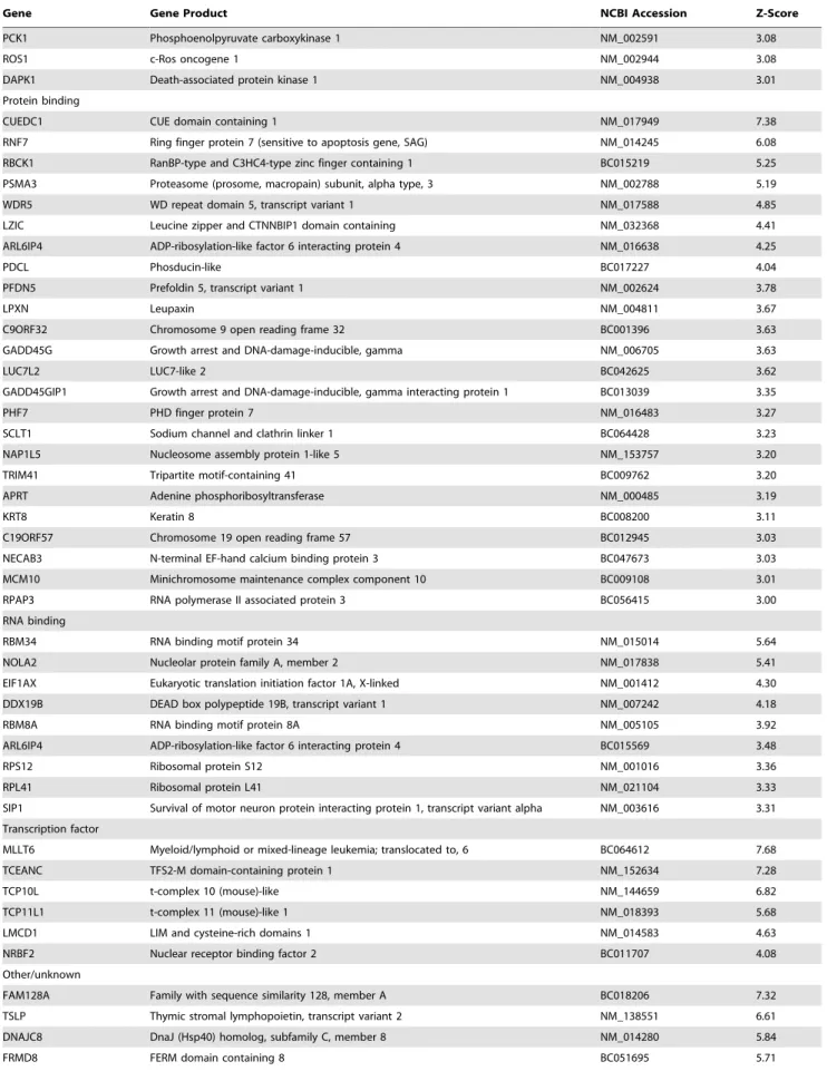
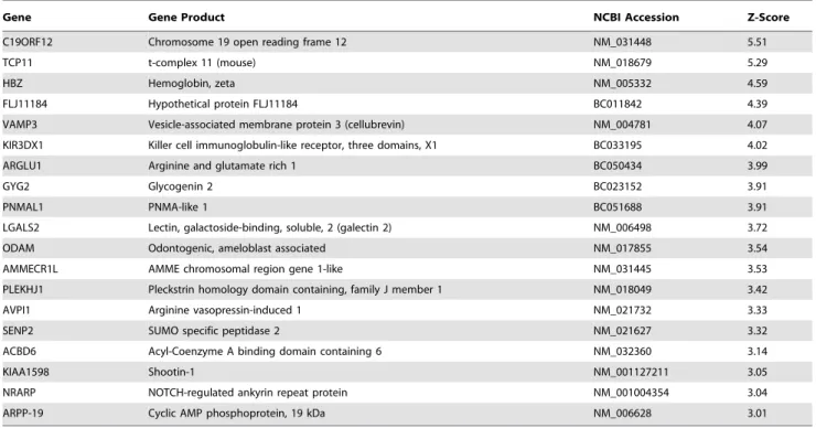
Table 1.Candidate NEMO interactors identified by protein microarray screening with full-length NEMO protein.
Gene Gene Product NCBI Accession Z-Score
Calcium binding
CETN3 Centrin EF-hand protein 3 BC005383 6.10
SYT1 Synaptotagmin I NM_005639 5.95
CPNE2 Copine II NM_152727 5.86
HPCAL1 Hippocalcin-like 1 NM_002149 4.20
MYL5 Myosin light chain 5, regulatory NM_002477 3.83
CALB1 Calbindin D28K NM_004929 3.61
TTYH2 Tweety homolog 2 BC004233 3.42
TPT1 Tumor protein, translationally-controlled 1 BC022436 3.40
DNA binding
H1F0 H1 histone family, member 0 BC029046 4.40
MCM7b
MCM7 minichromosome maintenance deficient 7 BC009398 3.38
CNOT7 CCR4-NOT transcription complex, subunit 7 BC060852 3.21
GTPase
CENTA2 Centaurin, alpha 2 BC033758 4.06
TBC1D7 TBC1 domain family, member 7 NM_016495 3.13
GNGT1 G protein,c-transducing activity polypeptide 1 NM_021955 3.04
SEPT9 Septin 9 BC054004 3.03
Kinase
IKBKBb
Inhibitor of kappa light polypeptide gene enhancer in B-cells, kinase beta NM_001556 8.41
PRKCB1 Protein kinase C type beta I X06318 8.33
TBK1b
Tank binding kinase 1 NM_013254 7.37
PRKCB2 Protein kinase C type beta II X07109 6.83
IKBKAb
Inhibitor of kappa light polypeptide gene enhancer in B-cells, kinase alpha NM_001278 6.52
LCK Leukocyte-specific protein tyrosine kinase M36881 5.97
CDK2 Cyclin-dependent kinase 2 NM_001798 5.34
FLT3 FMS-related tyrosine kinase 3 NM_004119 4.36
SRC2 Gardner-Rasheed feline sarcoma viral (v-fgr) oncogene homolog NM_005248 4.32
FLT3 D835Y FMS-related tyrosine kinase 3 (D835Y mutant) NM_004119 4.24
STK25 Serine/threonine kinase 25 NM_006374 4.21
PIM2 Pediatric index of mortality 2 NM_006875 4.13
EPHA4 Ephrin receptor A4 NM_004438 4.10
TEK TEK tyrosine kinase NM_000459 4.09
JAK2 Janus kinase 2 NM_004972 3.96
TEC Tyrosine kinase expressed in hepatocellular carcinoma NM_003215 3.92
FLT4 FMS-related tyrosine kinase 4 NM_182925 3.89
RPS6KB2 Ribosomal S6 kinase NM_003952 3.86
GRK4 G protein-coupled receptor kinase 4 NM_182982 3.84
PRKCN Protein kinase D3 NM_005813 3.66
MAP3K2 Mitogen-activated protein kinase kinase kinase 2 NM_006609 3.66
AKT1 v-akt murine thymoma viral oncogene homolog 1 BC000479 3.65
SGK1 Serum/glucocorticoid regulated kinase 1 NM_005627 3.62
ITK IL2-inducible T-cell kinase NM_005546 3.54
TYRO3 TYRO3 protein tyrosine kinase NM_006293 3.49
MERTK c-Mer proto-oncogene tyrosine kinase NM_006343 3.43
ALK Anaplastic lymphoma receptor tyrosine kinase NM_004304 3.42
JAK3 Janus kinase 3 NM_000215 3.36
RET RET proto-oncogene NM_020975 3.26
ROR2 Receptor tyrosine kinase-like orphan receptor 2 NM_004560 3.21
Gene Gene Product NCBI Accession Z-Score
PCK1 Phosphoenolpyruvate carboxykinase 1 NM_002591 3.08
ROS1 c-Ros oncogene 1 NM_002944 3.08
DAPK1 Death-associated protein kinase 1 NM_004938 3.01
Protein binding
CUEDC1 CUE domain containing 1 NM_017949 7.38
RNF7 Ring finger protein 7 (sensitive to apoptosis gene, SAG) NM_014245 6.08
RBCK1 RanBP-type and C3HC4-type zinc finger containing 1 BC015219 5.25
PSMA3 Proteasome (prosome, macropain) subunit, alpha type, 3 NM_002788 5.19
WDR5 WD repeat domain 5, transcript variant 1 NM_017588 4.85
LZIC Leucine zipper and CTNNBIP1 domain containing NM_032368 4.41
ARL6IP4 ADP-ribosylation-like factor 6 interacting protein 4 NM_016638 4.25
PDCL Phosducin-like BC017227 4.04
PFDN5 Prefoldin 5, transcript variant 1 NM_002624 3.78
LPXN Leupaxin NM_004811 3.67
C9ORF32 Chromosome 9 open reading frame 32 BC001396 3.63
GADD45G Growth arrest and DNA-damage-inducible, gamma NM_006705 3.63
LUC7L2 LUC7-like 2 BC042625 3.62
GADD45GIP1 Growth arrest and DNA-damage-inducible, gamma interacting protein 1 BC013039 3.35
PHF7 PHD finger protein 7 NM_016483 3.27
SCLT1 Sodium channel and clathrin linker 1 BC064428 3.23
NAP1L5 Nucleosome assembly protein 1-like 5 NM_153757 3.20
TRIM41 Tripartite motif-containing 41 BC009762 3.20
APRT Adenine phosphoribosyltransferase NM_000485 3.19
KRT8 Keratin 8 BC008200 3.11
C19ORF57 Chromosome 19 open reading frame 57 BC012945 3.03
NECAB3 N-terminal EF-hand calcium binding protein 3 BC047673 3.03
MCM10 Minichromosome maintenance complex component 10 BC009108 3.01
RPAP3 RNA polymerase II associated protein 3 BC056415 3.00
RNA binding
RBM34 RNA binding motif protein 34 NM_015014 5.64
NOLA2 Nucleolar protein family A, member 2 NM_017838 5.41
EIF1AX Eukaryotic translation initiation factor 1A, X-linked NM_001412 4.30
DDX19B DEAD box polypeptide 19B, transcript variant 1 NM_007242 4.18
RBM8A RNA binding motif protein 8A NM_005105 3.92
ARL6IP4 ADP-ribosylation-like factor 6 interacting protein 4 BC015569 3.48
RPS12 Ribosomal protein S12 NM_001016 3.36
RPL41 Ribosomal protein L41 NM_021104 3.33
SIP1 Survival of motor neuron protein interacting protein 1, transcript variant alpha NM_003616 3.31
Transcription factor
MLLT6 Myeloid/lymphoid or mixed-lineage leukemia; translocated to, 6 BC064612 7.68
TCEANC TFS2-M domain-containing protein 1 NM_152634 7.28
TCP10L t-complex 10 (mouse)-like NM_144659 6.82
TCP11L1 t-complex 11 (mouse)-like 1 NM_018393 5.68
LMCD1 LIM and cysteine-rich domains 1 NM_014583 4.63
NRBF2 Nuclear receptor binding factor 2 BC011707 4.08
Other/unknown
FAM128A Family with sequence similarity 128, member A BC018206 7.32
TSLP Thymic stromal lymphopoietin, transcript variant 2 NM_138551 6.61
DNAJC8 DnaJ (Hsp40) homolog, subfamily C, member 8 NM_014280 5.84
FRMD8 FERM domain containing 8 BC051695 5.71
of the screen. Other kinases with a high apparent affinity for NEMO included PKCb1, PKCb2, LCK, CDK2, FLT3 and another known NEMO binding partner, TBK1 (Table 1).
Further ontological analysis revealed that signal transduction proteins formed the largest biological process category, with 25% of the interactome, while 11% and 13% of interactors are involved in cell cycle and development, respectively (Figure 2B). Despite the well-characterized role of the NF-kB pathway in regulating cell survival, only 9% of interactors are involved in apoptosis and cell death-related processes, while 2% are related to stress responses. As expected, the majority of identified NEMO interactors localize to the cytoplasm and/or nuclear compartments (Figure 2C), consistent with the known subcellular distribution of NEMO.
NEMO also interacted with the E3 ubiquitin ligases SAG and RBCK1, which were previously reported to promote ubiquitina-tion of NF-kB signaling components IkBa and TAB2/3, respectively [20,21]. GST-NEMO also interacted with SYT1, a major neuronal synaptic calcium sensor [22]. NEMO was previously shown to interact with an extended variant of this protein, termed E-SYT1 [17].
NEMO Binds to Putative Interactors Expressed in Mammalian Cells
Based on our analysis of the list of putative NEMO interactors, we selected five proteins of particular interest to validate the microarray screen using GST pulldown and coimmunoprecipita-tion assays. Proteins were chosen based on their previously described roles related to NF-kB signaling. Calbindin D28K (CALB1) is 28 kDa neuronal calcium binding protein that is transcriptionally regulated by NF-kB following stimulation by neurotrophic growth factor [23]. Cyclin-dependent protein kinase
2 (CDK2) associates with NF-kB during the G1/S phase transition of the cell cycle [24], while sensitive to apoptosis gene 1 (SAG) is an E3 ubiquitin ligase that appears to target IkBafor degradation during the G1/S phase transition and in carcinogenesis [20,25]. The SUMO1-specific peptidase 2 (SENP2) was chosen due to the role of SUMOylation in regulating the cellular localization of NEMO [14,26]. Finally, synaptotagmin 1 (SYT1) was chosen due to a previous finding indicating that NEMO can bind to an extended SYT1 variant, E-SYT1 [17] and to the essential role of synaptotagmin 1 in synaptic Ca2+release [27,28,29].
Thus, HEK-293T cells were transfected with plasmids express-ing CALB1, CDK2, SAG, SENP2 or SYT1. Additionally, an IKKb expression vector was transfected as a positive control. Using GST or GST-NEMO as bait proteins (Figure 3A), we found that each of the five interactors was able to specifically bind GST-NEMO, while little or no binding was observed with the GST control (Figure 3B). These data suggested that the observed array binding between NEMO and the interactors was indeed specific. We next determined whether NEMO, when coexpressed with each of the five interactors, was able to form a complexin vivo using coimmunoprecipitation assays. Again, HEK-293T cells were transfected with expression vectors for NEMO and the five interactors, with IKKbor the parent vector serving as positive and negative controls, respectively. In mammalian cells the parent vector, pcDNA4-HisMaxA, expresses a 5.6 kDa irrelevant protein in place of NEMO. As expected, IKKbimmunoprecipitated only in the presence of NEMO (Figure 3C). Similarly, each of the five novel NEMO interactors also exhibited NEMO-specific immuno-precipitation (Figure 3D–H), indicating that these proteins are can form a complex with NEMO in vivo. In the case of SAG, coimmunoprecipitation was observed in the presence of 0.1%
NP-Gene Gene Product NCBI Accession Z-Score
C19ORF12 Chromosome 19 open reading frame 12 NM_031448 5.51
TCP11 t-complex 11 (mouse) NM_018679 5.29
HBZ Hemoglobin, zeta NM_005332 4.59
FLJ11184 Hypothetical protein FLJ11184 BC011842 4.39
VAMP3 Vesicle-associated membrane protein 3 (cellubrevin) NM_004781 4.07
KIR3DX1 Killer cell immunoglobulin-like receptor, three domains, X1 BC033195 4.02
ARGLU1 Arginine and glutamate rich 1 BC050434 3.99
GYG2 Glycogenin 2 BC023152 3.91
PNMAL1 PNMA-like 1 BC051688 3.91
LGALS2 Lectin, galactoside-binding, soluble, 2 (galectin 2) NM_006498 3.72
ODAM Odontogenic, ameloblast associated NM_017855 3.54
AMMECR1L AMME chromosomal region gene 1-like NM_031445 3.53
PLEKHJ1 Pleckstrin homology domain containing, family J member 1 NM_018049 3.42
AVPI1 Arginine vasopressin-induced 1 NM_021732 3.33
SENP2 SUMO specific peptidase 2 NM_021627 3.32
ACBD6 Acyl-Coenzyme A binding domain containing 6 NM_032360 3.14
KIAA1598 Shootin-1 NM_001127211 3.05
NRARP NOTCH-regulated ankyrin repeat protein NM_001004354 3.04
ARPP-19 Cyclic AMP phosphoprotein, 19 kDa NM_006628 3.01
a
The Z-score indicates the how far the average spot intensity for a particular putative interactor fell from the mean of the relevant protein microarray sector spot intensities, measured in standard deviations. A Z-score of greater than four standard deviations (P = 0.002) was deemed significant.
b
These proteins are known to interact directly with NEMO, based on the results of tandem affinity purification experiments [17]. doi:10.1371/journal.pone.0008799.t001
40 detergent, but not with 0.5% NP-40, while the remaining four proteins coimmunoprecipitated in the presence of 0.5% NP-40.
Finally, we used a two-hybrid system to validate the interactions between NEMO and the five interactors. A control experiment using two-hybrid luciferase vectors expressing NEMO and IkBa, a known NEMO interactor [17], showed a clear two-hybrid interaction between these two proteins when compared with the corresponding negative control (Figure 3I). Similarly, significant interactions were observed for each of the remaining NEMO interactors compared to their negative controls, with CDK2 appearing to have the highest affinity for NEMO on the basis of luciferase activity. A comparison between results obtained using the two-hybrid method and Z scores obtained from the original array screen did not reveal any obvious trend towards a high Z score and a strong two-hybrid signal, suggesting that array-based interactions do not correlate well to results obtained in a cellular environment.
NEMO Interactors Influence NF-kB Transcriptional Activation
To gain an understanding of how the identified interactors might contribute to the NF-kB pathway, we cotransfected HEK-293T cells with a secreted alkaline phosphatase (SEAP) NF-kB transcriptional reporter plasmid and expression plasmids for the five interactors. SEAP activity was then determined for cotrans-fected cells in the presence or absence of TNFa, a potent stimulator of NF-kB-dependent transcription. In the absence of TNFa we found that CALB1, CDK2 and SAG all significantly
induced reporter activity (Figure 4A). After TNFawas added, we again found that CALB1 and CDK2 overexpression increased expression of the reporter above normal levels, while SYT1 substantially reduced induction of the reporter (Figure 4B). We did not observe any significant influence of SENP2 overexpression on reporter activity under these experimental conditions.
We next determined if transient siRNA-mediated mRNA knockdown of the interactors led to any change in reporter activity. For these experiments we chose to look at only CDK2, SAG and SENP2 as these were expressed at readily detectable levels in HEK-293T cells (data not shown), making mRNA knockdown feasible. Cotransfection of the reporter plasmid with control or specific siRNAs against these three putative NEMO interactors led to a highly effective siRNA knockdown of more than 80% by specific siRNAs compared to control siRNAs, based on the results of RT-qPCR using gene-specific primers (Figure 4D). Assays of reporter gene activity following gene knockdown yielded modest but significant reductions of NF-kB transcriptional activation for all three knockdowns when cells were treated with TNFa, though no significant change was observed between control and specific siRNA treatments in the absence of this stimulator. Thus, at endogenous levels, CDK2, SAG and SENP2 can be considered as positive effectors of the TNFa-dependent NF-kB pathway.
Discussion
Signal transduction by the NF-kB pathway is typically considered as a ‘‘first response’’ mechanism as target gene activation by the NF-kB transcription factor occurs rapidly after stimulation by a wide variety of cellular stimuli. The position of NEMO within this pathway would therefore necessitate its ability to communicate with a large diversity of effectors to respond to these different stimuli. In this study, we have uncovered an exceptionally large number of NEMO interactors, most of which are novel and not previously known to communicate with the canonical NF-kB pathway.
While it is clear that many of the NEMO interactors identified here, including IKKaand IKKb, are physiologically relevant, we concede that a certain number of the array proteins may not interact with NEMOin vivo. This could be due an inaccessible subcellular localization, inappropriate temporal expression pat-terns between NEMO and the interactors, or an overabundance of NEMO and the interactors used during array screening [30]. Surprisingly, we also noted that several of the false positive hits obtained on both the control and NEMO arrays have been reported elsewhere to specifically bind other protein baits [31,32,33]. While the interactions reported in those works may well be authentic, the recurrence of a number of protein hits during ProtoArray screens with other protein baits in our laboratory suggests that certain arrayed proteins become artifi-cially promiscuous when arrayed.
Among the novel NEMO interactors identified, calbindin D28K and synaptotagmin 1 have well characterized neuronal functions. Calbindin D28K is commonly described as a neuronal calcium buffer that prevents the accumulation of toxic levels of calcium via four calcium binding domains [34,35]. In addition to this buffering role, however, calbindin D28K also appears to function as a calcium sensor by interacting with downstream effector proteins [34]. Neuronal calcium-sensing roles are also played by three other novel NEMO interactors identified here and elsewhere, namely calmodulin [17], hippocalcin-like 1 and synaptotagmin 1. Our observation that calbindin D28K overex-pression increased NF-kB activity points to calbindin D28K acting Figure 2. Cytoplasmic and nuclear signaling kinases dominate
the NEMO interactome.Gene ontologies were determined for each of the NEMO interactors and the results for each of the three standard ontological categories plotted as percentages. Genes belonging to more than one category were assigned to the category for which the gene has been best characterized.
as an inducer of the NF-kB pathway. This is in line with recent findings indicating that the nuclear export NEMO and its subsequent association with the IKK proteins is inducible by calcium [36]. We also found that overexpression of synaptotagmin 1 markedly diminished TNFa-dependent NF-kB activity, which would otherwise suggest that this protein is a repressor of the NF-kB pathway. We remain skeptical of this idea, however, because when synaptotagmin 1 is overexpressed in HEK-293 cells it localizes almost homogeneously throughout the cell membrane [37 and data not shown]. This may have had the unintended effect of disrupting the activity or structure of the TNF receptor and thus prevented proper activation of the NF-kB pathway upon TNFa addition. It also remains to be seen how synaptotagmin 1 impacts upon NF-kB activity in neurons and also whether or not NEMO is indeed able to localize to the neuronal synapse where synapto-tagmin 1 is normally present.
Another of the NEMO interactors, SAG, has been character-ized as a cellular antioxidant whose overexpression markedly reduces cell death following stroke or treatment with apoptosis inducers [38,39]. The E3 ligase activity of SAG is known to
promote ubiquitination and subsequent degradation of IkBa, resulting in activation of the NF-kB pathway [25]. It remains unclear how the SAG-NEMO interaction impacts upon this activity, though recent data from our lab suggests that overex-pression of SAG can also promote ubiquitination of NEMO (B. Fenner, unpublished results). Recent work has also revealed that SAG is transcriptionally induced under hypoxia by HIF1a[40]. We are currently investigating the impact of SAG on hypoxia-dependent induction of NF-kB.
Cyclin-dependent protein kinase 2, another of the novel NEMO interactors identified here, associates with NF-kB during the G1/S cell cycle transition [24]. We found that CDK2 is required for maximal NF-kB activation in response to TNFa stimulation, though we do not yet know whether this effect is mediated by NEMO, NF-kB or another unidentified intermediate.
SUMOylation of NEMO retains the protein inside the nucleus, and in response to certain cellular stresses NEMO is deSUMOy-lated, exits the nucleus and promotes activation of the NF-kB pathway [13,26,41,42,43]. It was therefore of special interest that we identified SENP2, a nuclear-envelope-associated SUMO Figure 3. Putative interactors bind to NEMO in GST pulldown, coimmunoprecipitation and mammalian two-hybrid assays.(A) Immunoblot analysis of GST and GST-NEMO proteins used as control and bait for the pulldown assay. Proteins were detected using anti-GST/HRP conjugate following SDS-PAGE and membrane transfer. (B) Results of GST pulldown assays showing binding of NEMO to putative interactors identified by protein array screening. Each of the interactors and IKKbeta, a known NEMO binder, were overexpressed in transiently transfected HEK-293T cells and the resulting lysates applied to immobilized GST or GST-NEMO. Following incubation and washing, the samples were resolved by SDS-PAGE and the proteins detected using appropriate antibodies. Input lanes were loaded with 5–10% of HEK-293T lysates to confirm protein expression. The size of relevant protein markers is shown beside the blot image. (C–H) Coimmunoprecipitation assays between NEMO and putative binders in HEK-293T cells. Plasmids encoding Xpress-tagged NEMO or the empty parent vector and tagged putative binders were used to transfect HEK-293T cells and the resulting cell lysates used for coimmunoprecipitation assays. For each putative binder, immunoblots are shown for detection of the binder using a tag- or protein-specific antibody, and for detection of Xpress-tagged NEMO. For IKKbeta and each of the five putative interactors, substantial coimmunoprecipitation occurred only in the presence immunoprecipitated NEMO. Input lanes contained 5–10% of the precleared input volume used prior to addition of anti-Xpress antibody. Binding and washing steps were performed in the presence of 0.5% NP-40 for all proteins except SAG, where 0.1% NP-40 was used. (I) NEMO interacts with CALB1, CDK2, SAG, SENP2 and SYT1 in a mammalian two-hybrid system. Empty two-hybrid vectors were cotransfected as a negative control. The MyoD/Id and NEMO/IkappaBalpha protein pairs were used as positive controls, while putative interaction partners cotransfected with empty complementing vector were used as negative controls. For each pair tested, a significant increase (n = 6; two-tailed T test; P#0.05) in luciferase activity was obtained in partner/NEMO experiments compared to partner/vector experiments (indicated by asterisks).
protease [44], as a NEMO interactor. Previous work indicates that NEMO can be deSUMOylated by SENP1 [14], another nuclear SUMO protease closely related to SENP2, and the activity of this protein may explain why we observed such a modest effect on NF-kB activity following efficient siRNA knockdown of SENP2. What is more intriguing though is why SENP2 associated with NEMO in what would presumably be the absence of SUMOylation during the microarray screen. Current models of deSUMOylation by SENPs do not suggest SUMO-independent recognition of a SUMOylated protein by SENPs [45,46]. It may therefore be of substantial interest to the field whether SENP2 can indeed deSUMOylate NEMOin vivoand how this reaction proceeds in relation to the SUMOylation status of NEMO.
We have identified several new NEMO protein interactors, and have putatively identified over a hundred novel interactors using protein microarrays. We expect that many researchers will be able to use this list to gain new insight into how the NF-kB pathway communicates with other signaling pathways. Future work from
our laboratory will focus on more detailed analyses of several of the interactors and how they communicate with NEMO and the NF-kB signaling pathways.
Materials and Methods
Preparation of Recombinant NEMO Probe
(Invitrogen) with a biotin:NEMO molar ratio of 9:1. Biotinylated recombinant NEMO was stored at280uC.
Protein Array Screening
ImaGenes UniPex colony macroarrays were screened using a previously described procedure [12]. Version 4.0 of the Invitrogen Protoarray was used for protein microarray screening experiments. After blocking the array in blocking buffer (PBS, 1% BSA, 0.1% Tween 20) for 1 h at 4uC, 10mg aliquots of biotinylated NEMO or GST control diluted in 120ml of probing buffer (PBS containing 0.5 mM DTT, 5 mM MgCl2, 5% glycerol, 0.05% Triton X-100 and Calbiochem protease inhibitor cocktail) were added to the array and the array covered and incubated at 4uC for a further 1.5 h. Arrays were then washed three times in ice cold probing buffer prior to the addition of streptavidin-alkaline phosphatase (Invitrogen) diluted in probing buffer to a final concentration of 0.25mg/ml. Arrays were incubated for 30 min on ice, washed three times in probing buffer and then dried for 2–3 h.
Arrays were imaged using a Perkin Elmer Scanarray Ex-pressHT system and the images analyzed using Invitrogen Prospector version 4 software. Significant interactions were identified based on a Z-score cutoff value of 3.0, with the data obtained from the biotin-GST experiment being subtracted from those of the biotin-NEMO experiment.
GST Pulldown and Coimmunoprecipitation
GST pulldowns using purified GST-NEMO and transfected HEK-293T cells expressing IKKb [47], CALB1 (Origene), CDK2-HA [48], SAG (Origene), SENP2-FLAG [49] or SYT1-FLAG (this study) were performed as described previously [12]. For coimmunoprecipitation, HEK293 cells were grown in 6-well plates containing DMEM supplemented with 10% FBS to a confluence of approximately 80%. After rinsing in serum-free DMEM, cells were transfected with 4mg of pcDNA4/HisMaxA-NEMO [12] and each of the abovementioned plasmids using Lipofectamine 2000 (Invitrogen). At 24 h post-transfection, cells were gently rinsed in ice-cold PBS and scraped into 0.5 ml of lysis buffer (20 mM Tris-HCl, pH 7.5, 137 mM NaCl, 0.5% NP-40, 0.5 mM DTT, 10% glycerol, 2 mM EDTA, Calbiochem protease inhibitor cocktail). The lysate was incubated with end-over-end mixing for 30 min at 4uC and insoluble material pelleted by centrifugation at 10,0006g for 20 min. Supernatants were collected and used for coimmunoprecipitation. A 1mg aliquot of mouse monoclonal anti-Xpress (Invitrogen) was added to 400ml (500–750mg total protein) of the cell lysates and the mixtures incubated with end-over-end mixing for 2 h at 4uC. The resulting immunocomplexes were precipitated by adding 25ml bed volume of protein A/G agarose (Santa Cruz Biotech) and further incubating with mixing at 4uC for 2 h. The agarose beads were then pelleted at 1,0006g for 3 min and washed three to five times
in chilled lysis buffer. Pellets were finally resuspended in 60ml of 26Laemmli sample buffer, boiled, and the supernatants collected. Aliquots of 20ml were used for SDS-PAGE followed by anti-FLAG, anti-Myc, anti-HA or anti-Xpress (Invitrogen) immuno-blotting and detection with Dura chemiluminescent substrate (Peirce).
Mammalian Two-Hybrid Assays
Protein-protein interactions were confirmed by two-hybrid analysis using the CheckMate/Flexi system (Promega) with HEK-293T cells. Two-hybrid complementation plasmids were generated by Pfu polymerase-mediated PCR using the above-mentioned plasmids as templates with cloning being performed using SgfI and PmeI restriction digestion of pFN10A (activation
domain) and pFN11A (binding domain). Plasmid sequences were validated by DNA sequencing. Firefly and Renilla luciferase activities were determined using a Dual-Luciferase kit (Promega) and expressed as normalized firefly/Renilla luciferase values.
NF-kB Reporter Ggene Assays
Transcriptional activation by NF-kB was monitored using a secreted alkaline phosphatase (SEAP) reporter plasmid, pNF-k B-SEAP, which contains fourkB consensus sequences upstream of SEAP [50]. HEK-293T cells were seeded in 24-well plates and grown overnight in antibiotic-free DMEM containing 10% FCS to approximately 60% confluence. Cells were then transfected with 400 ng of reporter plasmid and 1600 ng of the different expression plasmids using Lipofectamine 2000 (Invitrogen) and serum-free DMEM as recommended by the manufacturer. Medium was replaced with serum-containing medium at 4–6 h post-transfec-tion. At this point, cells were treated with TNFa(100 ng/ml) or left untreated for a further 20 h. Culture supernatants were then collected and heat-treated at 65uC for 5 min to inactivate endogenous alkaline phosphatase. Cells were washed in PBS and lysed directly using 50ml of Laemmli sample buffer for protein expression analysis by immunoblotting. SEAP was measured in 96-well plates by combining 10ml of supernatant with 190ml of Attophos substrate (Roche) and incubating plates at room temperature for 12 h prior to reading fluorescence with a BioTek Synergy HT plate reader (Ex485/Em528).
RNA Interference
HEK-293T cells were transfected with 400 ng of the NF-kB reporter plasmid as described above, along with 20 pmol of siRNAs directed against CDK2 (Ambion Silencer siRNA S204), SAG (Ambion Silencer siRNA S18473), SENP2 (Ambion Silencer siRNA S34009) or a nonspecific control (Ambion Silencer control siRNA). Pre-incubation of the siRNA and plasmid DNA with the Lipofectamine 2000 was reduced to 5 min to reduce siRNA degradation. Cells were incubated with the Lipofectamine 2000 complexes for 5 hours prior to replacement of the medium with DMEM containing 10% FBS and further incubated for 48 h. The medium was then replaced and the cells treated with TNFafor a further 24 h, at which time the SEAP reporter activity was determined as described above and cellular RNA prepared using a Qiagen RNeasy kit and Invitrogen RNase-free DNase.
Quantitative PCR
Amounts of CDK2, SAG, SENP2 and 18s rRNA were quantitated by RT-qPCR using a LightCycler RNA Master SYBR Green I kit (Roche) with gene-specific primers CDK2-FWD1 (59-GGAGAACTTCCAAAAGGTGG-39) and CDK2-REV1 (59-TCTCGGATGGCAGTACTGGG-39), SAG-FWD1 (59-CGACGTGGAAGACGGAGAGGAA-39) and SAG-REV1 (59-TGCAGATGGCGCACGTATCG-39), SENP2 FWD1 (59 -ATGTACAGATGGCTGGTTAG-39) and SENP2 REV1 (59 -TGGTCTTTTGGCTGGTATTT-39), or 18S FWD1 (59 -GTAACCCGTTGAACCCCATT-39) and 18S REV1 (59 -CCATCCAATCGGTAGTAGCG-39). Amounts of each mRNA were calculated relative to the 18S rRNA control and values expressed as percentages of the control siRNA sample for each siRNA knockdown.
Author Contributions
References
1. Skaug B, Jiang X, Chen ZJ (2009) The role of ubiquitin in NF-kappaB regulatory pathways. Annu Rev Biochem 78: 769–796.
2. Sen R, Baltimore D (1986) Multiple nuclear factors interact with the immunoglobulin enhancer sequences. Cell 46: 705–716.
3. Mankan AK, Lawless MW, Gray SG, Kelleher D, McManus R (2009) NF-kappaB regulation: the nuclear response. J Cell Mol Med 13: 631–643. 4. Vallabhapurapu S, Karin M (2009) Regulation and function of NF-kappaB
transcription factors in the immune system. Annu Rev Immunol 27: 693–733. 5. Yamaoka S, Courtois G, Bessia C, Whiteside ST, Weil R, et al. (1998) Complementation cloning of NEMO, a component of the IkappaB kinase complex essential for NF-kappaB activation. Cell 93: 1231–1240.
6. Poyet JL, Srinivasula SM, Lin JH, Fernandes-Alnemri T, Yamaoka S, et al. (2000) Activation of the Ikappa B kinases by RIP via IKKgamma/NEMO-mediated oligomerization. J Biol Chem 275: 37966–37977.
7. Cooke EL, Uings IJ, Xia CL, Woo P, Ray KP (2001) Functional analysis of the interleukin-1-receptor-associated kinase (IRAK-1) in interleukin-1 beta-stimu-lated nuclear factor kappa B (NF-kappa B) pathway activation: IRAK-1 associates with the NF-kappa B essential modulator (NEMO) upon receptor stimulation. Biochem J 359: 403–410.
8. Ea CK, Deng L, Xia ZP, Pineda G, Chen ZJ (2006) Activation of IKK by TNFalpha requires site-specific ubiquitination of RIP1 and polyubiquitin binding by NEMO. Mol Cell 22: 245–257.
9. Li H, Kobayashi M, Blonska M, You Y, Lin X (2006) Ubiquitination of RIP is required for tumor necrosis factor alpha-induced NF-kappaB activation. J Biol Chem 281: 13636–13643.
10. Wu CJ, Conze DB, Li T, Srinivasula SM, Ashwell JD (2006) Sensing of Lys63-linked polyubiquitination by NEMO is a key event in NF-kappaB activation. Nat Cell Biol 8: 398–406.
11. Cordier F, Grubisha O, Traincard F, Veron M, Delepierre M, et al. (2009) The zinc finger of NEMO is a functional ubiquitin-binding domain. J Biol Chem 284: 2902–2907.
12. Fenner BJ, Scannell M, Prehn JH (2009) Identification of polyubiquitin binding proteins involved in NF-kappaB signaling using protein arrays. Biochim Biophys Acta E-pub ahead of print.
13. Wu ZH, Shi Y, Tibbetts RS, Miyamoto S (2006) Molecular linkage between the kinase ATM and NF-kappaB signaling in response to genotoxic stimuli. Science 311: 1141–1146.
14. Mabb AM, Wuerzberger-Davis SM, Miyamoto S (2006) PIASy mediates NEMO sumoylation and NF-kappaB activation in response to genotoxic stress. Nat Cell Biol 8: 986–993.
15. Wu ZH, Miyamoto S (2008) Induction of a pro-apoptotic ATM-NF-kappaB pathway and its repression by ATR in response to replication stress. EMBO J 27: 1963–1973.
16. Sebban H, Yamaoka S, Courtois G (2006) Posttranslational modifications of NEMO and its partners in NF-kappaB signaling. Trends Cell Biol 16: 569–577. 17. Bouwmeester T, Bauch A, Ruffner H, Angrand PO, Bergamini G, et al. (2004) A physical and functional map of the human TNF-alpha/NF-kappaB signal transduction pathway. Nat Cell Biol 6: 97–105.
18. Chariot A, Leonardi A, Muller J, Bonif M, Brown K, et al. (2002) Association of the adaptor TANK with the I kappa B kinase (IKK) regulator NEMO connects IKK complexes with IKK epsilon and TBK1 kinases. J Biol Chem 277: 37029–37036.
19. Higashimoto T, Chan N, Lee YK, Zandi E (2008) Regulation of I(kappa)B kinase complex by phosphorylation of (gamma)-binding domain of I(kappa)B kinase (beta) by Polo-like kinase 1. J Biol Chem 283: 35354–35367. 20. Kim YS, Lee JY, Son MY, Park W, Bae YS (2003) Phosphorylation of threonine
10 on CKBBP1/SAG/ROC2/Rbx2 by protein kinase CKII promotes the degradation of IkappaBalpha and p27Kip1. J Biol Chem 278: 28462–28469. 21. Tian Y, Zhang Y, Zhong B, Wang YY, Diao FC, et al. (2007) RBCK1
negatively regulates tumor necrosis factor- and interleukin-1-triggered NF-kappaB activation by targeting TAB2/3 for degradation. J Biol Chem 282: 16776–16782.
22. Carr CM, Munson M (2007) Tag team action at the synapse. EMBO Rep 8: 834–838.
23. Wang HJ, Cao JP, Yu JK, Zhang LC, Jiang ZJ, et al. (2008) Calbindin-D28K expression induced by glial cell line-derived neurotrophic factor in substantia nigra neurons dependent on PI3K/Akt/NF-kappaB signaling pathway. Eur J Pharmacol 595: 7–12.
24. Chen E, Li CC (1998) Association of Cdk2/cyclin E and NF-kappa B complexes at G1/S phase. Biochem Biophys Res Commun 249: 728–734.
25. Gu Q, Bowden GT, Normolle D, Sun Y (2007) SAG/ROC2 E3 ligase regulates skin carcinogenesis by stage-dependent targeting of c-Jun/AP1 and IkappaB-alpha/NF-kappaB. J Cell Biol 178: 1009–1023.
26. Mabb AM, Miyamoto S (2007) SUMO and NF-kappaB ties. Cell Mol Life Sci 64: 1979–1996.
27. Yoshihara M, Montana ES (2004) The synaptotagmins: calcium sensors for vesicular trafficking. Neuroscientist 10: 566–574.
28. Koh TW, Bellen HJ (2003) Synaptotagmin I, a Ca2+sensor for neurotransmitter release. Trends Neurosci 26: 413–422.
29. Rizo J, Rosenmund C (2008) Synaptic vesicle fusion. Nat Struct Mol Biol 15: 665–674.
30. Levy ED, Landry CR, Michnick SW (2009) How perfect can protein interactomes be? Sci Signal 2: pe11.
31. Satoh J, Nanri Y, Yamamura T (2006) Rapid identification of 14-3-3-binding proteins by protein microarray analysis. J Neurosci Methods 152: 278–288. 32. Tong Y, Ben-Shlomo A, Zhou C, Wawrowsky K, Melmed S (2008) Pituitary
tumor transforming gene 1 regulates Aurora kinase A activity. Oncogene 27: 6385–6395.
33. Satoh J, Obayashi S, Misawa T, Sumiyoshi K, Oosumi K, et al. (2009) Protein microarray analysis identifies human cellular prion protein interactors. Neuropathol Appl Neurobiol 35: 16–35.
34. Schwaller B (2009) The continuing disappearance of ‘‘pure’’ Ca2+
buffers. Cell Mol Life Sci 66: 275–300.
35. Kojetin DJ, Venters RA, Kordys DR, Thompson RJ, Kumar R, et al. (2006) Structure, binding interface and hydrophobic transitions of Ca2+-loaded calbindin-D(28K). Nat Struct Mol Biol 13: 641–647.
36. Berchtold CM, Wu ZH, Huang TT, Miyamoto S (2007) Calcium-dependent regulation of NEMO nuclear export in response to genotoxic stimuli. Mol Cell Biol 27: 497–509.
37. Madziva MT, Bai J, Bhalla A, Chapman ER, Edwardson JM (2005) Effects of synaptotagmin reveal two distinct mechanisms of agonist-stimulated internali-zation of the M4 muscarinic acetylcholine receptor. Br J Pharmacol 144: 761–771.
38. Tan M, Gallegos JR, Gu Q, Huang Y, Li J, et al. (2006) SAG/ROC-SCF beta-TrCP E3 ubiquitin ligase promotes pro-caspase-3 degradation as a mechanism of apoptosis protection. Neoplasia 8: 1042–1054.
39. Yang GY, Pang L, Ge HL, Tan M, Ye W, et al. (2001) Attenuation of ischemia-induced mouse brain injury by SAG, a redox-inducible antioxidant protein. J Cereb Blood Flow Metab 21: 722–733.
40. Tan M, Gu Q, He H, Pamarthy D, Semenza GL, et al. (2008) SAG/ROC2/ RBX2 is a HIF-1 target gene that promotes HIF-1 alpha ubiquitination and degradation. Oncogene 27: 1404–1411.
41. Huang TT, Wuerzberger-Davis SM, Wu ZH, Miyamoto S (2003) Sequential modification of NEMO/IKKgamma by SUMO-1 and ubiquitin mediates NF-kappaB activation by genotoxic stress. Cell 115: 565–576.
42. Wuerzberger-Davis SM, Nakamura Y, Seufzer BJ, Miyamoto S (2007) NF-kappaB activation by combinations of NEMO SUMOylation and ATM activation stresses in the absence of DNA damage. Oncogene 26: 641–651. 43. Hay RT (2004) Modifying NEMO. Nat Cell Biol 6: 89–91.
44. Zhang H, Saitoh H, Matunis MJ (2002) Enzymes of the SUMO modification pathway localize to filaments of the nuclear pore complex. Mol Cell Biol 22: 6498–6508.
45. Mukhopadhyay D, Dasso M (2007) Modification in reverse: the SUMO proteases. Trends Biochem Sci 32: 286–295.
46. Au SW, Lam KH, Lam WL, Lam LS, Chan HY, et al. (2009) SUMO proteases: Redox regulation and biological consequences. Antioxid Redox Signal. 47. Mercurio F, Zhu H, Murray BW, Shevchenko A, Bennett BL, et al. (1997)
IKK-1 and IKK-2: cytokine-activated IkappaB kinases essential for NF-kappaB activation. Science 278: 860–866.
48. van den Heuvel S, Harlow E (1993) Distinct roles for cyclin-dependent kinases in cell cycle control. Science 262: 2050–2054.
49. Cheng J, Wang D, Wang Z, Yeh ET (2004) SENP1 enhances androgen receptor-dependent transcription through desumoylation of histone deacetylase 1. Mol Cell Biol 24: 6021–6028.