Ana Paula Candido dos Santos(a) Norberto Nobuo Sugaya(a) Décio dos Santos Pinto Junior(a) Celso Augusto Lemos Junior(a)
(a)Department of Stomatology, School of Dentistry, University of São Paulo, São Paulo, SP, Brazil.
Corresponding author: Ana Paula Candido dos Santos
Universidade de São Paulo, Faculdade de Odontologia, Departamento de
Estomatologia, Disciplina de Estomatologia Clínica
Av. Prof. Lineu Prestes, 2227 - Cidade Universitária
CEP: 05508-900 São Paulo - SP - Brasil E-mail: anapaulacs@usp.br
Received for publication on Nov 08, 2010 Accepted for publication on Feb 01, 2011
Fine needle aspiration biopsy in the
oral cavity and head and neck region
Abstract: The objective of the current study was to evaluate the sensi-tivity, speciicity and accuracy of ine needle aspiration biopsy (FNAB) of submucous nodules from the oral cavity and head and neck region as an auxiliary diagnostic tool. Fifty patients with nodule lesions in the oral cavity and the head and neck region were selected. All of them were submitted to FNAB and to either incisional or excisional biopsy. The di-agnoses from the FNABs were compared with the biopsy diagnosis as the gold standard. All the cases of FNAB were analyzed by a single oral pa-thologist prior to the biopsy diagnosis. The results showed that the sen-sitivity of FNAB was 75%, its speciicity was 96% and its accuracy was 58.8%. The false positive and false negative rates were 6.7% and 13.3%, respectively. The positive predictive value was 86% and the negative pre-dictive value was 93%. The inconclusive rate was 16/50. FNAB displayed a high success rate for identifying both malignant and benign lesions, but a low accuracy for making a inal diagnosis.
Descriptors: Biopsy, Needle; Biopsy, Fine-Needle; Pathology, Oral; Sensitivity and Speciicity.
Introduction
In 1930, two doctors from a New York hospital, Martin and Colley, and a technical developer, Ellis, conducted aspirations from several or-gans and conducted cytological studies on them.1-4
FNAB is primarily used for biopsies of palpable masses and deep le-sions with dificult access. It is also widely used in the head and neck regions, such as in the thyroid, lymph nodes, major salivary glands and others neoplasias.2, 4-7
In the oral cavity, the FNAB method is rarely used. The most com-mon masses biopsied in this manner are odontogenic tumors, intraos-seous lesions, minor salivary gland tumors, sublingual salivary glands and other oral regions.6, 8-12 However, some reports have described the relevance of FNAB for the diagnosis of oral cavity lesions and oropha-ryngeal lesions.8, 9
The most commonly described advantages of the FNAB method are the preoperative diagnoses of lesions, clinical follow-up, the ability to avoid unnecessary damage to crucial structures of the oral cavity, more comfort for the patient and a low risk of infection and tissue damage.8, 9
complete the procedure, and the dificulty involved in ixing the lesion.8
The sensitivity of FNAB in intraoral lesions may vary from 80% to 100%; speciicity varies from 80% to 100% and the accuracy varies from 60% to 100%. The false positive rate has been reported to vary from 0 to 3% and the false negative rate from 0 to 20%.8, 9
The most common diagnostic problem reported during the use of FNAB in the oral cavity is an in-suficient amount of material collected for analysis due to the dificulty of applying the technique in this region.8 The FNAB technique used in this study was the same as that described previously for use in other organs.2-4
Material and methods
The Ethics Committee of the School of Dentistry at São Paulo University approved the research. Fifty consecutive patients, who all fulilled the inclusion criteria with lesions in the oral cavity and head and neck region and who sought treatment at the Stoma-tology Clinic of Sao Paulo University, were selected. The inclusion criteria during the study were as fol-lows: both genders, all ethnicities, above 10 years old, without any comorbidity restrictions and on whom both a FNAB and a regular biopsy had been performed. The exclusion criteria were patients un-der 10 years old and those on whom only the FNAB had been performed without conirmation by a reg-ular biopsy.
After a detailed clinical examination and the es-tablishment of a differential diagnosis, patients with nodules in the oral cavity and head and neck region, regardless of etiology, were prepared for FNAB and, afterwards, for an incisional or excisional biopsy.
The FNAB was performed with a Franzen pis-tol (Medpej, Ribeirão Preto, Brazil), coupled to a 20 ml syringe (BD, São Paulo, Brazil), a 23 or 25 gauge needle (BD, São Paulo, Brazil; Terumo, São Paulo, Brazil) and 96° GL alcohol for the setting of the samples. The technique used was described by Zajicek.3, 4 Initially, the area was prepared in an aseptic manner and the area was anesthetized only if the biopsy was performed at the same surgical time.
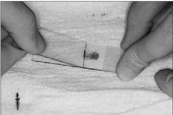
After that, the needle was inserted into the le-sion, a vacuum was applied and the operator made back and forth movements with the needle to obtain a large amount of cells for the smears. The pressure was then released and the needle removed from the lesion. The syringe was withdrawn from the gun and the needle was removed from the syringe. Most of the material collected was in the needle; subse-quently, after removing the needle from the syringe, it was illed with air and the needle was placed near the surface of a glass slide, on which the material collected was deposited.
The material was deposited onto six glass slides. Subsequently, the blade was ixed in 96° GL alcohol and sent to the Discipline of Oral Pathology, School of Dentistry, University of São Paulo. Hematoxylin-eosin was used both for the FNAB slides and the anatomic pathology slides.2, 4
The FNAB slides were evaluated by a pathologist without prior visualization of the anatomic pathol-ogy slides obtained by regular biopsy, but with a re-port of the patient’s data and the clinical diagnosis of the lesion.
The results of the FNAB samples and the results obtained from the regular biopsy were then com-pared to assess the speciicity, sensitivity and ac-curacy of the FNAB method. Figures 1 through 4 show the steps of the technique.
Results
Of the 50 patients examined and submitted to FNAB, 18 patients (36%) had benign neoplasms (BN), 13 patients (26%) had non-neoplastic prolif-erative lesions (NNPL), 11 patients (22%) had ma-lignant lesions (ML), seven patients (14%) showed an inlammatory process (IP) and one patient (2%) had reactive lesion (RL). These data are presented in Table 1.
Of the 50 patients, 39 presented benign lesions (18 BN, 13 NNPL, 7 IP and 1 RL) and 11 malignant lesions. The FNAB method diagnosed 26 true be-nign lesions, 6 true malignant lesions and 16 cases were inconclusive. There were two reported false negatives and one false positive.
The results of FNAB were consistent with the results of regular biopsy in 20 cases. Accuracy was calculated as the ratio of FNAB results compatible with those of regular biopsy, excluding the incon-clusive cases, with a score of 58.8%. Sensitivity was calculated by the ratio between true malignant FNAB and the sum of true negative and false nega-tive FNAB, with a result of 75%. Speciicity was cal-culated as the ratio between true benign FNAB and the sum of true positive and false positive FNAB,
with a result of 96%. The positive predictive value was calculated as the ratio between true malignant FNAB and the sum of true malignant and false posi-tive FNAB, with a score of 86%. The negaposi-tive pre-dictive value was calculated as the ratio between true benign FNAB and the sum of true negative and false negative FNAB, with a score of 93%. Data Figure 1 - Vaccum Pistol.
Figure 2 - Introducing the needle inside the lesion to do the back and forth movement.
Figure 3 - Preparation of the glass slide.
Table 1- Lesions presents in the oral cavity.
Lesions (%)
BN 36%
NNPL 26%
ML 22%
IP 14%
RL 2%
NNPL = non-neoplastic proliferative lesion, BN = benign neoplasm, ML = malignant lesion, IP = inflammatory process, RL = reactive lesion.
from the calculations of accuracy, sensitivity, speci-icity, positive predictive value and negative predic-tive value are presented in Tables 2 and 3.
Of the inconclusive cases, ive were non-neoplas-tic proliferative lesions, ive were benign neoplasms, three were malignant lesions and three were inlam-matory processes.
Discussion
In this study, FNAB was performed in submuco-sal nodules of the oral cavity and in nodules of the head and neck region. Of the 50 patients, 48 had oral lesions and only two cases presented lesions in the head and neck region. The reported cases of FNAB found in the literature involve, for the most part, lesions of the head and neck and thyroid and salivary gland tumors which are benign or malig-nant and arise from both infectious and inlamma-tory processes.2-4, 6, 7, 11, 13, 14
It is believed that preoperative FNAB in glan-dular lesions can guide treatment.15 Applying this concept to lesions from other origins, in this study it could be noted that, as a result of the sensitivity (75%) and speciicity (96%) of FNAB, treatment
can be guided by this exam.
All procedures performed in this study were done so in an outpatient clinic and were executed by a clinician, with or without local anesthesia. There were no contraindications in patients with comorbi-ties and the procedure was performed without com-plication, as is described in the literature.3
There are reports that FNAB in children should be performed under general anesthesia.16 This rec-ommendation was found to be unnecessary as, in this study, the procedure was well tolerated by pe-diatric patients.
We noted that in ibrous lesions, there was difi-culty with aspiration and the ability to obtain a suf-icient amount of material for the cytological analy-sis, which interfered with the interpretation by the pathologist.
Lesions with high blood content, presence of ne-crosis and ibers with scattered atypical cells also proved dificult to analyze cytologically, resulting in the 16 inconclusive cases of FNAB. In the literature, these criteria result in dificulty with the classiica-tion and diagnosis of tumors.17
The presence of lymphoid tissue can lead to the misdiagnosis of a particular lesion, raising the sus-picion of malignancy, which may lead to false posi-tives. The presence of fatty tissue can also lead to in-terpretative errors, which increases the suspicion of malignancy.2, 4 A case of false positive was detected in this study due to the presence of lymphoid con-tent analyzed in the sample smear.
The complexity of the architecture of the glandu-lar tissue, as well as the presence of mixed popula-tion in some glandular tumors, may lead to misin-terpretation of FNAB smears. This means that, at the time of collection, only cells with characteristics consistent with benignity can be aspirated.4 This ex-plains the presence of two false negative cases in the sample of glandular lesions. FNAB did not obtain material representative of the real origin of the le-sion, because only benign cells were obtained from the tumor tissue.
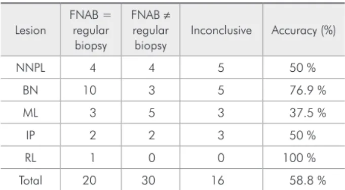
In the case of oral lesions, other disadvantages are reported, such as the small space available to perform the back and forth movement involved in the FNAB procedure and dificulty in ixing the le-Table 2 - Calculation of accuracy in all groups of lesions.
Lesion
FNAB = regular
biopsy
FNAB ≠ regular biopsy
Inconclusive Accuracy (%)
NNPL 4 4 5 50 %
BN 10 3 5 76.9 %
ML 3 5 3 37.5 %
IP 2 2 3 50 %
RL 1 0 0 100 %
Total 20 30 16 58.8 %
NNPL = non-neoplastic proliferative lesion, BN = benign neoplasm, ML = malignant lesion, IP = inflammatory process, RL = reactive lesion.
Table 3 - Calculation of sensitivity (75%), specificity (96%), positive predictive value (86%) and negative predictive value (93%) of FNAB for the 50 cases of the study.
Malignant lesions - regular biopsy
Benign Lesions - regular biopsy
Malignant lesions - FNAB 6 1
sion.8 In this study, however, there was no dificulty in immobilizing the lesion, but the issue of limited space was noted when performing the procedure in certain lesions.
The complications of this procedure are bleed-ing, infection, nerve injury, swelling and bruising of the area in which the procedure was performed and the possible spread of tumor cells throughout the body and in the path where the needle was in-serted.1, 4 In the present study, FNAB was performed with no complications besides mild discomfort and pressure during the procedure in patients without local anesthesia. There was no bleeding during or after FNAB. There was no instance of edema, he-matoma or infection in any of the patients.
The material collection for this study was done following recommendations from the literature;2, 4, 18 this was performed two to three times to gain the
maximum number of cells for analysis. During the aspiration of cysts, the cystic content must be fully drawn and then aspirated again to obtain material from the capsule. The large amount of blood con-tent must then be discarded to enable a better in-terpretation of the smears, as recommended by the literature.4, 19
According to literature, the use of cell block al-lows for the use of various stains and reactions, increasing the sensitivity and speciicity of the tech-nique.20 Cell block was performed in three cases where there was the presence of liquid content, thus helping in the diagnosis of these lesions showing the cells similar to the anatomic pathology slides.
The accuracy of the technique in oral lesions in the literature varies from 60 to 100%; sensitivity varies from 80 to 100% and speciicity varies from 80 to 100%.8, 9 In this study, for calculation purpos-es, the 16 inconclusive cases were excluded from the sample, a decision which coincides with that made in some previous studies.8, 9 The analysis of the results from the present study found that the accuracy of FNAB was 58.8%, the sensitivity was 75% and the speciicity was 96%. The low accuracy found can be explained by the 16 inconclusive samples. The lower sensitivity found in relation to the literature can also be explained by the fact that some cases of malig-nancy were found by the FNAB to be inconclusive
or as false negatives. The sensitivity rating found is similar to that presented in the literature.
Calculations of the positive predictive value and the negative predictive value of FNAB demonstrate the reliability of the test.5, 21 In this study, the posi-tive predicposi-tive value was 86% and the negaposi-tive pre-dictive value was 93%. The calculation of positive predictive value and negative predictive value has not been conducted elsewhere for oral lesions, but, for glandular lesions, the values were found to vary from 83% to 100% and 87.8% to 99%, respective-ly.1, 5, 13, 15
The rates of false positives and false negatives in this study were 6.7% and 13.3%, respectively. These igures agree with those reported in the litera-ture, which indicates that the false positive rate typi-cally varies from 0 to 3% and that the false negative rate varies from 0 to 20%.8, 9 An increase in the rate of false positives was observed, probably because of the high rate of inconclusive samples.
The high rate of inconclusive samples can be ex-plained by some factors that have been described previously in the literature. These are as follows:
• inexperience in the collection of cells,
• dificulty in interpreting the smears,
• poor or inadequate smears for interpretation and
• artifacts, such as necrosis and a high content of blood in some samples.17
Adequate clinical training must be conducted to ensure that the smears are of a satisfactory qual-ity for interpretation; the reduction of the number of artifacts in the sample is a must. As a result, the experience of the pathologist for interpreting these patterns in the cell smears should be considered.
The presence of a pathologist at the time of sam-ple collection, as well as during the staining for the rapid interpretation, may help the clinician at the time of aspiration. This can, therefore, help to mini-mize the rate of inconclusive cases.
serve to guide the type of treatment to be performed on certain lesions.
The limitations encountered in this study can be explained by previous arguments, but should not prevent the use of routine technique.
In clinical situations, where there is a suspi-cion of malignancy, the results of a negative FNAB should be evaluated with caution and a regular bi-opsy should be considered, as this remains the gold standard test.
When using FNAB, it is important that all the necessary preparations are used, that all the clinical features presented are analyzed, that the differential
diagnoses are made and that the outcome of all the investigations are considered. Studies with a similar methodology are inluenced by the experience of staff in using FNAB; so further studies should be conducted from which more meaningful values can be achieved.
Conclusion
FNAB displays a high sensitivity for identifying both malignant and benign lesions, but does not have a high degree of success in making the inal di-agnosis.
References
1.Costas A, Castro P, Martin-Granizo R, Monje F, Marron C, Amigo A. Fine needle aspiration biopsy (FNAB) for le-sions of the salivary glands. Br J Oral Maxillofac Surg. 2000 Oct;38(5):539-42.
2.Amedee RG, Dhurandhar NR. Fine-needle aspiration biopsy. Laryngoscope. 2001 Sep;111(9):1551-7.
3.Fulciniti F, Califano L, Zupi A, Vetrani A. Accuracy of fine needle aspiration biopsy in head and neck tumors. J Oral Maxillofac Surg. 1997 Oct;55(10):1094-7
4.Kline TS. Handbook of fine needle aspiration biopsy cytology. 2nd ed. New York: Churchill Livingstone; 1988. 492 p. 5.Florentine BD, Staymates B, Rabadi M, Barstis J, Black A.
The reliability of fine-needle aspiration biopsy as the initial diagnostic procedure for palpable masses: a 4-year experience of 730 patients from a community hospital-based outpatient aspiration biopsy clinic. Cancer. 2006 Jul 15;107(2):406-16. 6.Batra M, Wadhwa N, Mishra K. Cytologic diagnosis in benign
odontogenic tumor with abundant calcification: a case report. Acta Cytol. 2009 Jul-Aug;53(4):460-2.
7.Carrillo JF, Ramirez R, Flores L, Ramirez-Ortega MC, Arre-cillas MD, Ibarra M, et al. Diagnostic accuracy of fine needle aspiration biopsy in preoperative diagnosis of patients with parotid gland masses. J Surg Oncol. 2009 Aug 1;100(2):133-8. 8.Saleh HA, Clayman L, Masri H. Fine needle aspiration biopsy
of intraoral and oropharyngeal mass lesions. Cytojournal. 2008;5:4.
9.Shah SB, Singer MI, Liberman E, Ljung BM. Transmucosal fine-needle aspiration diagnosis of intraoral and intrapharyn-geal lesions. Laryngoscope. 1999 Aug;109(8):1232-7. 10.Vargas PA, da Cruz Perez DE, Mata GM, de Almeida OP,
Jones AV, Gerhard R. Fine needle aspiration cytology as an additional tool in the diagnosis of odontogenic keratocyst. Cytopathology. 2007 Dec;18(6):361-6.
11.David D, Clayman L, Saleh H. Value of fine-needle aspira-tion biopsy in initial evaluaaspira-tion of floor of the mouth masses:
report of a case of low-grade mucoepidermoid carcinoma. Diagn Cytopathol. 2010 Feb;38(2):81-4.
12.Gupta N, Gupta R, Bakshi J, Rajwanshi A. Fine needle as-piration cytology in a case of fibrous dysplasia of jaw. Diagn Cytopathol. 2009 Dec;37(12):920-2.
13.Christensen RK, Bjorndal K, Godballe C, Krogdahl A. Value of fine-needle aspiration biopsy of salivary gland lesions. Head Neck. 2010 Jan;32(1):104-8.
14.Granville LA, Laucirica R, Verstovsek G. Clinical significance of cultures collected from fine-needle aspiration biopsy. Diagn Cytopathol. 2008 Feb;36(2):85-8.
15.Cohen EG, Patel SG, Lin O, Boyle JO, Kraus DH, Singh B, et al. Fine-needle aspiration biopsy of salivary gland lesions in a selected patient population. Arch Otolaryngol Head Neck Surg. 2004 Jun;130(6):773-8.
16.Anne S, Teot LA, Mandell DL. Fine needle aspiration biopsy: role in diagnosis of pediatric head and neck masses. Int J Pediatr Otorhinolaryngol. 2008 Oct;72(10):1547-53. 17.Fathallah L, Tulunay OE, Feng J, Husain M, Jacobs JR,
Al-Abbadi MA. Histopathologic and cytopathologic diagnostic discrepancies in head and neck region: pitfalls, causes, and preventive strategies. Otolaryngol Head Neck Surg. 2006 Feb;134(2):302-8.
18.Safneck JR, Kutryk E, Chrobak A, Harper R, Ravinsky E. Fixation techniques for fine needle aspiration biopsy smears prepared off site. Acta Cytol. 2001 May-Jun;45(3):365-71. 19.Moatamed NA, Naini BV, Fathizadeh P, Estrella J, Apple SK.
A correlation study of diagnostic fine-needle aspiration with histologic diagnosis in cystic neck lesions. Diagn Cytopathol. 2009 Oct;37(10):720-6.
20.Dabbs DJ. The surgical pathologist’s approach to fine needle aspiration. Clin Lab Med. 1998 Sep;18(3):357-72, v. 21.Loong TW. Understanding sensitivity and specificity with the