Macrophages (MQ) orchestrate inflammatory and reparatory processes in injured connective tissues but their role during different phases of tendon healing is not known. We investigated the contribution of different MQsubsets in an equine model of naturally occurring tendon injury. Post mortem tissues were harvested from normal (uninjured), sub-acute (3–6 weeks post injury) and chronically injured (.3 months post injury) superficial digital flexor tendons. To determine if inflammation was present in injured tendons, MQsub-populations were quantified based on surface antigen expression of CD172a (pan MQ), CD14highCD206low (pro-inflammatory M1MQ), and CD206high (anti-inflammatory M2MQ) to assess
potential polarised phenotypes. In addition, the Lipoxin A4 receptor (FPR2/ALX) was used as marker for resolving
inflammation. Normal tendons were negative for both MQand FPR2/ALX. In contrast, M1MQpredominated in sub-acute injury, whereas a potential phenotype-switch to M2MQ polarity was seen in chronic injury. Furthermore, FPR2/ALX expression by tenocytes was significantly upregulated in sub-acute but not chronic injury. Expression of the FPR2/ALX ligand Annexin A1 was also significantly increased in sub-acute and chronic injuries in contrast to low level expression in normal tendons. The combination of reduced FPR2/ALX expression and persistence of the M2MQphenotype in chronic injury suggests a potential mechanism for incomplete resolution of inflammation after tendon injury. To investigate the effect of pro-inflammatory mediators on lipoxin A4(LXA4) production and FPR2/ALX expressionin vitro, normal tendon
explants were stimulated with interleukin-1 beta and prostaglandin E2. Stimulation with either mediator induced LXA4
release and maximal upregulation of FPR2/ALX expression after 72 hours. Taken together, our data suggests that although tenocytes are capable of mounting a protective mechanism to counteract inflammatory stimuli, this appears to be of insufficient duration and magnitude in natural tendon injury, which may potentiate chronic inflammation and fibrotic repair, as indicated by the presence of M2MQ.
Citation:Dakin SG, Werling D, Hibbert A, Abayasekara DRE, Young NJ, et al. (2012) Macrophage Sub-Populations and the Lipoxin A4Receptor Implicate Active Inflammation during Equine Tendon Repair. PLoS ONE 7(2): e32333. doi:10.1371/journal.pone.0032333
Editor:Martin Gerbert Frasch, Universite´ de Montre´al, Canada
ReceivedSeptember 29, 2011;AcceptedJanuary 25, 2012;PublishedFebruary 22, 2012
Copyright:ß2012 Dakin et al. This is an open-access article distributed under the terms of the Creative Commons Attribution License, which permits unrestricted use, distribution, and reproduction in any medium, provided the original author and source are credited.
Funding:This research article was funded by Biotechnology and Biological Sciences Research Council (BBSRC) UK and Ceva (France) grant number BB/F018258/1. The funders had no role in study design, data collection and analysis, decision to publish or manuscript preparation.
Competing Interests:SGD’s PhD is funded by Biotechnology and Biological Sciences Research Council (BBSRC) UK and Ceva (France). This does not alter the authors’ adherence to all the PLoS ONE policies on sharing data and materials. The authors have declared that no other competing interests exist.
* E-mail: [email protected]
Introduction
Pathologic changes in tendons due to repetitive use are a significant cause of morbidity in humans, with Achilles tendon injury rates of up to 56% amongst elite athletes [1,2]. The corresponding tensile region of the equine superficial digital flexor tendon (SDFT) is similarly susceptible to exercise induced tendinopathy [3], which may be exacerbated by sudden overstrain injury. The importance of inflammatory or degenerative processes contributing to tendon injuries is subject to debate. In humans, tendinopathy is described as a result of a primarily degenerative condition, as inflammatory cells are rarely observed in histological studies of over-strained or ruptured human tendons [4,5,6,7,8]. However, this may be attributable to factors such as the timing at which human patients present themselves for examination, which is usually at the later stages of injury (or with recurrent injury), and
the availability of tissues for examining at different times after injury, precluding study of the early phases of tendon repair. In naturally occurring equine tendinopathy, there is an initial transient phase (days) of clinically evident inflammation (heat, pain, swelling), which rapidly subsides [9]. The sub-acute phase (weeks) is characterised by the resolution of symptomatic inflammation and the onset of fibroplastic repair. In chronic injury the repair continues for many months but in both man and horse the normal architecture, composition and function of tendon are never restored after injury [6,10,11].
experiments, injury elicited a sequential pattern of inflammatory cell infiltration, consisting of a rapid and transient accumulation of neutrophils, followed by an increase in MQinfiltration between 1 and 28 days post injury in a rat model of Achilles tendon injury [13]. Similarly, Wong and co-workers documented temporal changes in inflammatory cell subsets in a murine immobilised surgical adhesion model of injury, reporting peak neutrophil and MQaccumulation 1–5 days and 21 days post surgery respectively [14]. The requirement of MQfor adult tissue repair is supported by the use of wound healing studies in murine MQ-knockout models, with impaired healing responses observed in MQdeplete wounds [15,16,17]. Although crucial for healing, MQinitially fulfil a different function by secreting pro-inflammatory agents in response to tissue damage, including interleukin-1-beta (IL-1b), Tumour Necrosis Factor alpha (TNFa), bioactive prostaglandins, reactive oxygen intermediates and many proteases [18]. These factors have been shown to act as important initiators of the tendinopathic cascade [19,20,21,22], which may drive matrix metalloproteinase (MMP) mediated catabolism of tendon extra-cellular matrix [21,22,23].
In order to identify MQ in normal and injured tendons we selected CD172a, as this antigen has been used to successfully identify MQ in ovine [24] and equine [25] tissues. The unique plasticity in function of MQcan in part be attributed to different subsets/phenotypes. MQhave the capacity to exist in at least two functionally distinct phenotypic states, differing in their cell surface receptor expression, effector function and cytokine production [26,27,28,29]. M1 polarised (classically activated) MQappear to show a pro-inflammatory response pattern, whilst M2 (alterna-tively activated) MQdampen inflammatory responses by produc-ing immunosuppressive cytokines such as IL-1 receptor antagonist (IL-1Ra), IL-10, IL-4 and IL-13 [28]. Expression of both M1 and M2 MQ markers is reported in circulating peripheral blood mononuclear cells [30,31]. M1MQsurface antigens include CD14 [32,33,34], CD16, CD32, CD64, CD80 and CD86 [28], but not the markers of M2MQ polarisation which include the mannose receptor (MR) CD206 and CD163 [28,30,34]. Therefore, M1MQexpress CD14, but not CD206 i.e. (CD14highCD206low), whereas M2MQ express CD206 but not necessarily CD14 (CD14lowCD206high) and so this combination of surface antigens permits identification of the predominating MQ phenotype. The temporal changes in MQ phenotype influence synthesis of the extracellular matrix of the injured tissue throughout the different phases of wound healing [16,35]. Furthermore a correlation between the degree of inflammation and development of subsequent fibrosis has been reported, which is thought to be a MQmediated progression [36].
Despite evidence suggesting that excessive inflammation causes damage to connective tissues by initiating the fibrotic repair that ensues, total inflammatory blockade may not necessarily be beneficial for wound healing processes [37,38]. An intriguing dimension to the physiology of inflammation is the identification of specialised pro-resolving mediators (SPM) encompassing lipoxins such as lipoxin A4 (LXA4) [39] acting upon FPR2/ALX
(previously termed FPRL1 or ALXR) [40] and more recently, the resolvins, protectins and maresins [41,42]. FPR2/ALX is expressed by monocytes and MQ[43] and is central to controlling the duration and magnitude of the inflammatory response by modulating leukocyte adherence and chemotaxis, and in so doing provides ‘endogenous stop signals’ for inflammation [44]. Although stereoselective for LXA4, FPR2/ALX is a
multi-recognition receptor [45], to which the anti-inflammatory protein Annexin A1 (ANXA1) also binds [46,47,48]. Annexin A1 is also implicated in the resolution of inflammation, with additional
important roles in phagocytosis and apoptosis [49,50]. These resolving pathways are programmed responses activated during inflammation, demonstrating critical roles in the switching of lipid mediators from the prostanoid to the lipoxin axis in order to promote resolution of inflammation and the return of tissues to their normal homeostatic state [39,44,51,52,53]. Despite the anticipated importance of MQ, SPMs and Annexin A1 in healing tendons, there is limited information documenting their presence in the early stages of tendon injury. The presence of SPMs has not been previously assessed in tendons, although studies in other inflamed connective tissues have emphasised their importance in resolving inflammation, including inhibition of neutrophil and MQ
recruitment and modification of vascular permeability [44]. Based on the similarities between tendon healing in horses and humans [54,55], studies in naturally occurring equine tendino-pathy may be physiologically representative of equivalent injury in humans. For example, aging of the tendon matrix is believed to be an important contributing factor to the development of tendino-pathy [56,57], yet the effects of aging are restricted in rodent models in contrast to those species displaying more evident aging phenotypes such as horse and man [58,59,60]. Moreover, the strain experienced during peak loading of the elastic energy storing tendons in equine and human Achilles tendonin vivofar exceed
those sustained by rodent models [54,61,62]. Furthermore, re-injury of chronically diseased tendons is common during rehabilitation of patients with tendinopathy, a scenario which is difficult to recapitulate in murine models of tendon injury. Thus analysis of injured equine tendon, particularly during the early phase of injury, presents a more appropriate and readily attainable source than the human counterpart, and analysis of naturally diseased tissues may be more representative than induced models of tendon injury [63,64]. Our analysis provides evidence for potential changes in MQ sub-populations, FPR2/ALX and Annexin A1 expression during stages of flexor tendon healing compared to normal (uninjured) tendons. Upregulation of FPR2/ ALX expression by tenocytes in sub-acute injury in vivo was
supported by in vitro experiments assessing the effect of
pro-inflammatory mediators on tendon.
Results
Macroscopic and microscopic analysis of injured equine tendons
Double immunostaining of SDFT reveals a shift in macrophage polarity
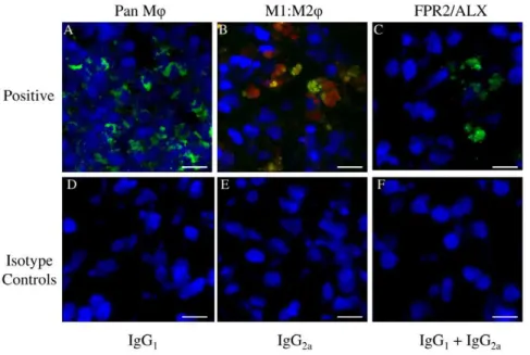
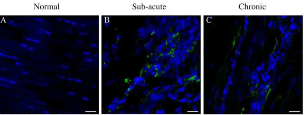
Images for the positive, negative and isotype controls for all antibodies were validated on cryosections of equine spleen and are shown in Fig. 3 A–F. Co-expression of CD14 and CD206 (demonstrated by yellow staining) was not identified in sub-acute or chronic injured tendons in contrast to equine spleen (Fig. 3 B). Immunostaining for the pan MQmarker CD172a on cryosections derived from normal, sub-acute and chronic injured SDFT’s (Fig.4 A–C) revealed a greater number of MQ in sub-acutely injured SDFT compared to normal SDFT (P= 0.008), with fewer MQ
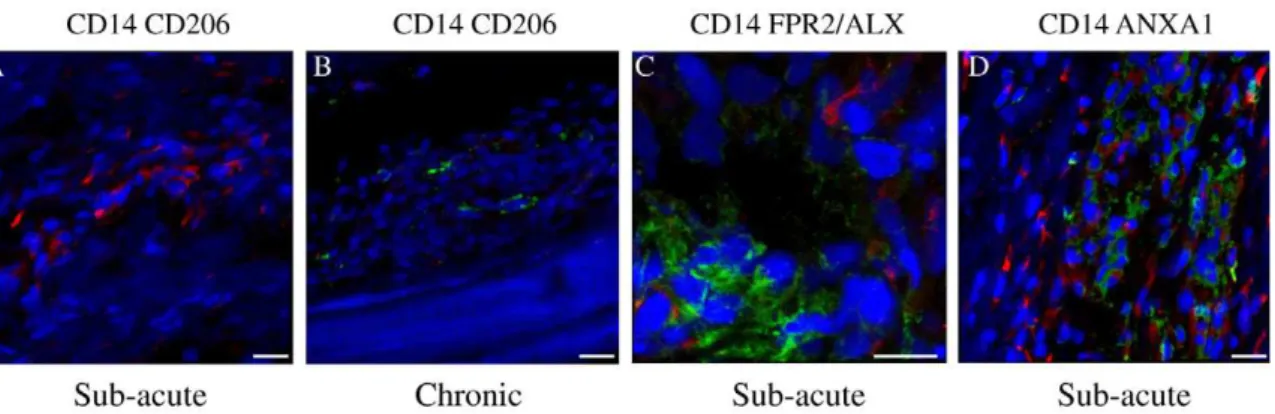
present in chronic injured tendons (Fig. 5A). Dual labelling using antibodies to CD14 and CD206 facilitated characterisation of MQ
sub-populations through the stages of tendon healing (Fig. 6 D–F). There was a significantly greater proportion of M1MQphenotype (CD14highCD206low) in sub-acute injury compared to chronic injured SDFT (P= 0.01) (Figs. 5B, 6E and 7A). In contrast, the
M2MQphenotype (CD14lowCD206high) was predominant during the chronic injury phase (Figs. 5C, 6F and 7B); these cells were located in the peri-vascular and endotenon regions. To facilitate direct comparison of MQ phenotypes during sub-acute and
chronic injury phases, a ratio of areas (mm2) of positive immuno-reactivity for M1:M2 MQwas derived. This showed predominance of the M1 polarised MQphenotype in sub-acute injury compared to chronic injury (P,0.001) (Fig. 5D).
Expression of FPR2/ALX is upregulated during the sub-acute phase of tendon injury
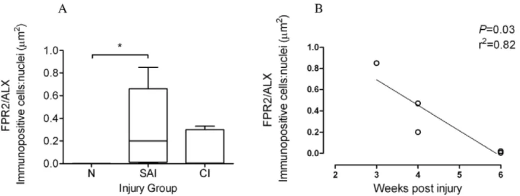
To assess whether changes in MQsubsets are accompanied by changes in FPR2/ALX expression, we next analysed expression of this protein in the same normal, sub-acute and chronic injured SDFT samples (Fig. 6 G–I). There was significantly greater FPR2/ ALX expression in sub-acute but not chronic injured SDFT compared to normal tendons (P= 0.01) (Fig. 8A). When separated
into the early and late stages of sub-acute injury, there was a significant decline in FPR2/ALX expression with increasing time after injury (P= 0.03, r2= 0.82 Fig. 8B), which may account for the
large range of FPR2/ALX expression across this group. FPR2/ ALX was expressed on cytoplasmic processes of tenocytes of sub-acutely injured tendons (Fig. 6H) but not by M1MQ, as verified by dual labelling for CD14 and FPR2/ALX on tendon sections (Fig. 7C).
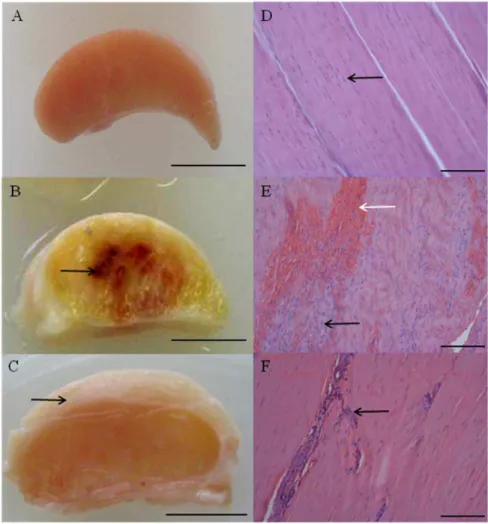
Figure 1. Typical macroscopic appearance of normal and injured equine flexor tendons.(A) Normal superficial digital flexor tendon (SDFT) from a 12 year old horse; (B) sub-acutely injured SDFT 3 weeks post injury from a 4 year old horse, exhibiting a haemorrhagic granular central core (arrow) and (C) chronically injured SDFT.3 months post injury with a thickened fibrosed paratenon (arrow) from a 12 year old horse. Scale bar for macroscopic images = 1 cm. Corresponding longitudinal histology sections stained with Haematoxylin and Eosin: (D) normal SDFT showing regular arrangement of collagen fibrils (arrow); (E) sub-acutely injured SDFT with marked cellular infiltration (black arrow) and haemorrhage (white arrow); (F) chronically injured SDFT with increased cellularity in peri-vascular regions (arrow). Histology scale bar = 12.5mm.
Figure 2. Haematoxylin and Eosin stained longitudinal histology sections of chronic injured SDFT (.3 months post injury) from a 7 year old horse (A, B and D).(A) Reactive fibroplasia with increased cellularity in peri-vascular region (arrows). Scale bar = 100mm. (B) Higher magnification of (A) showing presence of macrophages (arrowheads) in peri-vascular areas. Scale bar = 20mm. (C) Corresponding 3-dimensional reconstructed Z stack image of dual antibody labelling for CD14 (red) and CD206 (green). Blue represents Hoechst nuclear counter stain. White arrows show CD14lowCD206highM2MQlocated in peri-vascular endotenon regions. Scale bar = 20mm. (D) Histological appearance of more normal SDFT to the right of the image (straight arrow) in contrast to irregular arrangement of collagen fibrils on the left (dashed arrow). The linear interface between more normal and injured zones of tendon is demarcated by an area of increased cellularity containing macrophages (arrowhead). Scale bar = 50mm.
doi:10.1371/journal.pone.0032333.g002
Figure 3. Representative Z stack images of antibody control cryosections of equine spleen.Panel A–C represents positive controls for: (A) CD172a MQ(green); (B) Dual antibody labelling with CD14 (M1MQ red) and CD206 (M2MQgreen) showing co-expression of both markers (CD14highCD206high) in splenic monocytes/macrophages; (C) Lipoxin A
4 receptor (FPR2/ALX, green). Panel D–F represents negative controls, consisting of murine isotype matched primary control antibodies: (D) IgG1, (E) IgG2a, (F) IgG1and IgG2a. Blue represents Hoechst nuclear counter stain. Scale bar = 12mm.
Effect of pro-inflammatory mediators on FPR2/ALX expression
As FPR2/ALX was absent in normal SDFT, but present during sub-acute and chronic injury, we next assessed whether FPR2/ ALX expression was influenced by the presence of pro-inflammatory mediators that are known to be upregulated in tendon injury. We used anin vitrotendon explant model, whereby
macroscopically normal explants were stimulated with IL-1band PGE2, either alone or in combination, and analysed FPR2/ALX
expression over time. Representative images for FPR2/ALX expression 72 hours post stimulation and average data for tendon explants from the 3 horses are shown in Fig. 9. There was a
significant effect of time (P,0.001) and experimental condition
(P= 0.01) on FPR2/ALX expression. FPR2/ALX expression was
at baseline or low level between 0–72 hours in non-stimulated control tendon explants. Stimulation with IL-1b alone or in combination with PGE2 significantly increased FPR2/ALX
expression by tenocytes compared to non-stimulated controls at 24 hours (P= 0.049 andP= 0.02 respectively). In contrast there
was no significant effect of PGE2in isolation at this time point.
Maximal FPR2/ALX expression occurred 72 hours after stimu-lation with either IL-1bor PGE2compared to controls (P= 0.002
and P= 0.01 respectively). Combined stimulation with both
mediators also resulted in increased FPR2/ALX expression compared to controls at 72 hours (P= 0.02); however this was
Figure 4. Panel of representative 3-dimensional reconstructed Z stack immunofluorescent low magnification images of equine SDFT sections.Pan MQ(CD172a) staining is shown for is for (A) normal, (B) sub-acute and (C) chronic injured tendons. Immunopositive cells are green; blue represents Hoechst nuclear counter stain. Scale bar = 20mm.
doi:10.1371/journal.pone.0032333.g004
Figure 5. Box plots illustrating ratios of areas (mm2) of immunopositive cells: counterstained nuclei.(A) CD172a pan MQin normal (uninjured), sub-acute and chronic injured equine tendons; (B) CD14high(M1MQ) and (C) CD206high(M2MQ) expression in sub-acute and chronic injured tendons respectively. (D) Log transformed ratio of areas of positive immuno-reactivity for M1:M2 MQfrom dual labelled CD14 and CD206 sections of sub-acute and chronic injured tendons. SAI = sub-acute injury (n = 5), CI = chronic injury (n = 5), N = normal tendon (n = 5). All values represent median with maximum and minimum range. ***P,0.001, **P,0.01, *P,0.05.
not significantly different to expression observed with stimulating with either mediator alone.
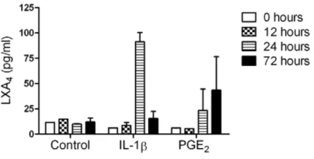
Effect of pro-inflammatory mediators on lipoxin A4
release in media
In addition to assessing the effect of pro-inflammatory mediators on FPR2/ALX expression in tendon explants in vitro, we also
investigated their effect upon the release of LXA4 into tissue
culture media. LXA4 production was at baseline or low level
(,15 pg/ml) between 0–72 hours in media from non-stimulated control explants. Stimulation with IL-1binduced maximal LXA4
release 24 hours post stimulation compared to controls (6-fold increase,P,0.001) but returned to baseline levels at 72 hours. In
contrast, LXA4levels increased maximally at 72 hours after PGE2
stimulation compared to controls (3-fold increase, P= 0.048)
(Fig. 10).
Figure 6. Panel of representative 3-dimensional reconstructed Z stack immunofluorescent images of equine SDFT sections.(A–C) CD172a MQ antibody, immunopositive cells are green; (D–F) Dual antibody labelling for CD14 (M1MQred) and CD206 (M2MQgreen) showing CD14highCD206lowMQin sub-acute injury (E) and CD14lowCD206highMQin chronic injury (F); (G–I) FPR2/ALX, immunopositive cells are green. Arrow in panel H shows FPR2/ALX expression on tenocyte cytoplasmic extensions. Panels A, D, & G are from normal uninjured tendon; B, E & H are from sub-acutely injured tendon; C, F & I are from chronically injured tendon. Blue represents Hoechst nuclear counter stain. Scale bar = 12mm.
doi:10.1371/journal.pone.0032333.g006
Figure 7. Panel of representative 3-dimensional reconstructed Z stack immunofluorescent images of injured SDFT sections.(A) Dual labelled CD14 (red) and CD206 (green) showing predominance of CD14highMQin sub-acute tendon injury. Scale bar = 20mm. (B) Dual labelled CD14 (red) and CD206 (green) showing predominance of CD206highMQin chronic tendon injury. Scale bar = 20mm. (C) Dual labelled CD14 (red) and FPR2/ ALX (green), showing FPR2/ALX expression by tenocytes but not M1MQin sub-acute tendon injury. Scale bar = 12mm. (D) Dual labelled CD14 (red) and Annexin A1 (green), showing Annexin A1 expression by tenocytes but not M1MQin sub-acute tendon injury. Scale bar = 20mm. Blue represents Hoechst nuclear counter stain.
Expression of Annexin A1 is upregulated during tendon injury
Annexin A1 is also a ligand for FPR2/ALX with additional roles in inflammation, resolution and apoptosis. To assess expression of Annexin A1 in tendon injuries, we next analysed expression of this protein in the same normal, sub-acute and chronic injured SDFT samples. Annexin A1 expression was restricted to tenocytes in injured tendons and not M1MQ, as co-expression of these markers was not observed (Fig. 7D). Annexin A1 expression was significantly increased in both sub-acute and chronic injured tendons compared to normal samples, which showed very low level expression (P,0.01) (Fig. 11).
Discussion
In the present study, we describe changes in MQsubsets, FPR2/ ALX and Annexin A1 expression in the sub-acute and chronic phases of equine flexor tendon injuries. Our study illustrates that
the number of MQ significantly increase in sub-acutely injured equine flexor tendons (3–6 weeks post injury) in contrast to their absence in normal uninjured tendon. When the injury progressed from sub-acute to the chronic injured stage, there was a potential shift from a pro-inflammatory M1MQ(CD14highCD206low) to an anti-inflammatory M2MQ (CD14lowCD206high) phenotype. Fur-thermore, tendon injury led to an up-regulation of FPR2/ALX in tenocytes in the sub-acute injury stage, which is likely to be mediated by the production of pro-inflammatory mediators by either tenocytes or infiltrating MQ. Annexin A1 expression was also significantly increased in sub-acute and chronic tendon injuries compared to normal tendons. Our study provides novel data illustrating components of inflammation are highly active during both the early and chronic stages of naturally occurring equine flexor tendon injury, which have not been described during the early phase of equivalent injury in man. Similarly, it is not possible to recapitulate chronic injury or re-injury using induced models of murine tendon injury, and may be more appropriate from a therapeutic perspective.
Figure 8. FPR2/ALX expression in normal and injured equine tendons.(A) Box plot illustrating ratio of areas (mm2) of immunopositive cells: counterstained nuclei of expression of the Lipoxin A4receptor (FPR2/ALX) in normal (N, n = 5), sub-acute (SAI, n = 5) and chronic injured (CI n = 5) equine tendons. Values represent median with maximum and minimum range. *P,0.05. (B) FPR2/ALX expression in sub-acutely injured tendons (n = 5) showing significant decline in FPR2/ALX protein expression with time after injury (P= 0.03, r2= 0.82).
doi:10.1371/journal.pone.0032333.g008
Figure 9. Panel of representative 2-dimensional images illustrating FPR2/ALX expression in tendon explants showing non-stimulated (vehicle only) control compared to explants non-stimulated with 5 ng ml21 IL-1b and 1.0
mM PGE2 either alone or in combination.Immunopositive staining is green, with Hoechst nuclear counter stain in blue. Scale bar = 12mm. Graph showing the effect of pro-inflammatory mediators on tendon FPR2/ALX expressionin vitro. Data represent average FPR2/ALX expression in tendon explants derived from 3 normal (uninjured) horses, whereby 2 replicates were analysed for each experimental condition and time point per horse. FPR2/ALX expression was determined at time points 0, 12, 24 and 72 hours after stimulation and compared to non-stimulated controls. Significant up-regulation of FPR2/ALX occurs 24 hours after stimulation with IL-1bor a combination of both mediators, and maximally 72 hours after stimulation with IL-1b, PGE2or combination of both mediators compared to controls. **P,0.01, *P,0.05. Error bars denote SEM.
The crucial role of MQas both effectors of tissue injury and repair are well documented in other healing connective tissues [65,66]. MQare responsible for executing specific functions during the diverse phases of wound repair as suggested by sub-optimal wound healing in MQ deficient injured sites [67] and murine knockout models [15,68]. Deficiency of MQ, particularly during the early stage of repair has profound adverse effects on healing, including reduced granulation tissue formation and impaired epithilialization [16]. The plasticity and functional polarisation of
tissue MQthat we observed in injured tendon was similar to that reported in skin repair [16]. Fully polarised M1 and M2 MQhave been described as the extreme phenotypes of a continual spectrum of functional states [28]. The greater proportion of M1MQ
polarised phenotypes (CD14highCD206low) in sub-acute compared to chronic injury was indicative of the highly pro-inflammatory status of recent injury in the sub-acutely injured tendon samples. The predominance of M2MQ (CD14lowCD206high) in chronic tendon injury suggests a potential phenotype or population change. Although we have not investigated the nature of the stimulus governing the fate of the M1MQ, this has been suggested to occur by either apoptosis or lymphatic clearance [69]. We observed M2MQin peri-vascular and endotenon regions, and at the interface between the previously injured area and adjacent more normal tendon, a site which is prone to re-injury due to disparate tissue biomechanical properties at this interface [11]. We hypothesise that M2MQin this region are actively surveying the healing tendon for ‘stress signals’ in order to protect this susceptible region from re-injury. It is likely that cyclical loading sustains an inflammatory response that M2MQfail to adequately resolve, resulting in the development and accumulation of localised collagen fibril damage (micro-lesions) during exercise rehabilitation. This innate failure to resolve inflammation may increase the propensity for fibrotic repair of adult connective tissues in contrast to the abilities of macrophage deficient foetal wounds that heal with minimal scar formation [16,36]. Thus the persistence of MQappears to be correlated with the deposition of scar tissue at the site of injury in tendons. We did not observe co-expression of CD14 and CD206 in the injured tendon sections analysed. This may reflect the presence of specific sub-sets of differentiated tissue MQ in tendon, because co-expression was Figure 10. The effect of pro-inflammatory mediators on release
of lipoxin A4 (LXA4) in media from tendon explants. Data represent average LXA4levels from 3 normal (uninjured) horses. LXA4 levels were determined at time points 0, 12, 24 and 72 hours after stimulation and compared to non-stimulated controls. There was a difference in the temporal response showing increased LXA4release 24 hours after stimulation with IL-1b (P,0.001) and 72 hours after stimulation with PGE2(P= 0.048) compared to non-stimulated controls. Values for significance were based on a linear model.
doi:10.1371/journal.pone.0032333.g010
Figure 11. Panel of representative 3-dimensional reconstructed Z stack immunofluorescent images of equine SDFT sections.
Annexin A1 (ANXA1) staining is shown for is for (A) normal, (B) sub-acute and (C) chronic injured tendons. Immunopositive cells are green; blue represents Hoechst nuclear counter stain. Scale bar = 20mm. Box plot shows significantly increased Annexin A1 expression in sub-acute and chronic injured tendons compared to low level expression in normal tendons. SAI = sub-acute injury (n = 5), CI = chronic injury (n = 5), N = normal tendon (n = 5). Values represent median with maximum and minimum range. *P,0.05.
inflammatory angiogenesis and neovascularisation [72], both of which are also prominent clinical features of early stage equine tendinopathy. FPR2/ALX has been reported to be expressed on circulating monocytes and MQ[40], but we were unable to show expression on M1MQ in dual labelled sections of sub-acutely injured tendons. The apparent absence of FPR2/ALX expression on M1MQmay be attributable to phenotypic changes that occur in the transition from infiltrating monocytes at injured sites to differentiated tissue MQ [73]. We speculate diminished FPR2/ ALX expression by MQmay contribute to the defective healing that occurs in injured tendons.
We also identified tenocytes expressing Annexin A1 (an additional ligand for FPR2/ALX) in sub-acute and chronic injured tendons. Annexin A1 has not been previously described in tendon; however it is reported to act as an endogenous anti-inflammatory mediator complimenting the actions of lipoxins, potentiating resolution of inflammation via FPR2/ALX [74]. Furthermore, as Annexin A1 is also implicated in promoting pro-apoptotic mechanisms [75], its presence in tendon may have an additional role in the identification of damaged tenocytes that are to be removed by MQ after injury. As Annexin A1 protein was significantly upregulated in both sub-acute and chronic phases of tendon injury, it would appear that inflammation and apoptosis continue for a prolonged period after initial injury and throughout tendon healing and remodelling.
Regulation of FPR2/ALX by pro-inflammatory mediators has not been previously reported in wound healing, although transcription of FPR2/ALX was shown to be upregulated by IL-13, IL-4, IL-6 and IL-1bin human enterocytes [76]. In our study, stimulation of normal tendon explants with IL-1b or PGE2
induced maximal release of LXA4at 24 and 72 hours respectively,
compared to non-stimulated controls. The differences in kinetics seen are potentially attributed to differences in receptor involve-ment and subsequent down-stream signalling between IL-1band PGE2 [77,78,79], or the class switching of arachadonic acid
derived eicosanoids from prostaglandins towards production of LXA4[52]. Alternatively, these data may suggest that IL-1bis a
more potent activator of LXA4than PGE2, as peak LXA4release
coincided with the observed upregulation of FPR2/ALX expres-sion at 24 hours and maximally at 72 hours after stimulation with IL-1b. Interestingly, combined stimulation with both mediators did not significantly increase FPR2/ALX expression compared to using either mediator in isolation. The lack of an additive effect of the two pro-inflammatory mediators may suggest maximal FPR2/ ALX transcription level is attained by stimulation with either mediator alone, and we speculate that this may be a mechanism to tightly regulate this gene.
M1MQare reported to secrete pro-inflammatory agents such as IL-1band PGE2[18,28]. In support of our in vitroexperimental
findings, the presence of M1MQalso coincided with FPR2/ALX up-regulation in naturally occurring early stage tendon injury.
the beneficial, yet enhance removal of the detrimental components of the cascade. It is possible that augmenting resolution by ‘tricking’ chronically inflamed tissues into a resolving pathway using potential new therapeutic targets such as lipoxins and other FPR2/ALX agonists [46,80,81] may reduce the propensities for fibrotic tendon repair and re-injury.
The categorisation of injury stage in this study was based on known clinical history and post mortem examination, which may not account for the presence of sub-clinical inflammatory processes that occur prior to the onset of clinical disease, either in older animals or those subjected to higher levels of exercise. We believe that our present study contributes to the evidence that whilst symptomatic inflammation is only subtle and transient in tendinopathies, the regulatory components of the inflammatory response in early stage tendinopathy are in fact highly active at the cellular level. Consequently, prolonged subclinical inflammation can potentially develop into symptomatic injury if sufficient extracellular matrix microdamage accumulates. The ability to determine the nature of the MQphenotype and FPR2/ALX status of sub-clinically injured tendon would provide valuable insight to the inflammatory processes occurring prior to the onset of clinical disease; however this is currently precluded by our inability to accurately identify sub-clinical injuries in vivo. Future research
should aim to identify parameters that determine the inflamma-tory and resolution status of injured tendonin vivo, such that
sub-clinical injury can be identified and treated prior to onset of clinical disease.
Materials and Methods
Ethics Statement
Ethics approval for the collection of post mortem equine tendons from an abattoir or local equine veterinary referral hospital for this study was sought and approved from the Ethics and Welfare Committee at the Royal Veterinary College (URN 2011 1117). No horses were euthanased for obtaining tissues for the purposes of this study and all tendons were harvested after informed consent had been obtained.
Collection and processing of equine tendons
aged based on historical information obtained from either the owner or referring veterinary surgeon prior to euthanasia of the horse. All harvested tendons were obtained from Thoroughbred or Thoroughbred cross breed horses aged between 4 and 16 years. Tendon pieces from the mid-tensional regions were embedded in optimal cutting temperature compound (OCT, Sakura Tissue-TekH, Alphen aan den Rijn, The Netherlands) and snap frozen in pre-chilled (280uC) n-hexane and stored at280uC until processed for histological or immunofluorescent analysis. Serial sections (8– 10mm thickness) were cut on a cryostat (Bright, Cambridge, UK)
and mounted onto poly-L-lysine coated slides (VWR, Lutterworth, UK) and allowed to dry for 2 hours at room temperature prior to storage at280uC.
Haematoxylin & Eosin staining of tendon cryosections
Tendon cryosections were fixed in chilled (4uC) acetone for 5 minutes and then allowed to air dry for 30 minutes. Sections were hydrated in Phosphate Buffered saline (PBS) and stained with Gills III Haematoxylin (Sigma Aldrich, Dorset, UK) for 3 minutes and then washed with running water for a further 3 minutes. Slides were stained with alcoholic Eosin (Sigma Aldrich,) for 40 seconds and rinsed with water. After dehydration and a final rinse using HistosolTM histological clearing agent (National Diagnostics, Hull, UK), slides were mounted using DPX mounting medium (BDH, Poole, UK).
Detection and characterization of macrophages, FPR2/ ALX and Annexin A1 in normal and injured equine tendons
Cryosections of equine spleen were used as positive control tissue to validate suitability and dilution of antibodies for use on tendon sections. Negative controls consisted of spleen and sub-acutely injured SDFT cryosections incubated with murine isotype matched primary control antibodies (Southern Biotech, Birming-ham, AL, USA). Subsequently, consecutive cryosections of normal and injured equine tendons were probed using mouse anti-human monoclonal antibodies to CD172a (pan MQ marker, IgG1,
VMRD, Pullman WA, USA), CD14 (M1 polarised MQ pheno-type, IgG2a, BioLegend, Cambridge, UK), CD206 (M2 polarised
MQ phenotype, IgG1, AbCam, Cambridge, UK), FPR2/ALX
(Lipoxin A4receptor, IgG1, AbCam, Cambridge) and Annexin A1
(IgG1, Abcam, Cambridge). Although CD172a is also a
granulo-cyte marker, we verified that granulogranulo-cytes were not present using Haematoxylin & Eosin stained cryosections of sub-acute and chronic injured tendons. After warming cryosections at room temperature for 30 minutes, sections were rehydrated in 1% PBS-Tween 20 (PBS-T) and blocked in PBS containing 5% normal goat serum (Sigma Aldrich) for 1 hour in a humid chamber at room temperature. This was followed by incubation with the primary antibody in PBS containing 5% normal goat serum for 2 hours, diluted either 1:100 or 1:50 (CD206). After 365 minute washes in PBS-T, sections were incubated with the respective goat anti-mouse secondary antibodies conjugated with either Alexa-FluorH 488 IgG, Alexa FluorH 594 IgG2a (Invitrogen, Paisley,
UK), or goat anti-mouse IgG1-R-PE (Southern Biotech), each
diluted 1:300 in PBS containing 5% normal equine serum (Sigma Aldrich) for 2 hours. To visualise the nuclei, slides were subsequently counterstained with 0.5mgml21 Hoechst 33342
(Invitrogen) for 20 minutes and washed in PBS-T. To quench tissue fluorescence, slides were incubated with 0.1% Sudan Black B (BDH, Poole, UK) in 70% ethanol for 20 minutes [82], washed in PBS-T and mounted using a solution of 80% glycerol in 0.5 mM Tris buffer (pH 7.2). Slides were stored at 4uC until image
acquisition. In order to identify the predominating MQ popula-tions in sub-acute and chronically injured tendon, dual staining using antibodies against CD14 and CD206 was performed. Due to the lack of commercially available antibodies to Lipoxin A4 for
immunofluorescence, Annexin A1 was chosen as an alternative ligand for FPR2/ALX. Dual staining was also performed using antibodies against CD14 and FPR2/ALX or CD14 and Annexin A1 to determine expression on tenocytes and M1MQ.
Effect of pro-inflammatory mediators on FPR2/ALX expression
As evaluation of injured tendon sections revealed differences in FPR2/ALX expression, we assessed the impact of pro-inflamma-tory mediators on FPR2/ALX expressionin vitro. Macroscopically
normal SDFTs from an equine abattoir were harvested from 3 Thoroughbred horses aged between 2 and 15 years (mean age 8 years). Tendons were cut into 0.4 cm3explants (300 mg630 mg) under aseptic conditions and incubated at 37uC and 5% CO2
under humidified atmosphere in 3 ml Dulbecco’s Modified Eagle Medium (PAA, Somerset, UK) containing 1% Penicillin and Streptomycin (PAA) without foetal calf serum. Samples were stimulated with either 5 ng ml21
human recombinant IL-1b
(Merck, CalbiochemH, Nottingham UK), 1.0mM PGE2 (Sigma
Aldrich) or a combination of 5 ng ml21IL-1band 1.0mM PGE2.
Non-stimulated (vehicle only) samples served as controls. Tendon explants and media were harvested immediately (time 0) and 12, 24 and 72 hours after stimulation. Media samples were stored at 280uC prior to processing for determination of LXA4 levels.
Tendon explants were snap frozen in chilled (280uC) n-hexane, embedded in OCT and stored at 280uC until processing for immunofluorescent analysis. Cryosections were cut and mounted onto poly-L-lysine coated slides as described above. Sections were allowed to dry for 2 hours at room temperature prior to storage at 280uC. Cryosections were probed with a 1:100 diluted primary mouse monoclonal FPR2/ALX antibody (Lipoxin A4 receptor,
IgG1, AbCam) and secondary goat anti-mouse IgG1 (Southern
Biotech), followed by Hoechst nuclear counter stain. Tissue fluorescence was quenched with Sudan Black B (BDH) and sections were mounted for imaging as before.
Image acquisition and analysis
Images for histology sections were acquired using a Leica DM4000B microscope with a DC500 camera (Leica Microsys-tems, Milton Keynes, UK). All immunofluorescent images were recorded on a Leica SP5 confocal microscope (Leica Micro-systems) using a 636oil immersion objective (NA = 1.4). For
2-dimensional images a minimum of 5 random high power field images were acquired per tissue section per horse (for analysis of normal and naturally diseased tendon), or per experimental condition for each time point (in vitroexplant experimental system).
Antigen expression was automatically quantified by measurement of the area of immunopositive cells in each image. As we expected this area to vary with the cellular density in the tendon, the area of immunopositive cells was standardised by expressing it as a ratio to the total area of counterstained nuclei, which was used as a measure of cellular density. Quantitative analysis was performed on 2-dimensional images which were acquired using a 636oil immersion objective for pan MQ, FPR2/ALX and Annexin A1 antibody treated cryosections. In order to determine the predominant MQpopulations by dual antibody labelling, a ratio of areas (mm2) of positive immuno-reactivity for CD14 (red) versus
determine release of the ligand LXA4. LXA4was separated from
the supernatant by passage through Bond Elut C18 columns (Agilent Technologies, CA, USA) followed by elution with methyl formate. Samples were evaporated to dryness in a stream of nitrogen and resuspended in extraction buffer from a LXA4
ELISA kit (Neogen Corp, Lexington, KY, USA). LXA4levels in
media samples were determined using this ELISA in duplicate wells according to the manufacturer’s instructions. The ELISA kit is specific for LXA4 showing minimal cross-reactivity [LXA4
100%, Lipoxin B41.0%, 15-hydroxyeicosatetraenoic acid (HETE)
0.1%, 5-HETE,0.1% and 12 HETE,0.1%].
Statistics
For normal and injured tendons, statistical analyses were performed on 2-dimensional confocal images using GraphPad Prism 5 (GraphPad Software Inc., San Diego, CA). Normality was tested using a Kolmogorov-Smirnov test. Kruskal-Wallis with post hoc Dunn’s multiple comparison tests were performed to determine differences between normal, sub-acute and chronic
points 0, 12, 24 and 72 hours. A linear model was used to assess LXA4levels in media to account for effects of horse, experimental
condition, time and interaction between condition and time. Linear contrast was then used on each data set to assess the differences between each experimental condition and time point.
P,0.05 was considered statistically significant.
Acknowledgments
We are grateful to Professor Kenneth Clark Smith for histological interpretation of injured equine flexor tendons, Dr Yu-Mei Chang for statistical advice and Marcus Head, David Rutherford and Andrew Crawford for assistance in sourcing injured tendons.
Author Contributions
Conceived and designed the experiments: SGD DW DREA RKWS JD. Performed the experiments: SGD. Analyzed the data: SGD AH NJY. Contributed reagents/materials/analysis tools: SGD DW AH NJY RKWS JD. Wrote the paper: SGD DW RKWS JD.
References
1. Kujala UM, Sarna S, Kaprio J (2005) Cumulative incidence of achilles tendon rupture and tendinopathy in male former elite athletes. Clin J Sport Med 15: 133–135.
2. Welsh RP, Clodman J (1980) Clinical survey of Achilles tendinitis in athletes. Can Med Assoc J 122: 193–195.
3. Avella CS, Ely ER, Verheyen KL, Price JS, Wood JL, et al. (2009) Ultrasonographic assessment of the superficial digital flexor tendons of National Hunt racehorses in training over two racing seasons. Equine Vet J 41: 449–454. 4. Jarvinen M, Jozsa L, Kannus P, Jarvinen TL, Kvist M, et al. (1997) Histopathological findings in chronic tendon disorders. Scand J Med Sci Sports 7: 86–95.
5. Jozsa L, Reffy A, Kannus P, Demel S, Elek E (1990) Pathological alterations in human tendons. Arch Orthop Trauma Surg 110: 15–21.
6. Kannus P, Jozsa L (1991) Histopathological changes preceding spontaneous rupture of a tendon. A controlled study of 891 patients. J Bone Joint Surg Am 73: 1507–1525.
7. Alfredson H, Lorentzon R (2002) Chronic tendon pain: no signs of chemical inflammation but high concentrations of the neurotransmitter glutamate. Implications for treatment? Curr Drug Targets 3: 43–54.
8. Astrom M, Rausing A (1995) Chronic Achilles tendinopathy. A survey of surgical and histopathologic findings. Clin Orthop Relat Res. pp 151–164. 9. Smith R, Schramme M (2003) Tendon injury in the horse: current theories and
therapies. In Practice 25: 529–539.
10. Silver IA, Brown PN, Goodship AE, Lanyon LE, McCullagh KG, et al. (1983) A clinical and experimental study of tendon injury, healing and treatment in the horse. Equine Vet J Suppl: 1–43.
11. Crevier-Denoix N, Collobert C, Pourcelot P, Denoix JM, Sanaa M, et al. (1997) Mechanical properties of pathological equine superficial digital flexor tendons. Equine Vet J Suppl: 23–26.
12. Millar NL, Hueber AJ, Reilly JH, Xu Y, Fazzi UG, et al. (2010) Inflammation is present in early human tendinopathy. Am J Sports Med 38: 2085–2091. 13. Marsolais D, Cote CH, Frenette J (2001) Neutrophils and macrophages
accumulate sequentially following Achilles tendon injury. J Orthop Res 19: 1203–1209.
14. Wong JK, Lui YH, Kapacee Z, Kadler KE, Ferguson MW, et al. (2009) The cellular biology of flexor tendon adhesion formation: an old problem in a new paradigm. Am J Pathol 175: 1938–1951.
15. Werner S, Grose R (2003) Regulation of wound healing by growth factors and cytokines. Physiol Rev 83: 835–870.
16. Lucas T, Waisman A, Ranjan R, Roes J, Krieg T, et al. (2010) Differential roles of macrophages in diverse phases of skin repair. J Immunol 184: 3964–3977. 17. Peters T, Sindrilaru A, Hinz B, Hinrichs R, Menke A, et al. (2005)
Wound-healing defect of CD18(2/2) mice due to a decrease in TGF-beta1 and myofibroblast differentiation. EMBO J 24: 3400–3410.
18. Nathan CF (1987) Secretory products of macrophages. J Clin Invest 79: 319–326.
19. Khan MH, Li Z, Wang JH (2005) Repeated exposure of tendon to prostaglandin-E2 leads to localized tendon degeneration. Clin J Sport Med 15: 27–33.
20. Langberg H, Skovgaard D, Karamouzis M, Bulow J, Kjaer M (1999) Metabolism and inflammatory mediators in the peritendinous space measured by microdialysis during intermittent isometric exercise in humans. J Physiol 515(Pt 3): 919–927.
21. Tsuzaki M, Guyton G, Garrett W, Archambault JM, Herzog W, et al. (2003) IL-1 beta induces COX2, MMP-IL-1, -3 and -IL-13, ADAMTS-4, IL-IL-1 beta and IL-6 in human tendon cells. J Orthop Res 21: 256–264.
22. Yang G, Im HJ, Wang JH (2005) Repetitive mechanical stretching modulates IL-1beta induced COX-2, MMP-1 expression, and PGE2 production in human patellar tendon fibroblasts. Gene 363: 166–172.
23. Thampatty BP, Li H, Im HJ, Wang JH (2007) EP4 receptor regulates collagen type-I, MMP-1, and MMP-3 gene expression in human tendon fibroblasts in response to IL-1 beta treatment. Gene 386: 154–161.
24. Herrmann-Hoesing LM, Noh SM, Snekvik KR, White SN, Schneider DA, et al. (2010) Ovine progressive pneumonia virus capsid antigen as found in CD163-and CD172a-positive alveolar macrophages of persistently infected sheep. Vet Pathol 47: 518–528.
25. Ibrahim S, Saunders K, Kydd JH, Lunn DP, Steinbach F (2007) Screening of anti-human leukocyte monoclonal antibodies for reactivity with equine leukocytes. Vet Immunol Immunopathol 119: 63–80.
26. Stein M, Keshav S, Harris N, Gordon S (1992) Interleukin 4 potently enhances murine macrophage mannose receptor activity: a marker of alternative immunologic macrophage activation. J Exp Med 176: 287–292.
27. Goerdt S, Orfanos CE (1999) Other functions, other genes: alternative activation of antigen-presenting cells. Immunity 10: 137–142.
29. Gordon S (2003) Alternative activation of macrophages. Nat Rev Immunol 3: 23–35.
30. Bouhlel MA, Derudas B, Rigamonti E, Dievart R, Brozek J, et al. (2007) PPARgamma activation primes human monocytes into alternative M2 macrophages with anti-inflammatory properties. Cell Metab 6: 137–143. 31. Smeekens SP, van de Veerdonk FL, Joosten LA, Jacobs L, Jansen T, et al. (2011)
The classical CD14 CD16 monocytes, but not the patrolling CD14 CD16 monocytes, promote Th17 responses to Candida albicans. Eur J Immunol 41: 2915–2924.
32. Satoh N, Shimatsu A, Himeno A, Sasaki Y, Yamakage H, et al. (2010) Unbalanced M1/M2 phenotype of peripheral blood monocytes in obese diabetic patients: effect of pioglitazone. Diabetes Care 33: e7.
33. Raife TJ, Lager DJ, Kemp JD, Dick FR (1994) Expression of CD24 (BA-1) predicts monocytic lineage in acute myeloid leukemia. Am J Clin Pathol 101: 296–299.
34. Zeyda M, Farmer D, Todoric J, Aszmann O, Speiser M, et al. (2007) Human adipose tissue macrophages are of an anti-inflammatory phenotype but capable of excessive pro-inflammatory mediator production. Int J Obes (Lond) 31: 1420–1428.
35. Brancato SK, Albina JE (2011) Wound macrophages as key regulators of repair: origin, phenotype, and function. Am J Pathol 178: 19–25.
36. Stramer BM, Mori R, Martin P (2007) The inflammation-fibrosis link? A Jekyll and Hyde role for blood cells during wound repair. J Invest Dermatol 127: 1009–1017.
37. Marsolais D, Cote CH, Frenette J (2003) Nonsteroidal anti-inflammatory drug reduces neutrophil and macrophage accumulation but does not improve tendon regeneration. Lab Invest 83: 991–999.
38. Magra M, Maffulli N (2006) Nonsteroidal antiinflammatory drugs in tendino-pathy: friend or foe. Clin J Sport Med 16: 1–3.
39. Serhan CN, Hamberg M, Samuelsson B (1984) Lipoxins: novel series of biologically active compounds formed from arachidonic acid in human leukocytes. Proc Natl Acad Sci U S A 81: 5335–5339.
40. Ye RD, Boulay F, Wang JM, Dahlgren C, Gerard C, et al. (2009) International Union of Basic and Clinical Pharmacology. LXXIII. Nomenclature for the formyl peptide receptor (FPR) family. Pharmacol Rev 61: 119–161. 41. Serhan CN, Hong S, Gronert K, Colgan SP, Devchand PR, et al. (2002)
Resolvins: a family of bioactive products of omega-3 fatty acid transformation circuits initiated by aspirin treatment that counter proinflammation signals. J Exp Med 196: 1025–1037.
42. Serhan CN, Yang R, Martinod K, Kasuga K, Pillai PS, et al. (2009) Maresins: novel macrophage mediators with potent antiinflammatory and proresolving actions. J Exp Med 206: 15–23.
43. Yang D, Chen Q, Le Y, Wang JM, Oppenheim JJ (2001) Differential regulation of formyl peptide receptor-like 1 expression during the differentiation of monocytes to dendritic cells and macrophages. J Immunol 166: 4092–4098. 44. Serhan CN, Takano T, Chiang N, Gronert K, Clish CB (2000) Formation of
endogenous ‘‘antiinflammatory’’ lipid mediators by transcellular biosynthesis. Lipoxins and aspirin-triggered lipoxins inhibit neutrophil recruitment and vascular permeability. Am J Respir Crit Care Med 161: S95–S101. 45. Serhan CN, Chiang N (2002) Lipid-derived mediators in endogenous
anti-inflammation and resolution: lipoxins and aspirin-triggered 15-epi-lipoxins. ScientificWorldJournal 2: 169–204.
46. Gilroy DW, Lawrence T, Perretti M, Rossi AG (2004) Inflammatory resolution: new opportunities for drug discovery. Nat Rev Drug Discov 3: 401–416. 47. Perretti M, Chiang N, La M, Fierro IM, Marullo S, et al. (2002) Endogenous
lipid- and peptide-derived anti-inflammatory pathways generated with gluco-corticoid and aspirin treatment activate the lipoxin A4 receptor. Nat Med 8: 1296–1302.
48. Parente L, Solito E (2004) Annexin 1: more than an anti-phospholipase protein. Inflamm Res 53: 125–132.
49. Perretti M, Croxtall JD, Wheller SK, Goulding NJ, Hannon R, et al. (1996) Mobilizing lipocortin 1 in adherent human leukocytes downregulates their transmigration. Nat Med 2: 1259–1262.
50. Lim LH, Pervaiz S (2007) Annexin 1: the new face of an old molecule. FASEB J 21: 968–975.
51. Serhan CN, Chiang N, Van Dyke TE (2008) Resolving inflammation: dual anti-inflammatory and pro-resolution lipid mediators. Nat Rev Immunol 8: 349–361. 52. Serhan CN (2010) Novel lipid mediators and resolution mechanisms in acute
inflammation: to resolve or not? Am J Pathol 177: 1576–1591.
53. Levy BD, Clish CB, Schmidt B, Gronert K, Serhan CN (2001) Lipid mediator class switching during acute inflammation: signals in resolution. Nat Immunol 2: 612–619.
54. Lui PP, Maffulli N, Rolf C, Smith RK (2011) What are the validated animal models for tendinopathy? Scand J Med Sci Sports 21: 3–17.
55. Dowling BA, Dart AJ, Hodgson DR, Smith RK (2000) Superficial digital flexor tendonitis in the horse. Equine Vet J 32: 369–378.
56. Moller A, Astron M, Westlin N (1996) Increasing incidence of Achilles tendon rupture. Acta Orthop Scand 67: 479–481.
57. Maffulli N, Waterston SW, Squair J, Reaper J, Douglas AS (1999) Changing incidence of Achilles tendon rupture in Scotland: a 15-year study. Clin J Sport Med 9: 157–160.
58. Dudhia J, Scott CM, Draper ER, Heinegard D, Pitsillides AA, et al. (2007) Aging enhances a mechanically-induced reduction in tendon strength by an active process involving matrix metalloproteinase activity. Aging Cell 6: 547–556.
59. Smith RK, Goodship AE (2008) The effect of early training and the adaptation and conditioning of skeletal tissues. Vet Clin North Am Equine Pract 24: 37–51. 60. Strocchi R, De Pasquale V, Guizzardi S, Govoni P, Facchini A, et al. (1991) Human Achilles tendon: morphological and morphometric variations as a function of age. Foot Ankle 12: 100–104.
61. Wilson AM, McGuigan MP, Su A, van Den Bogert AJ (2001) Horses damp the spring in their step. Nature 414: 895–899.
62. Ker RF, Wang XT, Pike AV (2000) Fatigue quality of mammalian tendons. J Exp Biol 203: 1317–1327.
63. Williams IF, McCullagh KG, Goodship AE, Silver IA (1984) Studies on the pathogenesis of equine tendonitis following collagenase injury. Res Vet Sci 36: 326–338.
64. Dahlgren LA, van der Meulen MC, Bertram JE, Starrak GS, Nixon AJ (2002) Insulin-like growth factor-I improves cellular and molecular aspects of healing in a collagenase-induced model of flexor tendinitis. J Orthop Res 20: 910–919. 65. Diez-Roux G, Lang RA (1997) Macrophages induce apoptosis in normal cells in
vivo. Development 124: 3633–3638.
66. Leibovich SJ, Wiseman DM (1988) Macrophages, wound repair and angiogenesis. Prog Clin Biol Res 266: 131–145.
67. van Amerongen MJ, Harmsen MC, van Rooijen N, Petersen AH, van Luyn MJ (2007) Macrophage depletion impairs wound healing and increases left ventricular remodeling after myocardial injury in mice. Am J Pathol 170: 818–829.
68. Nagaoka T, Kaburagi Y, Hamaguchi Y, Hasegawa M, Takehara K, et al. (2000) Delayed wound healing in the absence of intercellular adhesion molecule-1 or L-selectin expression. Am J Pathol 157: 237–247.
69. Lawrence T, Gilroy DW (2007) Chronic inflammation: a failure of resolution? Int J Exp Pathol 88: 85–94.
70. Chiang N, Serhan CN, Dahlen SE, Drazen JM, Hay DW, et al. (2006) The lipoxin receptor ALX: potent ligand-specific and stereoselective actions in vivo. Pharmacol Rev 58: 463–487.
71. Maderna P, Godson C (2009) Lipoxins: resolutionary road. Br J Pharmacol 158: 947–959.
72. Leedom AJ, Sullivan AB, Dong B, Lau D, Gronert K (2010) Endogenous LXA4 circuits are determinants of pathological angiogenesis in response to chronic injury. Am J Pathol 176: 74–84.
73. Lawrence T, Natoli G (2011) Transcriptional regulation of macrophage polarization: enabling diversity with identity. Nat Rev Immunol 11: 750–761. 74. Gavins FN, Sawmynaden P, Chatterjee BE, Perretti M (2005) A twist in
anti-inflammation: annexin 1 acts via the lipoxin A4 receptor. Prostaglandins Leukot Essent Fatty Acids 73: 211–219.
75. Arur S, Uche UE, Rezaul K, Fong M, Scranton V, et al. (2003) Annexin I is an endogenous ligand that mediates apoptotic cell engulfment. Dev Cell 4: 587–598.
76. Gronert K, Gewirtz A, Madara JL, Serhan CN (1998) Identification of a human enterocyte lipoxin A4 receptor that is regulated by interleukin (IL)-13 and interferon gamma and inhibits tumor necrosis factor alpha-induced IL-8 release. J Exp Med 187: 1285–1294.
77. Bourne HR, Lichtenstein LM, Melmon KL, Henney CS, Weinstein Y, et al. (1974) Modulation of inflammation and immunity by cyclic AMP. Science 184: 19–28.
78. Sims JE, Gayle MA, Slack JL, Alderson MR, Bird TA, et al. (1993) Interleukin 1 signaling occurs exclusively via the type I receptor. Proc Natl Acad Sci U S A 90: 6155–6159.
79. Bomsztyk K, Toivola B, Emery DW, Rooney JW, Dower SK, et al. (1990) Role of cAMP in interleukin-1-induced kappa light chain gene expression in murine B cell line. J Biol Chem 265: 9413–9417.
80. Gilroy DW (2010) Eicosanoids and the endogenous control of acute inflammatory resolution. Int J Biochem Cell Biol 42: 524–528.
81. Morris T, Stables M, Colville-Nash P, Newson J, Bellingan G, et al. (2010) Dichotomy in duration and severity of acute inflammatory responses in humans arising from differentially expressed proresolution pathways. Proc Natl Acad Sci U S A 107: 8842–8847.