O R I G I N A L A R T I C L E UDC: 616.314-76/-77 DOI: 10.2298/VSP1307653S
The significance of biometric parameters in determining anterior
teeth width
Zna
þ
aj biometrijskih parametara za odre
ÿ
ivanje širine prednjih zuba
Ljiljana Strajniü, Ivana Vuletiü, Predrag Vuþiniü
Clinic for Dentistry of Vojvodina, Faculty of Medicine, University of Novi Sad, Novi Sad, Serbia
Abstract
Background/Aim. An important element of prosthetic treat-ment of edentulous patients is selecting the size of anterior arti-ficial teeth that will restore the natural harmony of one’s dento-labial structure as well as the whole face. The main objective of this study was to determine the correlation between the inner canthal distance (ICD) and interalar width (IAW) on one side and the width of both central incisors (CIW), the width of cen-tral and lateral incisors (CLIW), the width of anterior teeth (ATW), the width between the canine cusps (CCW), which may be useful in clinical practice. Methods. A total of 89 subjects comprising 23 male and 66 female were studied. Their age ranged from 19 to 34 years with the mean of 25 years. Only the subjects with the preserved natural dentition were included in the sample. All facial and intraoral tooth measurements were made with a Boley Gauge (Buffalo Dental Manufacturing Co., Brooklyn NY, USA) having a resolution of 0.1mm. Results. A moderate correlation was established between the interalar width and combined width of anterior teeth and canine cusp width (r = 0.439, r = 0.374). A low correlation was established between the inner canthal distance and the width of anterior teeth and canine cusp width (r = 0.335, r = 0.303). The differ-ences between the two genders were highly significant for all the parameters (p < 0.01). The measured facial distances and width of anterior teeth were higher in men than in women. Conclu-sion. The results of this study suggest that the examined intera-lar width and inner canthal distance cannot be considered reli-able guidelines in the selection of artificial upper anterior teeth. However, they may be used as a useful additional factor com-bined with other methods for objective tooth selection. The fi-nal decision should be made while working on dentures fitting models with the patient’s consent.
Key words:
jaw, edentulous; dental prosthesis; anthropometry; esthetics, dental; anatomy.
Apstrakt
Uvod/Cilj. Važan element protetske terapije bezubih paci-jenata je odabir veliÿine prednjih veštaÿkih zuba koji ýe pov-ratiti prirodnu harmoniju dentolabijalnih struktura kao i harmoniju ÿitavog lica. Osnovni cilj ovog istraživanja bio je da se utvrdi korelacija izmeĀu interkantalnog rastojanja (IKTR) i interalarnog rastojanja (IAR) sa širinom oba cen-tralna sekutiýa (ŠCS), širinom centralnih i lateralnih sekutiýa (ŠCLS), širinom prednjih zuba (ŠPZ), širinom izmeĀu kvrži-ca oÿnjaka (ŠKO) koji bi mogli biti korisni u kliniÿkoj prak-si. Metode. Istraživanje je sprovedeno na 89 osoba sa oÿ u-vanom prirodnom denticijom, proseÿne starosti od 25 godi-na (19–34 godigodi-na). Bilo je 23 pacijegodi-nata muškog pola i 66 pacijenata ženskog pola. Sva merenja na licu i intraoralno na zubima izvršena su korišýenjem Bolejevog meraÿa (Buffalo Dental Manufecturing Co, Brooklyn NY, USA) sa precizno-šýu od 0,1 mm. Rezultati. UtvrĀena je umerena korelacija izmeĀu interalarnog rastojanja i širine prednjih zuba i širine kvržice oÿnjaka (r = 0,439; r = 0,374). UtvrĀena je niska ko-relacija izmeĀu interkantalnog rastojanja i širine frontalnih zuba i širine kvržice oÿnjaka (r = 0,335; r = 0,303). UtvrĀena je znaÿajna razlika za sve parametre meĀu polovima (p < 0,01). Merena facijalna rastojanja i širina prednjih zuba veýe su kod muškaraca nego kod žena. Zakljuÿak. Rezultati ove studije pokazuju da ispitivano interalarno i interkantalno ra-stojanje ne mogu biti pouzdani vodiÿi za selekciju prednjih gornjih veštaÿkih zuba. Ipak, ona se mogu koristiti u kom-binaciji sa ostalim metodama za objektivnu selekciju veštaÿ -kih zuba, a konaÿna odluka, svakako, treba da se donese na-kon probe modela proteza uz saglasnost pacijenta.
Kljuÿne reÿi:
Introduction
Loss of teeth, the anterior teeth in particular, leads to degradation of one’s physical appearance and a esthetic qualities which can create an inferiority complex with all its concequences, often resulting in psychological trauma.
An important element of prosthetic treatment of eden-tulous patients is determination of size, shape and color of artificial anterior teeth that will restore the natural dentola-bial harmony, as well as the dentofacial structure disturbed by teeth loss. The obligation and responsability are great for every doctor when restoring their patient’s disturbed appear-ance. Smile design has long been considered a doctor's indi-vidual subjective skill.
It is a fact that in everyday practice various methods and indicators are used when determining the size of artifi-cial anterior teeth for edentulous patients. Making the right choice is extremely important for both functional and physi-ognomic rehabilitation of these patients. It is therefore nec-essary to establish paramethers that are as objective as possi-ble in order to achieve optimal occlusion reconstruction in prosthetic treatment of edentulous patients.
Physiognomic prostethics develops one’s ability of ob-serving space and one’s sense of plastic restitution, necessary for all clinical and technical work. According to physiog-nomic standards, the visibility of anterior teeth is determined by their correlation with the upper and the lower lip. When talking and smiling, the visible row of anterior teeth repre-sents an individually different, wider or more narrow, hori-zontal stripe accentuated by its light colour. An especially prominent and specific detail of this stripe is its lower edge which represents the “incisive line”. The shape of this line and its specific setting are of great importance for facial ex-pression, as much as the shape of one's eyebrows or the hair-line. The position of the “incisive line” in relation to the up-per lip determines the visibility of the upup-per anterior teeth. When considering artificial teeth and the correlation between denture and physiognomy, most authors identify the idea of beauty with natural, non-intrusive, harmonious appearance and the position of artificial teeth in comparison with the en-tire face 1, 2.
Defining the ideal teeth size is a difficult task consid-ering the vast variety and individuality of features. In order to obtain the “magic numbers” clinical practitioners may ap-ply, mathematical theoremas were proposed, like the “golden ratio” based on elements of classic architecture and art. However, the first doctor who applied this formula to ante-rior teeth, Lombardi, discovered that it was too rigid for sto-matology. Preston’s 3 measurements confirm the unsustain-ability of the formula in this particular case. Numerous re-ports show that most beautiful smiles are not in correlation with the proportion of the golden ratio 4–6.
Patient’s morphological and constitution type, gender, age and individuality should be respected when considering the harmony of shapes, colour and size of every artificial tooth. The shape and size of anterior teeth need to be harmo-nized with the individual face type, especially their position and visibility while talking and smiling. They represent the
elements that, in the hands of a skillful prosthodontist can conjure up the patient's natural appearence, individual face expression and those tiny effects that are so specific and pre-cious to all of us.
Namely, there is little scientific data in dental literature that could be used as objective guidelines for defining the appropriate size and shape of artificial anterior teeth and their interrelationships. In addition, the selection of width is a bigger problem than the selection of teeth length, especially in edentulous patients, when data on preextractional meas-urements of natural teeth are not available.
By comparatively analyzing the width of upper anterior natural and artificial teeth in complete denture wearers, Baer and Reynolds 7 conclude in their research that people prefer their artificial teeth’s width to be less than their natural teeth’s width. They also find out that the difference in width of anterior teeth between men and women is 2 mm.
Authors of many recent studies suggest observing peo-ple’s facial measurements in order to obtain objective guide-lines for anterior teeth width selection, and measuring dis-tances between certain reference points of the face 5, 8–22. These points are, as a rule, easily located, although their ex-act position is often defined differently by different authors. Some use digital photography and photogrammetry in their research to accurately measure distances between facial landmarks and compare them with the width of anterior teeth 5, 14, 16, 17, 20.
The use of biometric guidelines represents a way of matching the width of anterior teeth in complete dentures as closely as possible to the original. In doing so, anthropomet-ric parameters obtained from one’s own population undoubt-edly play a significant role.
It is not pointless to state how bionorms based on for-eign populations can be applied to our population only for general assessement but that for more delicate analysis we must use data derived from our own population.
Studies on anthropometric facial charcateristics and the jaw complex, as well as studies on their interrelations with natural teeth have given us knowledge of their mutual indi-vidual harmony. A great number of conducted studies on the human face prove the existence of significant variations in parameters among different races, nations and populations, as well as among individuals. One of the basic characteris-tics and laws of nature is the existence of an immense num-ber of variations and intermediate forms, not uniformity or existence of a universal mold. Although all human faces are very similar, no two are the same.
There are several proposed anatomical parameters that would, in careful comparison with the widths of artificial teeth, lead to their correct selection. These anatomical pa-rameters are: bizygomatic width (BZW) 5, 10, 11, interpupillary distance (IPD) 5, 8, 10, 11, 16, intercommissural distance (ICMD) 19, 22, interalar width (IAW) 5, 8, 11, 14, 16, 19–22, which is defined as the distance between the widest points of the ala of the nose, inner canthal distance (ICD) 9, 10, 12–17, 22 which is defined as the distance between the medial angles of the palpebral fissures, width of the upper lip philtrum (PHULW) 9
This study was carried out to determine correlations and relationships between ICD and IAW with the mesiodistal width of upper anterior teeth, which may be useful in clinical practice. In relation with the main goal of this research, tasks are set on a representative sample to determine the average of inner canthal distance, interalar width, mesiodistal width of central incisors, mesiodistal width of the central and lat-eral incisors, mesiodistal width of six maxillary anterior teeth, and to conclude the significance of differences in the tested parameters between the genders, to compare the de-termined values of the mesiodistal tooth width with the measured facial distances.
Methods
A total of 89 Serbian adults, 23 males and 66 females, between the age of 19 and 34 (the average age of 25) with no facial or dental deformity were selected. Subjects included dental students of the Faculty of Medicine, as well as regular patients of the Clinic for Dentistry of Vojvodina in Novi Sad. The selection criteria included: being part of the Serbian population of Vojvodina and age in which the craniofacial growth and development and tooth growth are already com-pleted. All the subjects had a full complement of teeth with no history of orthodontic or prosthetic treatment, morpho-logical deformity or any form of major conservative restora-tion, abrasion or attrirestora-tion, diastema, postoperative periodon-tal treatment, signs of inflammation, hypertrophy or gingival recession, congenital or surgical defects of the face.
All measurements were made with a Boley Gauge (Buf-falo Dental Manufacturing Co., Brooklyn NY, USA) having a resolution of 0.1 mm. Each parameter was measured three times and the average value was taken into account. All measurements were taken by one person. The subjects were seated with their heads in an upright position and looking straight ahead. While nose width measurements were taken, the subjects were instructed to inhale and exhale deeply and briefly stop breathing, as to avoid measuring the ala of the nose widespread.
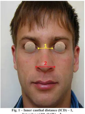
The following face measurements were taken: ICD, measured between the medial angles of the palpebral fissures and IAW, measured between the widest points of the ala of the nose (Figure 1).
Teeth measurements included: the width between the two proximal contact points for a given distance, the width of both central incisors (CIW) – the distance between proxi-mal contacts toward lateral incisors, the width of the central and lateral incisors (CLIW) – the distance between contact points of the lateral incisor and canine teeth, the width of anterior teeth (ATW) – the distance between contact points for canines and first premolars, the width between the canine cusp (CCW) – the distance between canine cusp (Figure 2).
Statistical analysis of the obtained data was performed by using computer programs Microsoft Excel 2000 and “SPSS 8.0 for Windows”. For each of the studied parameters following values were calculated: minimum value (min) and maximum value (max), mean value (ʉ), standard deviation (SD), standard error (SE), coefficient of variation (CV%) and
confidence interval (CI) representing the extent of those features that were found in 95% of cases within the selected sample.
Fig. 1 – Inner canthal distance (ICD) – 1, Interalar width (IAW) – 2.
Fig. 2 – Both central incisors width (CIW) – 1; Central and lateral incisor width (CLIW) – 2; Anterior teeth width
(ATW) – 3; Canine cusps width (CCW) – 4.
The interconnection between the measured parameters was determined by linear correlation analysis (Pearson’s cor-relation analysis) and summarized numerically with the lin-ear correlation coefficient (r).
Results
CIW for women ranged from 14.5 mm to 18 mm (ʉ = 16.3 mm). In females, the width of both central incisor ranged from 15.91 mm to 16.69 mm in 95% of cases. CLIW in females ranged from 25 mm to 32 mm in 95% of cases (ʉ = 27.9 mm). ATW in females ranged from 34 mm to 43.5 mm (ʉ = 37.1 mm). In females, ATW ranged 35.93 mm to 38.27 mm in 95% of cases. In females CCW ranged from 28.5 mm to 37 mm (ʉ = 32.1 mm), and the distance between canine cusps ranged from 30.93 mm to 33.27 mm in 95% of cases. IAW in females ranged from 26 mm to 37 mm (ʉ = 31.49 mm). In females, the interalar width ranged from 30.51 mm to 32.47 mm in 95% of cases. ICD in females ranged from 24 mm to 38 mm (ʉ = 30.47 mm). The inner canthal distance in 95% of cases in females ranged from 29.49 mm to 31.45 mm. All the results are showed in Table 1.
The width of both central incisors in males ranged from 15 mm to 19 mm (ʉ = 17 mm). In males, the width of both central incisor ranged from 15.6 mm to 18.37 mm in 95% of cases. CLIW in males ranged from 25.5 mm to 31 mm (ʉ = 28.8 mm). In males, the width of central and lateral incisor ranged from 27.42 mm to 30.17 mm in 95% of cases. ATW in males ranged from 35 mm to 42 mm (ʉ = 38.8 mm). In males, ATW ranged from 35.46 mm to 42.13 mm in 95% of cases. The distance between canines cusps ranged from 31 mm to 38 mm (ʉ = 33.7 mm) in males (the width between canines cusps ranged from 30.76 mm to 36.64 mm in 95% of
cases). IAW in males ranged from 31 mm to 40 mm (ʉ = 35.5 mm). The interalar width in males ranged from 32.37 mm to 38.63 mm in 95% of cases. The inner canthal distance ranged from 24 mm to 35 mm (ʉ = 32 mm) in males (the in-ner canthal distance ranged from 29.06 mm to 34.94 mm in 95% of cases). The results are showed in Table 1.
T-test analysis established a statistically significant dif-ference in the values of the measured parameters between the male and female subjects. By testing the statistical signifi-cance between genders a significant difference was estab-lished for the witdth of both central incisors (CIW, p = 0.003). A statistically significant difference between genders was also established for the parameter of width of central and lateral incisors (CLIW, p = 0.020). The tested difference between genders was highly significant for the parameters of ATW ( p = 0.0002). A significant diference between genders was established for the parameter of distance between ca-nines cusps (CCW, p = 0.001). A highly significant differ-ence between genders was established for the parameter of interalar width (IAW, p = 0.000). A significant difference between genders was also established for parameter of inner canthal distance (ICD, p = 0.01). The results are showed in Table 1.
The investigated parametral interconnection was ana-lyzed by linear correlative analyses (Pearson’s analysis) and numerically presented by the linear correlation coefficient (r) in Table 2. Linear correlative analysis resulted in a moderate correlation between interalar width and anterior teeth width
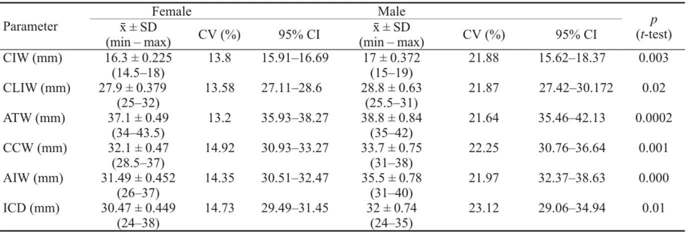
Table 1 The results of the investigated parameters in females and males
Female Male
Parameter ʉ ± SD
(min – max) CV (%) 95% CI
ʉ ± SD
(min – max) CV (%) 95% CI
p (t-test)
CIW (mm) 16.3 ± 0.225 13.8 15.91–16.69 17 ± 0.372 21.88 15.62–18.37 0.003
(14.5–18) (15–19)
CLIW (mm) 27.9 ± 0.379 13.58 27.11–28.6 28.8 ± 0.63 21.87 27.42–30.172 0.02
(25–32) (25.5–31)
ATW (mm) 37.1 ± 0.49 13.2 35.93–38.27 38.8 ± 0.84 21.64 35.46–42.13 0.0002
(34–43.5) (35–42)
CCW (mm) 32.1 ± 0.47 14.92 30.93–33.27 33.7 ± 0.75 22.25 30.76–36.64 0.001
(28.5–37) (31–38)
AIW (mm) 31.49 ± 0.452 14.35 30.51–32.47 35.5 ± 0.78 21.97 32.37–38.63 0.000
(26–37) (31–40)
ICD (mm) 30.47 ± 0.449 14.73 29.49–31.45 32 ± 0.74 23.12 29.06–34.94 0.01
(24–38) (24–35)
CIW – central incisors width; CLIW – central and lateral incisors width; ATW – anterior teath width; CCW – canine cusps width;
IAW – interalar width; ICD – inner canthal distance; min – minimal value; max – maximal value; ʉ – mean value; SD – standard deviation; CV– coefficient of variation; CI – confidence interval.
Table 2 Correlation coefficients between the investigated parameters
Parameter CIW CLIW ATW CCW AIW ICD
CIW 1 0.749 0.605 0.514 0.122 0.134
CLIW 0.749 1 0.738 0.690 0.235 0.232
ATW 0.605 0.738 1 0.787 0.439 0.335
CCW 0.514 0.690 0.787 1 0.374 0.303
AIW 0.122 0.235 0.439 0.374 1 0.410
ICD 0.134 0.232 0.335 0.303 0.410 1
and canine cusps width (r = 0.439 and r = 0.374, respec-tively). A low level of correlation between inner canthal dis-tance and anterior teeth width and canine cusps was estab-lished (r = 0.335 and r= 0.303, respectively).
Discussion
In case of absence of preextraction records, selection of upper anterior artificial teeth for edentulous patients is diffi-cult. A very important aspect in the upper anterior teeth se-lection for complete dentures is selecting the appropriate me-siodistal width of the six maxillary anterior teeth.
According to professional sources, a scientific and uni-versally accepted method for accurately determining the me-siodistal width of anterior artificial teeth has not yet been found 22.
Discussions on this topic are very present in the con-temporary professional literature 2, 7, 8, 11–22. Reviews of the recent scientific literature reveal studies that were carried out using different methodology and sample size, different face and natural teeth parameters 8, 10, 11–13, 15, 19, 20, casts 5, 16–18, 21, 22, or photographs 5, 14, 16, 17, 20, as well as various types of gauges which makes the comparison of results very difficult. Various face parameters such as bizygomatic 5, 10, 11, interpu-pillar 5, 8, 10, 11, 16, interalar 5, 8, 11, 14, 16, 19–22 and inner canthal distance 9, 10, 12–17, 22, intercommissural distance 19, 22, width of the upper lip filtrum 9 and nose lengt 20–22 have all been pro-posed as objective guidelines for solving this problem. In the most recent scientific literature, there are different views about the true value of these methods 5–22.
No theory is goad enough to help to select the size of artificial teeth, except when extracted natural teeth or casts of existe 19.
This study attempts to present the latest views and re-search in this area and investigate the possibility of using in-dividual biological parameters in prosthetic diagnosis and treatment of our population.
This research was carried out as an attempt to better understand and analyze biometric parameters of our popula-tion. Until now there have been no similar studies conducted on our population.
The purpose of this research was to establish weather the width of upper anterior teeth is in correlation with the interalar width and the distance between the medial angle of the palpebral fissure on a representative sample of our own population, as well as to determine interrelations between these parameters that could be useful in clinical treatment of our population.
By conducting statistical analysis of Arab population, al-el Sheikh and al-Athel 8 found a significant correlation between the interalar width (ƃ*ʉ = 35.54 mm, Ƃ† ʉ = 31.60 mm) and the combined width of anterior teeth (ƃʉ = 54.87 mm, Ƃʉ = 50.28 mm). The authors recommend to increase the measured value of interalar width by the statistically de-rived magnification factor (1.56). This method is suggested as a guideline for choosing the width of anterior artificial –––––––––
*ƃ – male; †Ƃ – female
teeth in combination with other methods. The existance of a significant difference in the examined parameters between the genders was also established.
After performing various facial and interalar measure-ments on members of Arab population, Al Wazan 10 deter-mined a significant correlation (p < 0.0001) between the inner canthal distance (ƃʉ = 32.94 mm, Ƃʉ = 31.91 mm) and the 4 anterior incisors (ƃʉ = 30.62 mm, Ƃʉ = 29.52 mm). No dif-ference in the inner canthal distance between genders was es-tablished 10. A low correlative coefficient between the interalar width (ƃʉ = 39.50 mm, Ƃʉ = 36.11 mm) and the intercanine distance was established. Researchers recommend using facial measurements as the initial step in determining the width of anterior artificial teeth for edentulous patients 11.
While exploring the interdependence of the inner can-thal distance (ƃʉ = 28.7 mm, Ƃʉ = 27.9 mm) and the width of central incisors (ƃʉ = 8.87 mm, Ƃʉ = 8.68 mm) Abdul-lah 12 and Abdullah et al. 13 measured facial parameters on a sample of Arab population and determined the possibility of using this distance as a guide for selection of central incisors but only after multiplying the obtained values with the coef-ficient of geometric progression (0.618) and then dividing the result by two.
While examining the correlation between interpupillar, bizygomatic and interalar distances (ƃʉ = 66.5 mm, Ƃʉ = 62.9 mm) in digital photographs and intercanine distance (ƃ ʉ = 60.6 mm, Ƃʉ = 62.8 mm) on casts on a Turkish popula-tion sample, Hasanreisoglu et al. 5 found a proportional rela-tionship between the intercanine distance and the interalar width in women and determined a significant difference in dimensions of the upper central incisors and canine teeth between the sexes (p < 0.05, p < 0.01). According to their re-sults interalar width can be used to determine the width of maxillary anterior artificial teeth, especially in women.
By analyzing facial and dental distances on a sample of Brazilian population in digital photographs and casts, Gomes et al. 14 conclude that the inner canthal distance (ƃʉ = 32.94 mm, Ƃʉ = 31.91 mm) and interalar width (ƃʉ = 34.78 mm, Ƃʉ = 33.76 mm) have a high correlation (p = 0.000) with intercanine distance, in photographs (ƃʉ = 43.10 mm, Ƃʉ = 41.77 mm) as well as in casts (ƃʉ = 54 mm, Ƃʉ = 53.50 mm). No difference in inner canthal distance was found be-tween the genders.
By analyzing statistical data obtained from an Indian population sample Tandale et al. 15 established a biometric ratio of 1 : 0.271 and 1 : 1.428 by comparing inner canthal distance (ƃʉ = 32.16 mm, Ƃʉ = 31.59 mm) and intraoral measurements of the width of four incisors (ƃʉ = 31.62 mm, Ƃʉ = 30.15 mm) and all the 6 anterior teeth (ƃʉ = 45.81 mm, Ƃʉ = 45.13 mm). Significant differences between gen-ders were found for all the measured parameters (p < 0.0001), except for inner canthal distance which showed no statistically significant difference between the genders. The authors conclude that the inner canthal distance can be used as a preliminary method for determining the width of the anterior teeth.
mm) and interalar width (ʉ = 39.36 mm) on digital photo-graphs. Individual widths of the 6 anterior teeth (central inci-sor ʉ = 8.54 mm, lateral incisor ʉ = 7.09 mm, canine ʉ = 7.94 mm) were measured on casts using a digital gauge. The authors conclude that by using regression analysis the width of anterior teeth can be predicted by combining analyzed fa-cial parameters.
On a Brazilian population sample Lucas et al. 17 used digital photography to measure the inner canthal distance (ʉ = 34.42 mm) and the distance between the maxillary canines tips (ʉ = 37.45 mm) and their distal surfaces (ʉ = 42.15 mm). They also measured the curved distance between the tips of maxillary canines (ʉ = 43.66 mm) and their distal surfaces (ʉ = 53.45 mm) on casts. A significant correlation was estab-lished between all determined variables (r = 0.476, r = 0.467, r = 0.285, r = 0.302). The authors conclude that inner canthal distance, when determined by photogrammetry, can be a re-liable guideline for selecting the anterior teeth 17.
Ibrahimagic et al. 19 conducted a research within Croa-tian population in order to determine the correlation between the width of upper incisors, the width between the upper ca-nines tips or incisor and canine width and the interalar width or the intercominsural width. The results show a statistically significant difference between males and females for all the measured variables (p < 0.01), and that the recorded values were higher among males. The obtained mean value for in-teralar width is 30.9 mm for females and 33.63 mm for males. The width between canines cusps in women is 31.021 mm and for males is 32.44 mm. Width of the nose approxi-mates to the width between the tips of canines (1.08 : 1). The calculated values for the studied population may help in choosing the size of upper anterior teeth and their prefer-ences in complete dentures.
Varjão and Nogueira 21 found out that the average value of interalar width for the white Brazilian male is 35.28 mm and the mean value of intercanine tooth width for the same population is 33.55 mm (measured on casts). The calculated Pearson's correlation coefficient was 0.238, which is a weak correlation between these two parameters. The authors con-clude that the method of measuring the width of the base of the nose is not an accurate guideline for selecting the width of artificial teeth.
While conducting research within Croatian population, Knezovic et al. 22 conclude that using facial measurements such as face, nose and upper lip length, inner canthal, inter-alar and intercomisural distance for selecting anterior artifi-cial teeth are generally inaccurate. The indexes of width/height were determined for central incisors, lateral in-cisors and canines. For interalar width (ƃʉ = 33.9 mm, Ƃʉ = 30.20 mm) a statistically significant difference related to gender (p < 0.0001) was obtained, while the inner canthal distance (ƃʉ = 15.41 mm, Ƃʉ = 15.31 mm) showed no dif-ference.
Comparing the results of a previous researches in re-lation to one’s own, differences rooted primarily in ethnic
and morphological characteristics are observed. Given the great individual variability in human physiognomy and values of morphological parameters, the use of inaccurate standards in diagnosis and treatment planning would not only lead to wrong conclusions about the existence and se-verity of deviations but also to unsatisfactory results of denture therapy, both in terms of esthetics and the aspect of planning the artificial occlusion complex. Therefore, the re-sults of specific relations of anatomic determinants and width of anterior teeth must be perceived as distinctive features of the population on which the study was per-formed.
Most studies, including ours, established the existance of significant differences in all values of facial and dental parametres between the genders, and show that male sub-jects have higher values than females. This research deter-mines that there is very little significant difference in inner canthal distance between genders while some studies find no difference in this parameter between the two sexes whatsoever 14, 15, 22. By taking measurements on samples of their own population, some authors do not find any signifi-cant correlation between facial and dental parameters and therefore conclude that inner canthal distance and interalar width are not reliable parameters for selecting the size of upper anterior teeth for dentures 21, 22. Considering that no research, including ours, confirms a high correlation be-tween facial and dental parameters, most authors rec-comend using this method as a guideline in choosing the width of anterior artificial teeth but only combined with other methods.
Conclusion
The analyzed values of facial and dental parameters in our population are moving in the biometric standards con-tained in the relevant literature. The determined differences arise from ethnic and morphological characteristics. A mod-erate correlation between the interalar width and anterior teeth width and canine cusps width was established. A low correlation between the inner canthal distance and width of anterior teeth and canine cusps width was established. By testing the statistical significance between genders signifi-cant differences for all the parameters was found. The meas-ured facial distances and anterior teeth width had higher val-ues for men than for women.
R E F E R E N C E S
1. Strajnic Lj. Determination of placement of anterior teeth in re-movable dental prostheses. Med Pregl 2002; 55(11î12): 490î4. (Serbian)
2. Rosenstiel SF, Ward DH, Rashid RG. Dentists' preferences of ante-rior toot proportion- a web-based study. J Prosthodont 2000; 9(3): 123î36.
3. Preston JD. The golden proportion revisited. J Esthet Dent 1993; 5: 247–51.
4. Ward DH. A study of dentists' preferred maxillary anterior tooth width proportions: comparing the recurring esthetic dental pro-portion to other mathematical and naturally occurring propor-tions. J Esthet Restor Dent 2007; 19(6): 324î37.
5. Hasanreisoglu U, Berksun S, Aras K, Arslan I. An analysis of max-illary anterior teeth: Facial and dental proportions. J Prosthet Dent 2005; 94(6): 530î38.
6. Ali Fayyad MA, Jamani KD, Agrabawi J. Geometric and mathe-matical proportions and their relations to maxillary anterior teeth. J Contemp Dent Pract 2006; 7(5): 62î70.
7. Baer ML, Reynolds MA. Comparison of anterior tooth width in natural and artificial dentitions. J Prosthodon 1992; 1: 84î7. 8. al-el-Sheikh HM, al-Athel MS. The relationship of interalar width,
interpupillary width and maxillary anterior teeth width in Saudi population. Odontostomatol Trop 1998; 21(84): 7î10.
9. Basker R.M., Davenport J. C. Prosthetic treatment of the Edentu-lous Patient. 4th ed. Oxford: Blackwell Publishing Company; 2002.
10. Al Wazzan KA. The relationship between intercanthal dimen-sion and the widths of maxillary anterior teeth. J Prosthet Dent 2001; 86(6): 608î12.
11. Al Wazzan KA, Al Haidan A, Al Madi EM, Al Murfarj A. The relationship between facial references and mesiodistal width of maxillary anterior teeth among Saudi patients. Alexandria Dent J 1995; 20(4): 39î45.
12. Abdullah MA. Inner canthal distance and geometric progression as a predictor of maxillary central incisor width. J Prosthet Dent 2002; 88(1): 16î20.
13.Abdullah MA, Stipho HD, Talic YF, Khan N. The significance of inner-canthal distance in prosthodontics. Saudi Dent J 1997; 9(1): 36î9.
14.Gomes VL, Gonçalves LC, do Prado CJ, Junior IL, de Lima Lucas B.
Correlation between facial measurements and the mesiodistal width of the maxillary anterior teeth. J Esthet Restor Dent 2006; 18(4): 196î205; discussion 205.
15.Tandale UE, Dange SP, Khalikar AN. Biometric relationship between intercanthal dimension and the widths of maxillary anterior teeth. J Indian Prosthodont Soc 2007; 7(3): 123î5. 16.Isa ZM, Tawfiq OF, Noor NM, Shamsudheen MI, Rijal OM.
Re-gression methods to investigate the relationship between facial measurements and widths of the maxillary anterior teeth. J Prosthet Dent 2010; 103(3): 182î8.
17.Lucas BL, Bernardino-Júnior R, Gonçalves LC, Gomes VL.
Distance between the medialis angles of the eyes as an ana-tomical parameter for tooth selection. J Oral Rehabil 2009; 36(11): 840î7.
18.Gonçalves LC, Gomes VL, De Lima Lucas B, Monteiro SB.
Correlation between the individual and the combined width of the six maxillary anterior teeth. J Esthet Restor Dent 2009; 21(3): 182î91.
19.Ibrahimagic L, Celebic A, Jerolimov V, Seifert D, Kardum-Ivic M, Filipovic I. Correlation between the size of maxillary frontal teeth, the width between alae nasi and the width between corners of the lips. Acta Stomatol Croat 2001; 35(2): 169î79.
20.Sülün T, Ergin U, Tuncer N. The nose shape as a predictor of maxillary central and lateral incisor width. Quintessence Int 2005; 36(8): 603î7.
21.Varjão FM, Nogueira SS. Nasal width as a guide for the selec-tion of maxillary complete denture anterior teeth in four racial groups. J Prosthodont 2006; 15(6): 353î8.
22.Knezovic Zlataric D, Kristek E, Celebic A. Analysis of width/lenght ratios of normal clinical crowns of the maxil-lary anterior dentition: correlation between dental propor-tions and facial measurements. Int J Prosthodont 2007; 20(3): 313î7.