Ital.J.anIm.ScI. vol. 6, (Suppl. 2), 688-690, 2007 688
Structural and ultrastructural
characterization of buffalo fetus (
Bubalus
bubalis
) ovarian germinative cells
F.c.a. carvalho
1, E. oba
1,2, a.v. mota
1, l.S. leal
11 Department of Animal Reproduction and Veterinary Radiology
– FmvZ/unESp, Botucatu/Sp- 18618-000, Brazil
2 Researcher of cnpq
Corresponding author: E. Oba. Department of Animal Reproduction and Veterinary Radiology – FMVZ/ UNESP. Distrito de Rubião Júnior, sem número - Botucatu/SP- 18618-000, Brazil - Tel. 55-14-3811-6249 - Fax: 55-14-3811-6249 - E-mail: [email protected]
ABSTRACT: The objective of the present study was to characterize ovogones, primary oocytes and preantral follicles of buffalo fetus in different ages of gestation. For this, 29 fetuses were collected from a slaughterhouse (Frigol, Brazil) and crown-rump lengths were measured to estimate the fetal age (0-3, 4-6, 7-10 months of gestation). The ovaries were removed and ovarian tissue was processed for classic histology and transmission eletron microscopy examination. The structural evaluation demonstrated that in the first period of the gestation (0-3 months) the buffalo fetus showed ovogones (in mitotic division) and in some cases, the primary oocytes surrounded by somatic cells. In the second period (4-6 months), it was verified that the preantral follicles were completely formed. In the last period (7o month to the end of gestation) the ovaries contained a large amount of preantral
follicles, and in some fetuses, antral follicles were observed. The ultrastructural analysis of the ovogones, primary oocytes and preantral follicles showed that these cells have few organelles and the quantity of mitochondria, endoplasmatic reticulum and apparatus Golgi complex is increased as the germinative cells passing from one stage to another.
Key words: Buffalo, Fetus, Ovarian follicles, Histology.
INTRODUCTION - Females of domestic species have a finite stock of germinal cells established during fetal life. There are thousands of primordial follicles in mammalian ova-ries, but almost all are eliminated in vivo by follicular atresia (Santos et al., 2006). The structural and ultrastructural studies of fetal germinative cells are very important for the development of techniques for culture of fetal ovarian follicles with applications in trangen-esis, conservation of extinct species and formation of genetic banks.
The aim of the present study was to characterize ovogones, primary oocytes and preantral follicles of buffalo fetus in different ages of gestation.
MATERIAL AND METHODS - For this experiment, 29fetuses were collected from a slaughterhouse (Frigol – Lençóis Paulista – SP – Brazil) and crown-rump lengths (Figure 1) were measured to estimate the fetal age (0-3, 4-6, 7-10 months of gestation, Table 1). Within
20 to 30 minutes after slaughter, the ovaries were removed and washed in 70% ethanol for 10 minutes and in 0.9% saline solution. The ovarian tissue was processed for classic histol-ogy and transmission eletron microscopy examination.
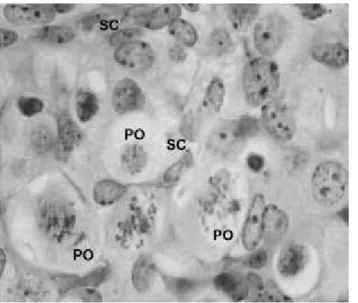
RESULTS AND CONCLUSIONS - The structural evaluation (Figure 2) demonstrated that in the first period of the gestation (0-3 months) the buffalo fetus showed ovogones (in mitotic division) and in some cases, the primary oocytes surrounded by somatic cells. Gos-den (1995) observed that ovine fetuses had primordial follicles at 74 days of gestation. In the second period (4-6 months) it was verified that the preantral follicles were completely formed. In the last period (7o month to the end of gestation) the ovaries contained a large
amount of preantral follicles, and in some fetuses, antral follicles were observed. These re-sults are not consistent with those of Santos et al. (2006). These researchers described that
buffalo fetuses had primordial licles at month 4, and antral fol-licles at month 6. The ultrastruc-tural analysis (Figure 3) of the ovogones, primary oocytes and preantral follicles showed that these cells have few organelles and the quantity of mitochondria,
Figure 2.
Histology of a buffalo fetus
ovary
(fetus with 11.5 cm).
po – primary oocytes Sc – somatic cells
PAS –H 100 x
Figure 1. The buffalo fetus was measured (crown-rump length) with a paquimeter to estimate the fetal age.
table 1. Determination of the fetal
age according to crown rump length (cm).
Gestation stage Mean of Crown rump
(months) length (cm)
1-2 2.5
2-3 7.3
3-4 14.0
4-5 21.8
5-6 29.1
6-7 37.3
Ital.J.anIm.ScI. vol. 6, (Suppl. 2), 688-690, 2007 689
endoplasmatic reticulum and apparatus Golgi complex is increased as the germinative cells passing from one stage to another. These results indicate that structure and ultrastructure of buffalo ovarian germinative cells are similar of those observed in bovine.
ACKNOWLEDGMENTS - The present study was supported by CNPq and FAPESP – Brazil.
REFERENCES -Santos, S. S. D., Biondi, F. C., Cordeiro, M. S., Miranda, M. S., Dantas, J. K., Figueiredo, J. R., Ohashi, O. M., 2006. Isolation, follicular density, and culture of pre-antral follicles of buffalo fetuses of different ages. Anim. Reprod. Sci., 95: 1-15. Gosden, R. G. 1995. Ovulation 1: Oocyte development throughout life. In: Grudzinskas, J. G., Yovich, J. L. Gametes , 119-149.
Figure 3.
Eletron micrography of a buffalo fetus ovogone
(fetus with 9.0 cm).
n – nucleus n – nucleolus m – mitochondria v – vesicle
G- apparatus Golgi complex 9.750 x
Ital.J.anIm.ScI. vol. 6, (Suppl. 2), 688-690, 2007 690