Cell Proliferation by Monocyte-Derived Macrophages
from Diabetic Patients
Te-Chuan Chen1, Mao-Ling Sung2, Hsing-Chun Kuo3,4, Shao-Ju Chien5, Chia-Kuang Yen2,
Cheng-Nan Chen6*
1Division of Nephrology, Kaohsiung Chang Gung Memorial Hospital and Chang Gung University College of Medicine, Kaohsiung, Taiwan,2Department of Cardiology, St. Martin De Porres Hospital, Chiayi, Taiwan,3Institute of Nursing and Department of Nursing, Chang Gung University of Science and Technology, Chronic Diseases and Health Promotion Research Center, CGUST, Taoyuan, Taiwan,4Research Center for Industry of Human Ecology, Chang Gung University of Science and Technology, Taoyuan, Taiwan,5Division of Pediatric Cardiology, Department of Pediatrics, Kaohsiung Chang Gung Memorial Hospital, Chang Gung University College of Medicine, Kaohsiung, Taiwan,6Department of Biochemical Science and Technology, National Chiayi University, Chiayi, Taiwan
Abstract
Macrophage accumulation in the arterial wall and smooth muscle cell (SMC) proliferation are features of type 2 diabetes mellitus (DM) and its vascular complications. However, the effects of diabetic monocyte-derived macrophages on vascular SMC proliferation are not clearly understood. In the present study, we investigated the pro-proliferative effect of macrophages isolated from DM patients on vascular SMCs. Macrophage-conditioned media (MCM) were prepared from macrophages isolated from DM patients. DM-MCM treatment induced HASMC proliferation, decreased p21Cip1and p27Kip1 expressions, and increased microRNA (miR)-17-5p and miR-221 expressions. Inhibition of either miR-17-5p or miR-221 inhibited DM-MCM-induced cell proliferation. Inhibition of miR-17-5p abolished DM-MCM-induced p21Cip1down-regulation; and inhibition of miR-221 attenuated the DM-MCM-induced p27Kip1 down-regulation. Furthermore, blocking assays demonstrated that PDGF-CC in DM-MCM is the major mediators of cell proliferation in SMCs. In conclusion, our present data support the hypothesis that SMC proliferation stimulated by macrophages may play critical roles in vascular complications in DM patients and suggest a new mechanism by which arterial disease is accelerated in diabetes.
Citation:Chen T-C, Sung M-L, Kuo H-C, Chien S-J, Yen C-K, et al. (2014) Differential Regulation of Human Aortic Smooth Muscle Cell Proliferation by Monocyte-Derived Macrophages from Diabetic Patients. PLoS ONE 9(11): e113752. doi:10.1371/journal.pone.0113752
Editor:Jozef Dulak, Faculty of Biochemistry, Poland
ReceivedJuly 14, 2014;AcceptedOctober 28, 2014;PublishedNovember 19, 2014
Copyright:ß2014 Chen et al. This is an open-access article distributed under the terms of the Creative Commons Attribution License, which permits unrestricted use, distribution, and reproduction in any medium, provided the original author and source are credited.
Data Availability:The authors confirm that all data underlying the findings are fully available without restriction. All relevant data are within the paper. Funding:Grants CZRPG880253, CMRPF6A0073, CMRPG891581, CMRPG6C0071, CMRPG6C0011 and EZRPF6C0011 from Chang Gung Memorial Hospital-Kaohsiung Medical Center, Chang Gung Memorial Hospital, and Chang Gung University of Science and Technology, Chia-Yi Campus, Taiwan, and by the National Science Council, Taiwan (NSC101-2320-B-415-003-MY3, NSC102-2314-B-750-001, NSC101-2622-B-255-001-CC3, NSC102-2313-B-255-002). The funders had no role in study design, data collection and analysis, decision to publish, or preparation of the manuscript.
Competing Interests:The authors have declared that no competing interests exist. * Email: [email protected]
Introduction
Diabetes mellitus (DM) is associated with an increased risk for atherothrombotic complications such as peripheral artery disease, coronary artery disease, and myocardial infarction [1,2]. It is well documented that macrophage accumulation is a common feature of type 2 diabetes and its complications [3,4]. Recruitment of monocytes from the peripheral blood to the intima of the vessel wall and the monocytes’ subsequent differentiation into macro-phages are critical events in atherogenesis. In addition, patholog-ical environments, such as hyperglycemia, may promote macro-phage activation and secretion within DM tissues. It has been demonstrated that high-glucose (HG) treatments of human monocytes lead to the increased expression of inflammatory cytokine and chemokine genes [5]. Our previous study also reported that the macrophage inflammatory protein (MIP)-1aand 1b released by HG-treated monocyte-derived macrophages are major mediators for the induction of E-selectin expression in vascular endothelial cells (ECs) [6]. However, the effect of monocyte-derived macrophages isolated from diabetic patients
on vascular smooth-muscle-cell (SMC) activation has not been completely understood.
The proliferation and phenotypic changes of vascular SMCs are key events in the development of atherosclerosis and its complications [7]. During the development of atherosclerotic plaque, SMCs migrate from the media into the intimal layer of the arterial wall, where they proliferate and produce an extracellular matrix (ECM), resulting in the formation of intimal hyperplasia [8]. Growth factors, inflammatory cytokines, and chemokines have been implicated as factors that mediate such SMC proliferation [9]. Among these factors, the platelet-derived growth factor (PDGF) family possesses the most potent mitogenic effects for SMC [10]. The G1-to-S phase transit of the cell cycle requires activation of the cyclin-cyclin-dependent kinase (CDK) complex. The cyclin-dependent kinase inhibitors (CKI) such as p21Cip1and p27Kip1 have been shown to regulate the activity of these complexes in the G1 phase [11]. p21Cip1 and p27Kip1 are
environment can activate vascular SMCs and cause vascular dysfunction [13]. In addition, it has also been found that hyperglycemic conditions have mitogenic effects on vascular SMCs [14]. Therefore, examining the proliferative response of vascular SMCs to hyperglycemia may potentially extend our understanding of the pathogenic mechanisms of vascular compli-cations in diabetes.
The functions of SMCs may be modulated by their interaction with other vascular cells such as monocyte-derived macrophages. It has been reported that interactions between monocytes and vascular SMCs may contribute to monocyte retention within the vasculature [15]. Moreover, macrophages can release a wide range of regulatory factors that are growth mediators to affect cell proliferation. Previous studies have indicated that human mono-cyte-derived macrophages have an inhibitory effect on SMC growth [16,17]. Evidence also shows that palmitate-stimulated macrophages promote SMC proliferation via the secretion of bone morphogenetic protein (BMP) 2 and BMP4 [18]. Although there is considerable research on the role of macrophages in the development of atherosclerotic lesions, the contribution of macrophages under hyperglycemic conditions to SMC prolifera-tion remains unclear.
SMCs in the media of the artery are encompassed by a network of ECM such as fibrillar collagen. Culturing SMCs on fibrillar collagen promotes the maintenance of the SMC contractile phenotype and exerts an anti-proliferative effect [19]. Since proliferation and accumulation of SMCs are believed to play important roles in the progression of macrophage-rich lesions to fibroatheromas [4], it was hypothesized that monocyte-derived macrophages differentiated from diabetic patients may alter gene expression and affect SMC proliferation. We found that the SMC proliferation induced by macrophages in hyperglycemia is mediated through the up-regulation of microRNA (miR)-17-5p and miR-221. This study presents evidence for a novel mechanism in which miRNAs act synergistically to induce SMC proliferation.
Methods
Materials
All culture materials were purchased from Gibco (Grand Island, NY, USA). Rat tail type I collagen was purchased from BD Biosciences (San Diego, CA). Mouse monoclonal antibodies (mAbs) against p21Cip1 and rabbit polyclonal antibodies (pAbs) against p27Kip1were purchased from Cell Signaling Technology (Beverly, MA). mAbs against osteopontin (OPN) matrix gla protein (MGP) were purchased from Santa Cruz Biotech (Santa Cruz,
CA, USA). Other chemicals of reagent grade were obtained from Sigma (St Louis, MO).
Subjects
The Ethics Committees of St. Martin De Porres Hospital (Chiayi City, Taiwan) approved the study protocol, and written informed consents were obtained from all patients before enrollment. The study group consisted of 13 patients with type 2 diabetes and 9 healthy control subjects. The patients had a mean (6SEM) age of 53.468.5 years, body mass index of 31.462.5 kg/ m2, fasting glucose of 185612.4 mg/dL, triglyceride level of 182.9616.1 mg/dL, LDL cholesterol level of 94.367.8 mg/dL, and hemoglobin A1Cof 7.661.1%. All patients were treated with
glyburide and metformin. None of the patients was primarily insulin dependent. In addition, we recruited 9 volunteers, who were admitted to the St. Martin De Porres Hospital for the purpose of routine physical examinations, as the control subject group. They had a mean (6SEM) age of 49.267.9 years, body
mass index of 22.761.3 kg/m2, fasting glucose of 84.661.7 mg/ dL, triglyceride level of 147.168.7 mg/dL, LDL cholesterol level of 95.263.8 mg/dL, and hemoglobin A1Cof 4.760.4%. None of
the normal subjects had infectious or inflammatory conditions, or cardiac, renal, or pulmonary decompensated diseases. Volunteers who smoked cigarettes, used alcohol or were under medications (hormonal replacement therapy, nonsteroidal anti-inflammatory drugs, corticosteroids, and anticoagulant drugs) were excluded from this normal subject group.
Human monocyte isolation
Human monocytes were isolated as previously described [6]. Peripheral blood mononuclear cells (PBMCs) were isolated by Histopaque 1077 density-gradient centrifugation. Monocytes were purified from PBMCs by negative selection using the magnetic-activated cell sorting (MACS) monocyte isolation kit (Miltenyi Biotech, Auburn, CA); PBMCs were first treated with FccR blocking reagent (human IgG), followed by a hapten/antibody mixture (mixture of hapten-conjugated monoclonal anti-CD3, anti-CD7, anti-CD19, anti-CD45RA, anti-CD56, and anti-IgE antibodies). After treatment with MACS antihapten magnetic microbeads conjugated to monoclonal antihapten antibody, the labeled cells were passed over a MACS column, and the effluent was collected as the negative fraction representing enriched monocytes (.95% purity).
Preparation of macrophage-conditioned medium (MCM) Differentiation of monocyte-derived macrophages from diabetes patients or normal control subjects was achieved by culturing the freshly isolated monocytes (56105 cells/mL) in RPMI-1640
medium supplemented with 10% autologous serum. After 4 days in culture, monocyte-derived macrophages were incubated for another 48 h in fresh serum-free RPMI medium. The conditioned medium was collected, centrifuged, filtered, and defined as diabetic (DM)-MCM and normal control (NC)-MCM.
Cell culture
Human aortic SMCs (HASMCs) were obtained commercially (Clonetics, Palo Alto, CA) and maintained in F12K medium supplemented with 10% FBS. Cells at passages 3 to 6 were used. Growth of the cells was arrested by incubating in F12K medium with 0.5% FBS for 48 hours before use.
Collagen matrices
Fibrillar collagen (0.1%) was prepared by mixing 4 mg/ml rat tail type I collagen (25%), 0.1 M NaOH (5%), 26F12K medium (40%), FBS (10%), and complete medium (F12K with 10% FBS; 20%). The mixture (0.15 ml/cm2) was allowed to form fibrillar collagen matrices for at least 1 h at 37uC. SMCs were cultured on the surface of fibrillar collagen as described [19].
MTT assay and flow cytometric analysis for cell proliferation
Cells were cultured on type I fibrillar collagen in 96-well plates. Cell proliferation was determined by 3-(4,5-dimethylthiazol-2-yl)-2,5-diphenyltetrazolium bromide (MTT) assay. After the incuba-tion period, MTT soluincuba-tion was added to each well to a final concentration of 0.5 mg/mL, and the mixture was incubated at 37uC for 3 hours to allow MTT reduction. The formazan crystals were dissolved by adding dimethylsulfoxide (DMSO) and absor-bance was measured at 570 nm with a spectrophotometer.
(Becton Dickinson), and the data were analyzed by using a mod-fit cell cycle analysis program [19].
Real-time quantitative PCR
Total RNA preparation and the RT reaction were carried out as described previously [6]. Regular real-time quantitative PCR was performed to confirm PCR array results. PCRs were performed using an ABI Prism 7900HT according to the manufacturer’s instructions. Amplification of specific PCR prod-ucts was detected using the SYBR Green PCR Master Mix (Applied Biosystems). In addition, the designed primers in this study were: p21Cip1 forward primer, 59-CTGAA AGATG GACGC TCAAT-39; p21Cip1 reverse primer, 59-CGTTT CA-GAA GCCAG AAGAG-39; p27Kip1forward primer, 59-CTGAA AGATG GACGC TCAAT-39; p27Kip1 reverse primer, 59 -CGTTT CAGAA GCCAG AAGAG-39; OPN forward primer, 59- TTGCA GCCTT CTCAG CCAA-39; OPN reverse primer, 59- GGAGG CAAAA GCAAA TCACT G-39; MGP forward primer, 59- GCTCA ATAGG GAAGC CTGTG AT-39; MGP reverse primer, 59- TTTCT TCCCT CAGTC TCATT TGG-39; 18S rRNA forward primer, 59-CGGCG ACGAC CCATT CGAAC-39, 18S rRNA reverse primer, 59-GAATC GAACC CTGAT TCCCC GTC-39. Quantification was performed using the 22DDCtmethod [6].
Western blot analysis
SMCs were lysed with a buffer containing 1% NP-40, 0.5% sodium deoxycholate, 0.1% SDS, and a protease inhibitor mixture (PMSF, aprotinin, and sodium orthovanadate). The total cell lysate (50mg of protein) was separated by SDS-polyacrylamide gel
electrophoresis (PAGE) (12% running, 4% stacking) and analyzed by using the designated antibodies and the Western-Light chemiluminescent detection system (Bio-Rad, Hercules, CA), as previously described [19].
Inhibition of miR-17-5p and miR-221
Anti-miR inhibitors for miR-17-5p, miR-221, and correspond-ing negative controls were purchased from Ambion, Life Technologies (Austin, TX, USA). The SMCs were then transfect-ed with the miRNA inhibitors or miRNA inhibitor negative control by Oligofectamine Transfection Reagent from Invitrogen, Life Technologies, in accordance with the manufacturer’s procedure. The final concentration for miRNA inhibitor was 200 nmol/L. The transfection efficiency of miRNA inhibitors was further verified by Real-time quantitative PCR assay. After transfection with the miR-17-5p and miR-221 inhibitors, the expression levels of miR-17-5p and miR-221 were decreased by 75.3% and 78.7%, respectively.
ELISA for PDGF-BB and PDGF-CC
The levels of PDGF-BB and PDGF-CC in the MCM were determined by using sandwich ELISA (sensitivity 18 pg/mL; R&D) according to manufacturer’s protocols, as previously described [19].
Statistical analysis
The results are expressed as mean6standard error of the mean (SEM). Statistical analysis was performed by using an independent Student t-test for two groups of data and analysis of variance (ANOVA) followed by Scheffe’s test for multiple comparisons.P
values less than 0.05 were considered significant.
Results
The effect of DM-MCM on SMC proliferation
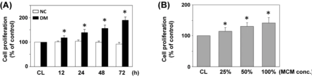
The effects of monocyte-derived macrophages from DM patients on cell proliferation were studied by treating SMCs cultured on fibrillar collagen with DM-MCM (vs. NC-MCM). SMCs were stimulated with the two types of MCM at different concentrations for 24 h, or 16DM-MCM for the times indicated. Cell proliferation was analyzed by MTT assays. As shown in Figure 1A, proliferation of SMC was significantly increased (38%, 56% and 89% after cultivation for 24, 48 and 72 h, respectively) when cultivated with DM-MCM. SMCs cultured with NC-MCM had limited proliferation capacities (Figure 1A). SMCs treated with DM-MCM at different concentrations for 24 h showed a significantly higher growth rate when compared to the control cells (Figure 1B). Table 1 summarizes the flow cytometry analysis of cell distribution in the cell cycle phases. SMCs stimulated with DM-MCM for 24 and 48 h had marked decreases of cells in the G0/G1phases and increases in the S-phase (Table 1).
DM-MCM decreased p21Cip1and p27Kip1and increased synthetic differentiation marker expression in SMCs
We examined the effects of DM-MCM on the expression of cell-cycle regulatory proteins p21Cip1 and p27Kip1. As shown in Figure 2B, SMCs with DM-MCM stimulation decreased the expressions of p21Cip1and p27Kip1. However, there was no effect on p21Cip1and p27Kip1expression in SMCs cultured with NC-MCM (Figure 2A). Figure 2C shows that DM-NC-MCM induced decreases in the mRNA expression of p21Cip1and p27Kip1after 12 h of DM-MCM stimulation.
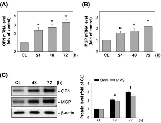
We also examined the effect of DM-MCM on the expression of synthetic differentiation markers in SMCs. SMCs were cultured on fibrillar collagen, and the mRNA and protein expression of OPN and MGP was analyzed at various time points after cultivation. Real-time PCR analysis indicated the time-dependent increase in the levels of mRNA expression for OPN (Figure 3A) and MGP (Figure 3B) in SMCs on fibrillar collagen. The cultivation of SMCs by DM-MCM also caused significant increases in the OPN and MGP expression at 72 h on fibrillar collagen (Figure 3C).
miR-17-5p and miR-221 are involved in the regulation of HASMC proliferation
Recent studies have demonstrated that miRNAs are involved in regulating SMC gene expression and proliferation [20]. We therefore examined the effects of DM-MCM on the expression of miRNAs. Stimulation of SMCs with DM-MCM increased the expression of the miR-17-5p (Figure 4A) and miR-221 (Fig-ure 4B), in a time-dependent manner.
To determine whether DM-MCM-induced cell proliferation was mediated by the up-regulation of miR-17-5p and miR-221, SMCs were pretreated with miR-17-5p and miR-221 inhibitors and subsequently stimulated with DM-MCM. The DM-MCM-induced SMC proliferation was significantly inhibited by pretreat-ments with miR-17-5p and miR-221 inhibitors (Figure 4C). The DM-MCM-induced decrease in the percentage of cells in the G0/
G1phases and the increase in the percentage of cells in the S-phase
were also significantly inhibited by miR-17-5p and miR-221 inhibitors (Figure 4D).
whereas miR-221 inhibitor inhibited the DM-MCM-induced expression of p27Kip1.
DM-MCM induced SMC proliferation was mediated by PDGF-CC
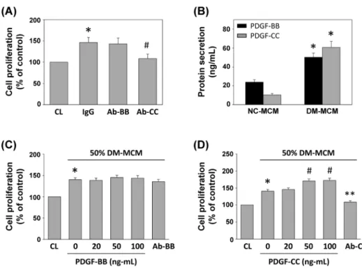
The effect of DM-MCM on SMC proliferation suggests that macrophages under a DM environment may release soluble mediators and exert paracrine effects on SMCs to induce cell proliferation. PDGF was identified in a search for serum factors that stimulate SMC proliferation [10]. As shown in Figure 5A, the incubation of SMCs with PDGF-CC neutralizing antibody, but not PDGF-BB, significantly inhibited DM-MCM-induced SMC proliferation. To confirm these results, the protein levels of PDGF-BB and PDGF-CC in NC- and DM-MCM were analyzed by ELISA. As shown in Figure 5B, culturing of monocyte-derive macrophages caused significant increases of PDGF-BB and PDGF-CC protein secretion in macrophages isolated from diabetic patients.
To further characterize the effect of the BB or PDGF-CC in MCM on SMC proliferation and to determine the optimal PDGF concentrations, we examined the responses of SMC to 50% DM-MCM with different concentrations of the PDGF-BB and PDGF-CC (0–100 ng/mL) and their neutralizing antibodies. The results shown in Figure 5D indicated that PDGF-CC promoted SMC proliferation, and the effect was significantly inhibited by neutralizing antibody against PDGF-CC, while PDGF-BB had no significant effect on SMC proliferation (Figure 5C). Figure 5D also indicated that the optimal concentration of PDGF-CC was 50 ng/ mL.
Discussion
Diabetic patients have an increased susceptibility to the development of atherosclerosis [21]. Atherosclerotic lesions in patients with diabetes are characterized by excessive macrophage infiltration, suggesting that the accumulation of monocyte-derived macrophages into the vasculature may be augmented under hyperglycemic conditions [3]. In addition, the migration and proliferation of SMCs are believed to play important roles in the progression of macrophage-rich lesions to fibroatheromas, and diabetic conditions have been shown to enhance this process [22]. It has been suggested that the macrophage-SMC coexistence as neighbors within the vessel walls induces regulatory signals involved in atherogenesis [23]. There is also evidence that hyperglycemia may enhance the interaction of macrophages and SMCs [24]. However, the pathophysiological mechanism of macrophage-SMC interaction remains poorly understood. The present study characterized the roles of two major monocyte-derived macrophage-induced mediators from diabetic patients in the regulation of p21Cip1 and p27Kip1expressions by HASMCs. Several lines of evidence from the current study indicate that the effect of macrophages from DM patients on SMC proliferation was mediated by the down-regulation of expressions of p21Cip1 and p27Kip1and that this down-regulation was mediated through the differential regulation of the miR-17-5p and miR-221. First, stimulation by DM-MCM induced decreased expression of p21Cip1 and p27Kip1, as well as increased cell proliferation. Second, stimulation of SMCs by DM-MCM induced expression of miR-17-5p. Pretreatment of the cells with miR-17-5p inhibitor suppressed the DM-MCM-induced down-regulation of p21Cip1
Figure 1. Effect of DM-MCM on cell viability of HASMCs.Bar graphs represent folds of controls (CL) SMCs, mean6standard error of the mean (SEM) of 5 independent experiments. *P,0.05 versus CL SMCs. (A) SMCs were kept as CL or stimulated with NC-MCM or DM-MCM at the indicated time periods, or (B) stimulated with different concentrations of DM-MCM for 24 h. Cell proliferation was assayed by the MTT test.
doi:10.1371/journal.pone.0113752.g001
Table 1.Cell cycle analysis of SMCs stimulated with NC- and DM-MCM.
% cells (mean±SEM)
Duration (h) G0/G1 S-phase G2/M
Control 0 87.462.1 6.560.8 6.161.2
NC-MCM 24 92.362.7 5.160.6 2.661.0
48 92.961.9 3.960.4 3.260.8
72 90.362.6 5.660.6 4.161.1
DM-MCM 24 72.164.8* 19.262.4* 8.761.4
48 70.664.3* 22.362.7* 7.161.3
72 65.766.6* 27.964.2* 6.460.9
SMCs were kept as controls or on fibrillar collagen with NG- or HG-MCM treatment. Cells were analyzed for DNA content by flow cytometry to show percentages in G0/
G1, synthetic, or G2/M phases of cell cycle. Data are mean6SEM from three independent experiments.
*P,0.05 vs. control cells.
Figure 2. Effects of DM-MCM on p21Cip1and p27Kip1expression.Bar graphs represent folds of CL SMCs, mean
6SEM from 3 independent experiments. *P,0.05 versus CL SMCs. (A) SMCs were kept as CL or stimulated with NC-MCM or DM-MCM for the times indicated. Protein expressions were determined by Western blot analysis. Expression levels of p21Cip1and p27Kip1are presented as band densities (normalized tob-actin) relative to CL. (B) mRNA expressions were determined by real-time PCR analysis and normalized to 18S rRNA.
doi:10.1371/journal.pone.0113752.g002
Figure 3. Effects of DM-MCM on synthetic differentiation marker expression in SMCs.Bar graphs represent folds of CL SMCs, mean6SEM from 3 independent experiments. *P,0.05 versus CL SMCs. (A, B) DM-MCM induced mRNA expressions of osteopontin (OPN) and matrix gla protein (MGP) in SMCs. SMCs were kept as controls (CL) or stimulated with DM-MCM for the times indicated, and the mRNA expressions of OPN (A) and MGP (B) were determined using real-time PCR analysis and normalized to 18S rRNA. (C) SMCs were kept as CL or stimulated with DM-MCM for the times indicated. Protein expressions of OPN and MGP were determined by Western blot analysis. Expression levels of OPN and MGP are presented as band densities (normalized tob-actin) relative to CL.
and proliferation of the SMCs. Third, stimulation of SMCs with DM-MCM also induced expression of miR-221. Pretreatment of the cells with miR-221 inhibitor decreased the DM-MCM-induced down-regulation of p27Kip1and SMC proliferation.
It is believed that extravasation of macrophages in the arterial wall promotes formation of atherosclerotic lesions. It has also been suggested that diabetes-elicited SMC proliferation occurs second-ary to the increased macrophage infiltration into the arterial wall [22]. The effects of HG levels on the proliferative capacity of arterial SMCs are controversial. While several studies have demonstrated that HG levels can stimulate SMC proliferation [14,25], others have found no stimulatory effect [26,27]; yet another study has shown an inhibitory effect of HG levels on SMC proliferation [28]. By culturing HASMCs on fibrillar collagen that promotes the maintenance of SMC in a non-proliferative phenotype [19], we have been able to elucidate the factors that signal and control the modulation of HASMC proliferation. The present findings in HASMC cultures showed that treatment with DM-MCM had a stimulatory effect on the proliferation of HASMCs. During cell cycle progression, p21Cip1 and p27Kip1 have been shown to mediate cell cycle arrest by inhibiting Cdk activities [29]. In this study, mRNA and protein expressions of p21Cip1 and p27Kip1were down-regulated in DM-MCM-stimu-lated HASMCs. Our data also demonstrated that treatment of SMCs with DM-MCM increased the percentage of cells in the S-phase, while the percentage of cells in the G0/G1 phases
decreased.
Several studies have revealed the effects of miRNAs on the modulation of functions of SMCs [20,30,31], but the mechanism underlying the DM-MCM-induced proliferation of SMC remains largely unclear. Different miRNAs have been implicated in the regulation of the mitogenic response in SMCs [31,32]. miR-17-5p is one of the critical miRNAs for cell proliferation [33], and its up-regulation has been shown to modulate p21Cip1 expression in cancer cells [34]. miR-221 has also been implicated in the regulation of SMC proliferation and neointimal hyperplasia [35], and it has been reported to play an important role in the regulation of p27Kip1down-regulation in SMCs [36]. The present
study demonstrated that DM-MCM stimulates SMC proliferation through at least two miRNAs, as indicated by the DM-MCM-induction of increases in miR-17-5p and miR-221, which may be involved in the mitogenic action. We further showed that the inhibition of miR-17-5p inhibited the DM-MCM-induced down-regulation of p21Cip1 but had no effect on p27Kip1, while the inhibition of miR-221 affected the down-regulation of p27Kip1but had no effect on p21Cip1. It has previously been shown that the differential regulation of cell-cycle regulatory proteins occurs by different mechanisms [37]. On the basis of these studies and our findings, it is reasonable to propose that DM-MCM may stimulate SMC proliferation through the differential regulation of p21Cip1 and p27Kip1expressions regulated by different miRNAs.
It has been reported that a variety of regulatory molecules with the ability to regulate SMC or stem cell function in a paracrine fashion are released from macrophages [18,38]. It is thus
Figure 4. miR-17-5p and miR-221 are involved in the regulation of HASMC proliferation.(A, B) HASMCs were kept as controls (CL) or stimulated with DM-MCM for the times indicated. Relative miR-17-5p (A) and miR-221 (B) levels were determined through real-time PCR in HASMCs and normalized to U6 snRNA from 3 independent experiments. *P,0.05 versus CL SMCs. (C–E) SMCs were kept as CL or pretreated with miR-17-5p inhibitor (17-5p inh), miR-221 inhibitor (221 inh), or miRNA control inhibitor (CL inh) for 24 h, and then stimulated with DM-MCM for 24 h. The results are mean6SEM from 3 independent experiments. *P,0.05 versus CL.#
P,0.05 versus CL inhibitor (CL inh)-treated SMCs with DM-MCM stimulation. Cell proliferation was assayed by the MTT test (C). The distribution of the cell cycle was analyzed by flow cytometry (D). Protein expressions were determined by Western blot analysis. Expression levels of p21Cip1and p27Kip1are presented as band densities (normalized tob-actin) relative to CL (E).
postulated that the elicited SMC growth in atherosclerotic lesions in diabetic conditions is due to the secretion of mediators from macrophages, and that macrophages may be directly affected by the diabetic environments. In our previous study, we observed that MIP-1a and 1b are significantly increased only in macrophages after differentiation under an HG environment [6]. In addition, it has been shown that levels of IGF-I in macrophage-rich regions in lesions of atherosclerosis from diabetic pigs were increased compared to non-diabetic controls [39]. Our present data demonstrated that MCM from DM patients significantly en-hanced SMC proliferation, and this induction of cell proliferation by DM-MCM was inhibited following the neutralization of the PDGF-CC in DM-MCM. Hence, we suggest that the accumula-tion of macrophages in the arterial wall thereby increases the PDGF-CC level and contributes to SMC proliferation.
Taken together, our findings contribute new information about the mechanisms by which DM macrophages induce SMC proliferation. Treatment of SMCs with DM-MCM resulted in differential regulation of SMC–proliferation-mediated miRNAs. Activation of the miR-17-5p led to down-regulation of p21Cip1,
whereas activation of the miR-221 led to decreased expression of p27Kip1. These findings provide insights into the mechanisms underlying the interplay between hyperglycemic macrophages with SMCs in modulating SMC function and gene expression, which may well be involved in the development of vascular complications in patients with diabetes.
Study limitations
There are some inherent limitations to this study. Although this study demonstrated that monocyte-derived macrophages differen-tiated from diabetic patients may affect SMC proliferation, the overall number of controls and patients was small. This association may therefore be correspondingly under- or over-estimated.
Author Contributions
Conceived and designed the experiments: TCC CNC. Performed the experiments: TCC MLS HCK SJC CKY. Analyzed the data: TCC MLS CNC. Contributed reagents/materials/analysis tools: MLS HCK. Wrote the paper: CNC.
References
1. Giacco F, Brownlee M (2010) Oxidative stress and diabetic complications. Circ Res 107: 1058–1070.
2. Rask-Madsen C, King GL (2013) Vascular complications of diabetes: mechanisms of injury and protective factors. Cell Metab 17: 20–33 3. Liang CP, Han S, Senokuchi T, Tall AR (2007) The macrophage at the
crossroads of insulin resistance and atherosclerosis. Circ Res 100: 1546–1555.
4. Kanter JE, Johansson F, LeBoeuf RC, Bornfeldt KE (2007) Do glucose and lipids exert independent effects on atherosclerotic lesion initiation or progression to advanced plaques? Circ Res 100: 769–781.
5. Shanmugam N, Reddy MA, Guha M, Natarajan R (2003) High glucose-induced expression of proinflammatory cytokine and chemokine genes in monocytic cells. Diabetes 52: 1256–1264.
Figure 5. PDGF-CC is the major factor underlying DM-MCM-induced SMC proliferation.(A) Prior to culturing under control conditions (CL) or stimulation with DM-MCM, the DM-MCM and SMCs were pre-incubated with isotype-matched IgG or neutralizing antibodies against PDGF-BB (Ab-BB) or PDGF-CC (Ab-CC) individually for 2 h, and then stimulated with DM-MCM for 24 h. Cell proliferation was assayed by the MTT test from 3 independent experiments. *P,0.05 versus CL.#
P,0.05 versus IgG-treated SMCs under DM-MCM stimulation. (B) The expression levels of PDGF-BB and PDGF-CC in MCM were determined by sandwich ELISA from 3 independent experiments. *P,0.05 versus NC-MCM. (C, D) SMCs were kept as CL or stimulated with 50% DM-MCM for 48 h. The proliferation of SMCs was obviously promoted by PDGF-CC at 50 and 100 ng/mL, and the promoting effect was inhibited by the neutralizing antibody against PDGF-CC (Ab-CC) (D). PDGF-BB has no significant effect on promoting SMC proliferation (C). Cell proliferation was assayed by the MTT assay from 3 independent experiments. *P,0.05 versus CL.#
P,0.05 versus 50% DM-MCM-treated SMCs. **P,0.05 versus 50% DM-MCM-treated SMCs.
6. Chen TC, Chien SJ, Kuo HC, Huang WS, Sheen JM, et al. (2011) High glucose-treated macrophages augment E-selectin expression in endothelial cells. J Biol Chem 286: 25564–25573.
7. Lacolley P, Regnault V, Nicoletti A, Li Z, Michel JB (2012) The vascular smooth muscle cell in arterial pathology: a cell that can take on multiple roles. Cardiovasc Res 95: 194–204.
8. Owens GK, Kumar MS, Wamhoff BR (2004) Molecular regulation of vascular smooth muscle cell differentiation in development and disease. Physiol Rev 84: 767–801.
9. Tedgui A, Mallat Z (2006) Cytokines in atherosclerosis: pathogenic and regulatory pathways. Physiol Rev 86: 515–581.
10. Raines EW (2004) PDGF and cardiovascular disease. Cytokine Growth Factor Rev 15: 237–254.
11. Ahuja P, Sdek P, MacLellan WR (2007) Cardiac myocyte cell cycle control in development, disease, and regeneration. Physiol Rev 87: 521–544.
12. Findeisen HM, Gizard F, Zhao Y, Qing H, Heywood EB, et al. (2011) Epigenetic regulation of vascular smooth muscle cell proliferation and neointima formation by histone deacetylase inhibition. Arterioscler Thromb Vasc Biol 31: 851–860.
13. Bornfeldt KE, Tabas I (2011) Insulin resistance, hyperglycemia, and athero-sclerosis. Cell Metab 14: 575–585.
14. Jeong IK, Oh da H, Park SJ, Kang JH, Kim S, et al. (2011) Inhibition of NF-kB prevents high glucose-induced proliferation and plasminogen activator inhibitor-1 expression in vascular smooth muscle cells. Exp Mol Med 43: 684–692. 15. Cai Q, Lanting L, Natarajan R (2004) Growth factors induce monocyte binding
to vascular smooth muscle cells: implications for monocyte retention in atherosclerosis. Am J Physiol Cell Physiol 287: C707–714.
16. Proudfoot D, Fitzsimmons C, Torzewski J, Bowyer DE (1999) Inhibition of human arterial smooth muscle cell growth by human monocyte/macrophages: a co-culture study. Atherosclerosis 145: 157–165.
17. Schubert SY, Benarroch A, Ostvang J, Edelman ER (2008) Regulation of endothelial cell proliferation by primary monocytes. Arterioscler Thromb Vasc Biol 28: 97–104.
18. Chung JH, Jeon HJ, Hong SY, Lee da L, Lee KH, et al. (2012) Palmitate promotes the paracrine effects of macrophages on vascular smooth muscle cells: the role of bone morphogenetic proteins. PLoS One 7: e29100.
19. Chen CN, Li YS, Yeh YT, Lee PL, Usami S, et al. (2006) Synergistic roles of platelet-derived growth factor-BB and interleukin-1bin phenotypic modulation of human aortic smooth muscle cells. Proc Natl Acad Sci U S A 103: 2665– 2670.
20. Robinson HC, Baker AH (2012) How do microRNAs affect vascular smooth muscle cell biology? Curr Opin Lipidol 23: 405–411.
21. Oso´rio J (2010) Diabetes: Severe hypoglycemia associated with risk of vascular events and death. Nat Rev Cardiol 7: 666.
22. Askari B, Renard CB, Bornfeldt KE (2002) Regulation of smooth muscle cell accumulation in diabetes-accelerated atherosclerosis. Histol Histopathol 17: 1317–1328.
23. Matsumoto T, Kobayashi T, Kamata K (2010) Diabetic conditions act as matchmaker for monocytes and vascular smooth muscle cells. Am J Physiol Heart Circ Physiol 298: H731–733.
24. Febbraio M, Hajjar DP, Silverstein RL (2001) CD36: a class B scavenger receptor involved in angiogenesis, atherosclerosis, inflammation, and lipid metabolism. J Clin Invest 108: 785–791.
25. Yu S, Xi Z, Hai-Yan C, Ya-Li C, Shao-Hu X, et al. (2012) Interferon regulatory factor-1 as a positive regulator for high glucose-induced proliferation of vascular smooth muscle cells. J Cell Biochem 113: 2671–2678.
26. Suzuki LA, Poot M, Gerrity RG, Bornfeldt KE (2001) Diabetes accelerates smooth muscle accumulation in lesions of atherosclerosis: lack of direct growth-promoting effects of high glucose levels. Diabetes 50: 851–860.;
27. Indolfi C, Torella D, Cavuto L, Davalli AM, Coppola C, et al. (2001) Effects of balloon injury on neointimal hyperplasia in streptozotocin-induced diabetes and in hyperinsulinemic nondiabetic pancreatic islet-transplanted rats. Circulation 103: 2980–2986.
28. Peiro´ C, Lafuente N, Matesanz N, Cercas E, Llergo JL, et al. (2001) High glucose induces cell death of cultured human aortic smooth muscle cells through the formation of hydrogen peroxide. Br J Pharmacol 133: 967–974. 29. Tanner FC, Boehm M, Akyu¨rek LM, San H, Yang ZY, et al. (2000) Differential
effects of the cyclin-dependent kinase inhibitors p27Kip1 , p21Cip1
, and p16Ink4 on vascular smooth muscle cell proliferation. Circulation 101: 2022–2025 30. Li P, Zhu N, Yi B, Wang N, Chen M, et al. (2013) MicroRNA-663 Regulates
Human Vascular Smooth Muscle Cell Phenotypic Switch and Vascular Neointimal Formation. Circ Res 113: 1117–1127.
31. Sun Y, Chen D, Cao L, Zhang R, Zhou J, et al. (2013) MiR-490-3p modulates the proliferation of vascular smooth muscle cells induced by ox-LDL through targeting PAPP-a. Cardiovasc Res 100: 272–279.
32. Liu X, Cheng Y, Chen X, Yang J, Xu L, et al. (2011) MicroRNA-31 regulated by the extracellular regulated kinase is involved in vascular smooth muscle cell growth via large tumor suppressor homolog 2. J Biol Chem 286: 42371–42380. 33. Cloonan N, Brown MK, Steptoe AL, Wani S, Chan WL, et al. (2008) The miR-17-5p microRNA is a key regulator of the G1/S phase cell cycle transition. Genome Biol 9: R127.
34. Ballarino M, Jobert L, Dembe´le´ D, de la Grange P, Auboeuf D, et al. (2013) TAF15 is important for cellular proliferation and regulates the expression of a subset of cell cycle genes through miRNAs. Oncogene 32: 4646–4655. 35. Liu X, Cheng Y, Zhang S, Lin Y, Yang J, et al. (2009) A necessary role of
miR-221 and miR-222 in vascular smooth muscle cell proliferation and neointimal hyperplasia. Circ Res 104: 476–487.
36. Davis BN, Hilyard AC, Nguyen PH, Lagna G, Hata A (2009) Induction of microRNA-221 by platelet-derived growth factor signaling is critical for modulation of vascular smooth muscle phenotype. J Biol Chem 284: 3728–3738. 37. Chiang JK, Sung ML, Yu HR, Chang HI, Kuo HC, et al. (2011) Homocysteine induces smooth muscle cell proliferation through differential regulation of cyclins A and D1 expression. J Cell Physiol 226: 1017–1026.
38. Lee MJ, Kim MY, Heo SC, Kwon YW, Kim YM, et al. (2012) Macrophages regulate smooth muscle differentiation of mesenchymal stem cells via a prostaglandin F2a-mediated paracrine mechanism. Arterioscler Thromb Vasc Biol 32: 2733–2740.