Abstract
Objective: To investigate the variability in the establishment of the midaxillary line as external reference point (ERP), by different healthcare workers, for the measurement of central venous pressure in children.
Methods: Descriptive and correlational study carried out in a pediatric intensive care unit of a teaching hospital. During the establishment of the midaxillary line as ERP for central venous pressure measurement, five assessments of the same patient made by healthcare workers and one assessment made by a trained evaluator were compared. A total of 120 assessments were made by 44 healthcare workers, 17 (38.6%) by nursing assistants and nursing technicians, 16 (36.3%) by nurses and 11 (25.1%) by physicians, in addition to 24 assessments made by the trained evaluator. The data were analyzed using the chi-square test, ANOVA, Kruskal-Wallis test and t test. Significance level was set at 5%.
Results: There was statistically significant difference between the assessments made by healthcare workers and by the evaluator (p < 0.001). The comparison of the variability in the measurements made by healthcare workers revealed that 56 (46.7%) measurements were lower than those obtained by the evaluator (range from -0.5 to -9), 44 (36.7%) were higher (range from 0.5 to 4) and 20 (16.7%) were concordant (zero variability). Professional category did not influence the concordance between the ERPs (p = 0.899), or the variability observed (p = 0.778). However, the measurements made by professionals with greater experience in intensive care tended to differ more sharply from those made by the evaluators.
Conclusion: The indications of the midaxillary line as ERP presented variations when measured by the healthcare team and by the trained evaluator. Variability was not influenced by professional category, and the more experienced the healthcare worker, the greater the probability for underestimation of the ERP. According to the results of this study, such situations may compromise both the efficacy of this procedure and patient safety.
J Pediatr (Rio J). 2006;82(5):389-94: Central venous pressure, pediatric nursing, pediatric intensive care, hemodynamic monitoring.
O
RIGINALA
RTICLE389 Copyright © 2006 by Sociedade Brasileira de Pediatria
doi:10.2223/JPED.1526
1. Enfermeira Especialista em Cuidados Intensivos Pediátricos, Universidade Federal de São Paulo (UNIFESP), São Paulo, SP, Brasil.
2. Doutora. Professora adjunta, Departamento de Enfermagem, UNIFESP, São Paulo, SP, Brasil.
3. Enfermeira Especialista em Cuidados Intensivos Pediátricos, UNIFESP, São Paulo, SP, Brasil. Mestranda, Departamento de Enfermagem, UNIFESP, São Paulo, SP, Brasil. Bolsista CAPES.
4. Doutor e livre-docente. Professor adjunto livre-docente, Departamento de Pediatria, UNIFESP, São Paulo, SP, Brasil.
5. Enfermeira Especialista em Cuidados Intensivos Pediátricos, UNIFESP, São Paulo, SP, Brasil.
Manuscript received Sep 30 2005, accepted for publication Jun 07 2006.
Suggested citation: Belela AS, Pedreira ML, Peterlini MA, Kusahara DM, Carvalho WB, Gentil GC. Variability in the establishment of an external reference point for central venous pressure measurement in children. J Pediatr (Rio J). 2006;82:389-94.
Variability in the establishment of an external reference point
for central venous pressure measurement in children
Aline S. C. Belela,1 Mavilde L. G. Pedreira,2 Maria Angélica S. Peterlini,2 Denise M. Kusahara,3 Werther B. Carvalho,4 Gisele C. Gentil5
Introduction
In pediatric intensive care units (PICU), monitoring of cardiac function and hemodynamic status is a crucial activity, since it allows maintaining adequate tissue
perfusion and assessing the efficacy of treatment in restoring the vital functions of critically ill children.1,2
During cardiovascular failure, central venous catheterization allows the assessment of the patients clinical conditions by monitoring the different pressures in the circulatory system, such as the mean right atrial pressure or central venous pressure (CVP), which corresponds to the end-diastolic pressure or right ventricular filling pressure, in the absence of tricuspid valve stenosis. It provides important clinical information to the establishment of goal-oriented treatment, with minimum risks,3-7 being often used to help determine
drug interventions and fluid replacement.8-11
levels range from 0 and 6 mmHg (mean of 3 mmHg) in children, but these levels change considerably according to breathing patterns. When CVP is measured using the water column method, the values obtained in centimeters of water (cmH2O), are assessed by using the ratio of 1 mmHg to 1.36 cmH2O as a conversion parameter.4
The accurate measurement of CVP basically depends on three factors: placement of the patient in a neutral supine position, proper insertion and permeability of the central venous catheter tip, and selection of an external reference point (ERP) to determine the equivalence with atmospheric pressure (zero level).13
A study about the effect of elevated supine position on the CVP of children submitted to heart surgery showed that pressure levels do not vary in horizontal (0º) and elevated (30°) supine positioning.14 With regard to the
position of the central venous catheter tip, Hayashi et al.10
noted that the catheter tip should be located between the third (T3) and fifth (T5) thoracic vertebrae, which anatomically correspond to the position of the superior vena cava, so that reliable CVP readings can be obtained in children.
Furthermore, the water column or the electronic transducer should be positioned at the same height as the zero point, i.e., the equivalence between hydrostatic pressure of the venous system and atmospheric pressure.15
Despite the fact that few studies have investigated zero point in humans, the right atrium is used for this purpose, as ERP.5,16 The correct determination and constant use of
the same ERP are crucial so that accurate CVP measurements can be obtained.4
Several methods have been described in the literature for determination of the ERP, and midaxillary line (MAL) is one of the ERPs most widely used in clinical practice,17
although some authors have argued that it has a better accuracy when used as an indicator of the phlebostatic level, described in 1945 by Winsor & Burch.18,19 Another
important aspect that should be considered is the lack of uniformity for the selection of ERP and the criteria for its determination.17,20,21 Thus, the aim of the present study
was to investigate the variability in the determination of the MAL, by different healthcare workers, as ERP for CVP measurement in children.
Methods
Descriptive and correlational study carried out at a PICU of a teaching hospital, level IA, according to the Critical Care Society.22 The data were collected between
August and November 2004 after the study was approved by the local Research Ethics Committee, protocol 0309/04.
A convenience sample of 44 healthcare workers was gathered. The healthcare workers were directly involved
in intensive care, and agreed to participate in the study, accepting the terms described in the consent form. Of these healthcare workers, 17 (38.6%) were nursing technicians and nursing assistants, 16 (36.3%) were nurses and 11 (25.1%) were physicians.
The following variables were used to describe the healthcare team: professional category, age, length of time since graduation, and amount of experience in intensive care.
The MAL as ERP was determined in 13 children admitted to the PICU during the data collection period. All of these children met the inclusion criteria.
Inclusion of the children was based on their capacity to tolerate changes in positioning, absence of anatomical thoracic cage abnormalities and presence of clinical condition at the time of data collection, in such a way that the study would not interfere with the care provided. All children were placed in the supine position at 30°, and their ages and weight were recorded. The elevated supine position was precisely determined using a device developed by Pedreira et al.23 The device consists of three rulers, two
of which are perpendicular and distally attached to one another, whereas one of them is movable, allowing for the accurate determination of the headboard angle.
A numerical scale for CVP measurement by the water column method was used. The water column was fixed onto the vertical pole, located at the bedside, from which the saline bottle was suspended. The healthcare worker was instructed about the study objectives and then asked to determine the MAL in the patient, indicating it on the numerical scale as ERP. This reference was made using a ruler with a bubble level, for CVP measurement, which is of regular use in healthcare centers.
An evaluator determined the MAL in the same patient by using a metric tape and a ruler, placed on the midpoint between the anterior and posterior axillary folds. The evaluator then checked the MAL values against the numerical scale. The values were recorded in a data collection tool. Before data collection, two professionals were trained and asked to follow strictly the method for determination of ERP using the selected measurement tools.
The same patient was submitted to six MAL measurements so that their variability could be assessed. In every five healthcare workers, one evaluator determined the ERP, as described. Of 13 children, seven participated more than once in the study, but the measurements were always made by different professionals. Thus, there were 24 groups with five measurements made by healthcare workers and one with measurements made by an evaluator.
chi-Professional category Variability in measurements (discrepancy between healthcare worker and researcher)
Mean SD Q1 Q2 Q3 Minimum Maximum n
(1st quartile) (median) (3rd quartile)
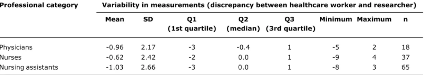
Physicians -0.96 2.17 -3 -0.4 1 -5 2 18
Nurses -0.62 2.42 -2 0.0 1 -9 4 37
Nursing assistants -1.03 2.66 -3 0.0 1 -8 3 65
Measured by Measurement (cm H2O)
Mean SD Minimum Maximum
Healthcare workers 22.2 5.8 8 34
Evaluators 23.1 6.5 10 32
Variability -0.89 2.51 -9 4
Table 1 - Variability in the establishment of the midaxillary line as external reference point for central venous pressure measurements between healthcare workers and evaluators
SD = standard deviation. Paired t test: p < 0.001.
Table 2 - Variability in the establishment of the midaxillary line as external reference point for central venous pressure measurements between healthcare workers and evaluators according to professional category
SD = standard deviation. Kruskal-Wallis test: p = 0.778.
square test was used to determine the association between categorical variables, and the ANOVA and the Kruskal-Wallis test were used for a comparative analysis. The paired t test was used for the mean variation in each group studied. Statistical significance was established at 5%.
Results
A total of 144 descriptions of MAL as ERP were obtained, 120 of which were identified by healthcare workers, whereas 24 were made by the two evaluators.
The ages of the 13 children who participated in the study averaged 4.6 years (minimum of 7 months and maximum of 11.2 years) and their weights ranged from 5.8 to 41 kg (mean of 17.2 kg).
Among the 120 determinations of MAL as ERP made by the healthcare workers, 56 (46.7%) were lower than those calculated by the evaluator (range from 0.5 to -9), of which 23 (41.1%) were 1 to 2 cm lower, 20 (16.7%) were concordant (range = 0) and 44 (36.7%) were higher (range from 0.5 to 4), whereas 42 (95.4%) were 1 to 2 cm higher than the measurements obtained by the evaluator. Thirty-five (29.1%) MAL measurements showed a difference greater than 2 cm (adjusted upward or downward).
As shown in Table 1, there was a statistically significant difference between the measurements made by the healthcare workers and by the evaluators (p < 0.001). The difference between the measurements corresponded to 0.89 (±2.51), indicating that the measurements made by healthcare workers were, on average, significantly lower than those obtained by the evaluators.
Of the 44 healthcare workers, 17 (38.6%) were nursing assistants and nursing technicians, and performed 65 MAL measurements; 16 (36.3%) were nurses, who carried out 37 measurements, and 11 (25.1%) were physicians, who performed 18 measurements.
As shown in Table 2, there was no statistically significant difference (p = 0.778) regarding the variability in MAL measurements according to professional category.
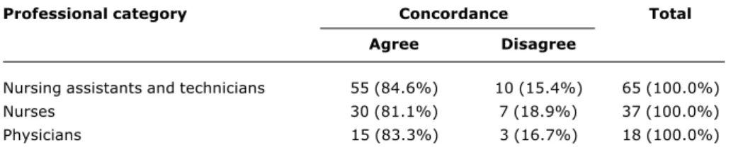
The influence of professional category on the concordance between measurements was further investigated, but no statistically significant difference was observed (p = 0.899), as shown in Table 3.
With regard to working experience, mean length of time since graduation was 6.2 (±7.1) years, and the amount of experience in intensive care averaged 3.5 (±4.9) years. There was statistically significant negative correlation between experience in intensive care and the variation in the measurements (r = -0.26, p = 0.005), showing that, the greater the working experience, the larger the negative discrepancy from the measurements made by the evaluator.
Discussion
Professional category Concordance Total
Agree Disagree
Nursing assistants and technicians 55 (84.6%) 10 (15.4%) 65 (100.0%)
Nurses 30 (81.1%) 7 (18.9%) 37 (100.0%)
Physicians 15 (83.3%) 3 (16.7%) 18 (100.0%)
Table 3 - Concordance for determination of the MAL as ERP for CVP measurement, according to professional category and evaluators
CVP = central venous pressure; ERP = external reference point; MAL = midaxillary line. Chi-square test: p = 0.899.
in the selection and determination of an ERP. Several points are available for this measurement, but the MAL is one of the most widely used ones.17
Given that CVP measurements obtained from different ERPs cannot be compared, the aim of this study was to assess the variability in the indication of the MAL, performed by different healthcare workers.
There was a variation of up to 9 cm in the determination of ERP in a child, suggesting that, even when the same right atrium reference point is used, comparison between CVP measurements is not reliable, due to the failure to correctly determine the MAL, use of different methods for the determination of this site, or positioning of rulers. Therefore, this points out situations that interfere with the efficacy of the procedure, with possible compromise of medical treatment.
No agreement exists in the literature about an acceptable variation in hemodynamic pressure measurement errors, which should then be established by an institutional protocol. In clinical practice, a variation of up to 2 cm (adjusted upward or downward) is acceptable for CVP measurements. However, of 120 measurements made by healthcare workers, 29.1% were greater than 2 cm in relation to the MAL measurements made by the evaluator, showing the necessity for educational intervention to indicate the correct location of the MAL, or the development of technological devices that can help healthcare workers perform more accurate measurements.
A small variation of 1 to 2 cm H2O in CVP measurements in children can bring about remarkable changes in clinical management. Thus, the variability shown in Table 1 indicates possible situations that can affect patient safety, such as inefficient fluid administration, incorrect titration of vasoactive drugs, and the amount of fluids infused. A
study on this issue revealed that the variability in the selection of ERP could result in differences of up to 6 mmHg in CVP measurements in some patients, which would change nursing care and medical treatment.17
There is some statistically significant difference between the measurements made by healthcare workers and those made by the evaluators. This may be related to the fact that the evaluators were trained to take anatomical criteria into account when determining the ERP (midpoint between the anterior and posterior axillary folds measured with a metric tape), whereas healthcare workers determined the MAL by only observing the anatomical region, based on their experience and clinical practice.
Drake20 conducted a similar study, using two methods,
in order to assess the variability in the determination of ERP between ICU nurses. In the first method, the healthcare worker determined the ERP by using his/her clinical experience, describing the method for its determination. In the second method, the nurse used the criteria established by the researcher. There was variability in the determination of ERP regardless of the method used, due to the subjective determination of this point.
No statistically significant difference was found regarding the variability in measurements based on professional category, nor concordance between professional category and the measurements obtained by the evaluators. Nevertheless, when we separately analyzed professional category, according to the amount of experience in PICU, and then compared it to the measurements obtained by the evaluators, we noted that the measurements made by nursing assistants and nursing technicians with greater experience in PICU varied more remarkably, with statistically significant difference. These findings are different from those obtained by Drake,20
References
1. Bigatello LM, George E. Hemodynamic monitoring. Minerva. Anestesiol.2002;68:219-25.
2. Tibby SM, Murdoch IA. Monitoring cardiac function in intensive care. Arch Dis Child. 2003;88:46-52.
3. Blot F, Laplanche A. Accuracy of totally implanted ports, tunnelled, single- and multiple-lumen central venous catheters for measurement of central venous pressure. Intensive Care Med. 2000;26:1837-42.
4. Lough ME. Introduction to hemodynamic monitoring. Nurs Clin North Am.1987;22:89-110.
5. McGee SR. Physical examination of venous pressure: a critical review. Am Heart J.1998; 136:10-7.
6. Potger KC, Elliott D. Reproducibility of central venous pressures in supine and lateral positions: a pilot evaluation of the phlebostatic axis in critically ill patients. Heart Lung.1994;23: 285-99.
7. Sykes MK. Clinical measurement and clinical practice. Anaesthesia. 1992;47:425-32.
8. Baumann UA, Marquis C, Stoupis C, Willenberg TA, Takala J, Jakob SM. Estimation of central venous pressure by ultrasound. Resuscitation. 2005;64:193-9.
9. Tobias JD, Johnson JO. Measurement of central venous pressure from a peripheral vein in infants and children. Pediatr Emerg Care.2003;19:428-30.
10. Hayashi Y, Maruyama K, Takaki O, Yamauchi J, Ohnishi Y, Kuro M. Optimal placement of CVP catheter in paediatric cardiac patients. Can JAnaesth.1995;42:479-82.
11. Pittman JA, Ping JS, Mark JB. Arterial and central venous pressure monitoring. Int Anesthesiol Clin. 2004;42:13-30. 12. Clutton-Brock TH, Hutton P. Central venous and pulmonary
artery catheterization. In: Hutton P, Prys-Roberts C. Monitoring in anesthesia and intensive care. Philadelphia: WB. Saunders; 1994. p. 145-54.
13. Oliveira Filho GR, Bernal REJ, Pivatto SL, Tomasi AT, Soares LF, Helayel PE. A articulação acrômio-clavicular como ponto de referência alternativo para o nível flebostático. Rev Bras Anestesiol.2001;51:511-7.
14. Callow LB, Pieper B. Effect of backrest on central venous pressure in pediatric cardiac surgery. Nurs Res.1989;38:336-8. 15. Seth R, Magner P, Matzinger F, Walraven CV. How far is the sternal angle from the mid-right atrium? J Gen Intern Med. 2002;17:861-5.
16. Knell PJW. Central venous pressure measurement. A device for continuously indicating zero. Anaesthesia.1980;35:991-2. 17. Bartz B, Maroun C, Underhill S. Differences in midanteroposterior
level and midaxillary level in patients with a range of chest configurations. Heart Lung.1988;17:308.
18. Winsor T, Burch GE. Phlebostatic axis and phlebostatic level, reference levels for venous pressure measurements in man. Proc Soc Exp Biol Med. 1945;58:165-9.
19. Keckeisen M. Monitoring pulmonary artery pressure. Crit Care Nurse.2004;24:67-70.
20. Drake JJ. Locating the external reference point for central venous pressure determination. Nurs Res.1974;23:475-82. 21. Debrunner F, Bühler F. Normal central venous pressure,
significance of reference point and normal range. Br Med J. 1969;3:148-50.
the measurements made by healthcare workers with greater experience; that author, though, studied only healthcare workers who had a college degree.
For Courtois,24 the variability in the selection and
determination of ERP is related to the lack of information of healthcare workers about the effect of hydrostatic pressure on the measurements obtained, based on fluid-filled systems. Therefore, sometimes the importance of correct positioning of the transducer or of the identification of the zero point on the water column is overlooked.
Moreover, it is important that the determination of ERP and positioning of the pressure transducer or water column be quickly and easily implemented,24 highlighting
the necessity for standard and specific criteria for the determination of the selected ERP, so that it can be easily and consistently located. Thus, the authors highlight that continuing educational programs in PICU should include this topic, which, albeit simple, considering the numerous technological resources used in the care of severely ill children, can contribute to patient safety b y p r o v i d i n g a r e l i a b l e c o m p a r i s o n b e t w e e n measurements. This allows a more effective assessment of the clinical status of patients, resulting in more appropriate treatment and care.
To provide more accuracy in the determination of an ERP for CVP measurements in children, Belela et al.25 developed a device that helps nurses to determine the phlebostatic level, allowing comparative studies between CVP measurements in children. The device consists of a base positioned on the posterior surface of the patients chest, attached to a ruler with markings, forming a 90-degree angle. A movable board is adapted to this ruler, which, when leaning against the anterior surface of the chest, provides the anteroposterior measurement of the rib cage. The midpoint of this measurement, at the fourth intercostal space, determines the phlebostatic level.25 A
limitation of this study was the necessity to use a larger sample of healthcare workers and to corroborate the concordance between the measurements made by the trained evaluators, indicating whether the use of rulers can or cannot provide greater reliability in the determination of the MAL. Even though it is not within the goals of this study, demonstrating a relationship between age, weight, chest circumference and variability of CVP measurements would allow a more accurate assessment of the clinical importance of such variability, and other technological resources could be used, such as magnetic transthoracic bioimpedance.
Since it is the duty of nurses to reference, zero, measure, and record CVP measurements,6 and once the
tendency of the obtained values is more important than the isolated measurement, all healthcare workers must use the same ERP and the same criteria to determine it, such that the comparison between measurements can
be accurate and reliable, resulting in efficient treatment.20,26
Correspondence: Mavilde L. G. Pedreira.
Rua General Calado, 158/41, Jardim Anália Franco CEP 03334-060 São Paulo, SP Brazil
Tel.: +55 (11) 9709.2429 Fax: +55 (11) 5549.4305 E-mail: [email protected] 22. Rosenberg DI, Moss MM, American College of Critical Care
Medicine of the Society of Critical Care Medicine. Guidelines and levels of care for pediatric intensive care units. Crit Care Med. 2003;32:2117-27.
23. Pedreira MLG, Rocha PK, Kusahara DM, Peterlini MAS, Caravalho WB. Raised decubitus: accuracy of nursing professionals and students estimates of degrees of elevation. IntensiveCareMed. 2003;29(Suppl 1):61.
24. Courtois M, Fattal PG, Kovacs SJ Jr, Tiefenbrunn AJ, Ludbrook PA.Anatomically and physiologically based reference level for measurement of intracardiac pressures. Circulation.1995;92: 1994-2000.
25. Belela ASC, Pedreira MLG, Peterlini MAS, Gentil GC. Dispositivo para localização do nível flebostático para a medida de pressão venosa central em crianças. ScientiaMedica. 2004;14 (Supl. 1): 59.