Assessment of fluid responsiveness in patients
under spontaneous breathing activity
Avaliação da responsividade a volume em pacientes sob
ventilação espontânea
INTRODUCTION
One of the most frequent interventions in an intensive care setting is luid replacement. Recent trials emphasize that excessive volume, given un-necessarily, may be harmful to the patient, and that assessment of volume responsiveness is fundamental for intensivists.(1,2)
Volume responsiveness may be deined as increased systolic volume (SV) with consequent increased cardiac output (CO) from an established volume infusion which would provide better oxygen supply to the tissue. However, this response to volume testing will only take place when both ventricles operate in the ascending phase of the Frank-Starling curve, i.e., in a preload dependence status.(3)
In the last decade, with improved knowledge and practical application of physiology and heart-lung interaction,(4-6) along with critical patient
Fernando José da Silva Ramos1,
Luciano Cesar Pontes de Azevedo2
1. Resident of Intensive Care from Hospital Sírio-Libanês. São Paulo (SP), Brazil.
2. Researcher from the Instituto de Ensino e Pesquisa do Hospital Sírio-Libanês. São Paulo (SP), Brazil; Physician from the Intensive Care Unit of Hospital Sírio-Libanês. São Paulo (SP), Brazil.
ABSTRACT
To assess luid responsiveness in patients under spontaneous breathing activity ventilation remains a challenge for intensive care physicians. Much of the knowledge on heart-lung interac-tions and dynamic indexes of luid responsiveness may not be useful for these patients. Historically, the most frequently used variables to guide luid responsiveness on this population have been the static preload indexes. How-ever, more recently, dynamic indexes from less invasive devices are being of-ten used, even though their usefulness on spontaneously-breathing subjects remains controversial. he purpose of this article was to review evidences on the assessment of luid responsiveness in patients under spontaneous venti-lation. A search in literature showed poor evidence for use of static
vari-ables, such as illing pressures and ventricular end-diastolic volumes. Dy-namic indexes, such as pulse pressure variation and other indexes had not been appropriately tested during spon-taneous ventilation. Favorable results were found with central venous pres-sure variation and with transthoracic echocardiography or transesophageal Doppler dynamic indexes, especially when associated to passive lower limb elevation. We conclude that although central venous pressure variation and echocardiography variables could aid bedside clinicians in assessing luid re-sponsiveness during spontaneous ven-tilation, more studies on this subject are deinitely required
Keywords: Fluid shifts; Fluid therapy/ methods; Blood volume determination/ methods; Stroke volume; Tidal volume; Hemodynamic
Received from Hospital Sírio-Libanês. São Paulo (SP), Brazil.
Author for correspondence:
Luciano César Pontes de Azevedo Hospital Sírio Libanês – Instituto de Ensino e Pesquisa
Rua Cel. Nicolau dos Santos, 69 - Bela Vista
CEP: 01308-060 - São Paulo (SP), Brazil.
monitoring techniques, new volume responsiveness assessment methods were described, called dynamic methods. Described as such are pulse pressure variation (PPV),(7) systolic pressure variation (SPV),(8) systolic
volume variation (SVV),(9) in addition to techniques
using echocardiography to evaluate superior and
infe-rior vena cava collapsibility.(10) he dynamic evaluation
methods have good accuracy to predict luid respon-siveness, with much higher predictive values than static
measurements.(11) However, an important limitation of
these methods is that indexes and measurements were validated for speciic groups of patients under sedation and volume controlled mechanical ventilation, with no respiratory efort and no arrhythmias. Other stud-ies that tried to reproduce these results in diferent set-tings, did not reach the same results.(12-14)
In spontaneous breathing patients, or in those un-der mechanical ventilation with respiratory efort, luid responsiveness assessment still requires additional stud-ies,(15,16) as the current intensive care trend is to
main-tain the patient with the mildest sedation and weaning
from mechanical ventilation as soon as possible.(17)
his review aims to summarize the main evidences on luid responsiveness assessment in the spontaneous breathing patient, didactically dividing the static mea-surement studies from those with dynamic methods.
METHODS
A search was conducted in the Pubmed database us-ing the key words: luid responsiveness, spontaneous breathing, preload and echocardiography. Articles in english deemed relevant for this review were selected.
STATIC VARIABLES
his item will describe the main evidences of luid responsiveness in spontaneous breathing patients as as-sessed by central venous pressure (CVP), pulmonary wedge pressure (PWP), right ventricular end-diastolic volume (RVEDV) and left ventricle end-diastolic vol-ume (LVEDV).
Central Venous Pressure
CVP is the pressure measured in the right atrium or superior vena cava by a central or pulmonary artery catheter, and is one of the most assessed hemodynamic
parameters in an intensive care unit (ICU).(18) he
Sur-viving Sepsis Campaign,(19) a standardization of care to
the septic patient based on the study by Rivers et al.,(20)
recommend that, in the initial management of severe sepsis and septic shock patient, CVP be used as an he-modynamic parameter of volume resuscitation.
Michard & Teboul(11) in a review of luid
respon-siveness in the ICU evaluated ive CVP-related stud-ies. Although this analysis involved both patients under spontaneous breathing and under mechanical ventila-tion, only two of the ive studies had a relationship be-tween low CVP values before volume testing and luid responsiveness.(21,22)
A recent systematic review on CVP selected twenty four trials with total 830 patients, also with a mixed population (spontaneous and mechanical ventilation) concluding that no satisfactory data were available for
use of CVP as luid responsiveness parameter.(23)
A single study performed with healthy patients un-der spontaneous breathing (22) also failed to establish
a relationship between CVP baseline value and luid responsiveness. hese authors could not establish any relation between baseline CVP and volume indexes.
Pulmonary artery wedge pressure
PAWP is measured by a pulmonary artery catheter and tends to reflect left atrial pressure. For a long time it was used as a volume marker. However, recent stud-ies showed that PAWP is a poor predictor of fluid re-sponsiveness and failed to establish a relation between the baseline value and responsiveness to volume
ex-pansion.(11,14) Furthermore, in these studies the
popu-lation was not comprised only of spontaneous breath-ing patients, but included a majority of mechanical ventilation patients.
Kumar et al.,(24) also evaluated the PAWP value in
healthy spontaneous breathing patients, and also in this group no relation between initial PAWP and luid re-sponsiveness was found, nor a relationship to RVEDV or systolic volume.
Ventricular end-diastolic volumes
With development of the pulmonary artery catheter and the possibility of checking the ventricular end-di-astolic volume, RVEDV measurement was believed to become useful to predict the hemodynamic response after a volume expansion. However, few studies were able to correlate baseline RVEDV values and luid re-sponsiveness. Only two trials, conducted by the same group, were able to establish a relation between the ini-tial value and considerable CO increase.(25,26) According
to these studies, a baseline RVEDV value lower than
re-sponsiveness with 64% accuracy, while values above
138 mL/m2 were related to a 100% failure of response.
Criticism to these trials comes from the use of mixed populations and also due to a considerable gap between the values 90-138 mL/m2, where responsiveness to a
volume ratio could not be established.
RVEDV analysis by pulmonary artery catheter and
cardiac scintigraphy(21) did not show a relationship
be-tween baseline values and the luid responsiveness pre-diction. A recent literature review also failed to ind any study favorable to RVEDV use for volume responsive-ness evaluation.(15)
DYNAMIC VARIABLES
When assessing dynamic volume responsiveness in spontaneous breathing patients, evidences related to
CVP variation (∆CVP), PPV and methods using
tran-sthoracic echocardiogram and esophageal doppler will be reviewed.
Central venous pressure variation
he irst studies testing the hypothesis of ∆CVP to
predict volume response in spontaneous breathing pa-tients were issued in the nineties by Magder et al.(27,28)
he rationale is that patients with suicient inspiratory capacity to cause a 2 mmHg reduction of PAWP drop and presenting, in this respiratory cycle, a CVP varia-tion decreasing more than 1 mmHg would be in a state of preload dependence and therefore would be luid responsive. he authors showed that a CVP decrease of more than 1 mmHg has a 77% positive predictive value (PPV) and 81% negative predictive value (NPV) in the identiication of responsive patients. In this trial, patients were in the immediate postoperative period of heart surgery, and under spontaneous breathing or
were ventilator disconnected for ∆CVP measurement.
Furthermore, they were monitored by the pulmonary artery catheter. Only patients with suicient inspira-tory capacity to cause a PWCP decrease above 2 mmHg were included.
However a recent study tried to reproduce the above
mentioned indings, with diferent results.(29) he
pop-ulation studied was under spontaneous breathing or under mechanical ventilation with support pressure,
and the authors assessed CVP, PAWP, ∆CVP and PPV.
Surprisingly, ∆CVP was less accurate than CVP. In this
study a high speciicity was seen for volume responsive subjects when CVP was below 5 mmHg.
Noteworthy are some issues justifying the diferent
results among studies.(30) 1. he last study did not check
the patients’ inspiratory capacity; 2. he patients could
make a respiratory efort, confounding ∆CVP
assess-ment; 3. CVP measurement was made at the middle axillary line, diferent from the irst study where these measurements were performed at a point 5 cm below the sternum, which may bring about an up to 3 mmHg diference on mean CVP value; 4. Assessment of the CVP value may have been performed at a non-appro-priate point of its curve.
Pulse pressure variation
The role of PPV for predicting volume responsive-ness in spontaneous breathing patients is not yet fully understood. Although current data show that this is not a good parameter for volume replacement in this group of patients, three studies on the subject are noteworthy.(29,31,32)
Monnet et al.,(31) evaluated the ability of PPV to
pre-dict luid responsiveness in two groups, one with con-trolled mechanical ventilation and the other with spon-taneous breathing or respiratory efort. PPV was com-pared to aortic blood low variation and passive lower limb elevation (PLLE) and 500 mL saline infusion, to conirm results of volume responsiveness. Among spon-taneous breathing and sinus rhythm patients, PLLE maneuver for PPV assessment had a speciicity of 75% and speciicity of 46% , results below those of aortic blood low variation.
Another study evaluated PPV in patients under pressure support ventilation or ventilation with a face mask, showing a PPV predictive value below CVP and PAWP.(29)
he third and more recent study evaluated 32 spon-taneous breathing patients. Findings were better than the previous, with a sensitivity of 63% and a speciicity of 92% for PPV above 12%. When testing PPV after a forced respiratory cycle, the PPV cutof value increased to 33%, while accuracy was signiicantly decreased. Noteworthy, the responsiveness criterion was an in-crease of over 15% in the cardiac index (CI), identiied by calculating variables reached from aortic
transtho-racic echocardiography Doppler analysis.(32)
ECHOCARDIOGRAPHY AND ESOPHAGEAL DOPPLER VARIABLES
here is a growing interest in this method for volume
dynamic and volume responsiveness assessments.(10)
his test may be conducted by a transesophageal meth-od, using Doppler installed in the esophageal region, and capture aortic low velocity (AFV), or a transtho-racic echocardiogram with data such as diameter, aor-tic area (AA) and aoraor-tic velocity-time integral (VTI), allowing to calculate SV by the formula: SV = VTI x AA.(33) Echocardiography can also be used for volume
evaluation by the superior and inferior vena cava diam-eter variation indexes, however these are only validated for mechanical ventilation patients.(10)
Esophageal Doppler
A rapid diagnosis is fundamental in the ICU, and echocardiography is a very useful tool in this setting. As it is easily performed and not invasive, transthorac-ic echocardiography is the most widely used method. However, in up to 40% of the cases it fails to obtainr appropriate images and data, mainly in obese patients, those with chest wall deformities, subcutaneous em-physema, surgical drains and wounds. Esophageal dop-pler allows good quality register of aortic low velocity along the descending thoracic aorta based on a nomo-gram (taking into account weight, height and age) for aortic area estimation, allowing CO calculation. he probe may remain for some days and allows instant CO measurements.(34) Dark & Singer validated the
esopha-geal doppler as a reliable CO monitoring method for critically ill patients.(35)
An important study that used esophageal Doppler to assess luid responsiveness, considered that an AFV increase above 10% induced by PLLE could predict lu-id responsiveness with a sensitivity of 97% and a speci-icity of 94%. hese sensitivity and specispeci-icity values are higher when compared to PPV in the same patient
group.(31) However, in spontaneous breathing patients,
this method is often not feasible due to the probe size, extremely uncomfortable for many patients.
Transthoracic Echocardiogram
When assessing fluid responsiveness by transtho-racic echocardiography, two studies are worthy of
mention.(36,37) In these, PLLE was also evaluated as for
induction of sufficient hemodynamic changes in the evaluated parameters.
Lamia et al.(36) evaluated 24 spontaneous breathing
patients, 14 of them under pressure support ventila-tion. In this study the authors, in addition to transtho-racic echocardiography assessment regarding SV
chang-es after PLLE, further studied the role of changchang-es in measurements such as left ventricle end-diastolic area versus a volume expansion. he PLLE efect predicts an index of SV above 15% with 77% sensitivity and 100% speciicity. However, analysis of other indexes did not disclose satisfactory values.
he second transthoracic echocardiography study also assessed PLLE efect on SV and CO changes and the ability to detect these changes by transthoracic echocardiography. hirty four patients were recruited, all under spontaneous breathing. A patient was con-sidered responsive if PLLE was able to promote a 12% change on SV or CO. Results of SV changes were a sensitivity of 69% and a speciicity of 89% , while for CO change sensitivity was 63% and speciicity 89%, practically showing equivalence of these variables.(37)
COMMENTS
Identiication of volume responsive patients is dif-icult, especially in those under spontaneous breathing. It is estimated that 40 to 72% of patients have increased
SV when faced with volume expansion.(11) On the other
hand, critical patients receiving excessive luids, unnec-essary at late resuscitation animation stage, may have potentially preventable clinical complications.(38,39)
Many luid responsiveness studies were performed in patients under sedation and controlled mechanical ventilation, so dynamic volume assessment parameters are only accurately validated for this population.(7-11)
Few studies focused on volume assessment in spontane-ous breathing patients.
his review corroborates other studies and re-views,(11,15,16) showing that static measurements of
pres-sures or volumes, are not good predictors of luid re-sponsiveness and should not be used.
Regarding dynamic evaluation methods, PPV is much less accurate when results are compared to those of controlled mechanical ventilation patients,(8,29,31,32)
although a study found satisfactory results when pa-tients made no respiratory efort.(32)
Based on Magder studies,(27,28,30)∆CVP continues to
be the dynamic method with best outcomes, although
the paper by Heenen(29) shows conlicting data. he
technical diferences between the Magder and Heenen studies must be remembered, also that measurements according to the Magder,(27,30) technique may be a
pa-tients have a suicient inspiration to cause a 2 mmHg PWCP decrease. Many patients, especially those with acute respiratory distress cannot be disconnected from the ventilator for measurement.
he use of echocardiography as a luid respon-siveness assessment method appears to be promising, mainly when associated with PLLE. PLLE is advanta-geous because it allows dynamic assessment, avoiding unnecessary luid infusion. Both esophageal Doppler and transthoracic echocardiography appear to be useful tools, accurate for volume assessment in critically ill pa-tients, with the additional advantage that transthoracic echocardiography is non-invasive. he greatest obstacle for these methods is that these devices are not available
full time for many ICUs and they require an appropri-ately trained operator.(40)
hus we can conclude that luid responsiveness as-sessment in spontaneous breathing patients calls for ad-ditional studies, and that current evidence shows that static parameters should be avoided. Furthermore, best
results were found with ∆CVP evaluation and with
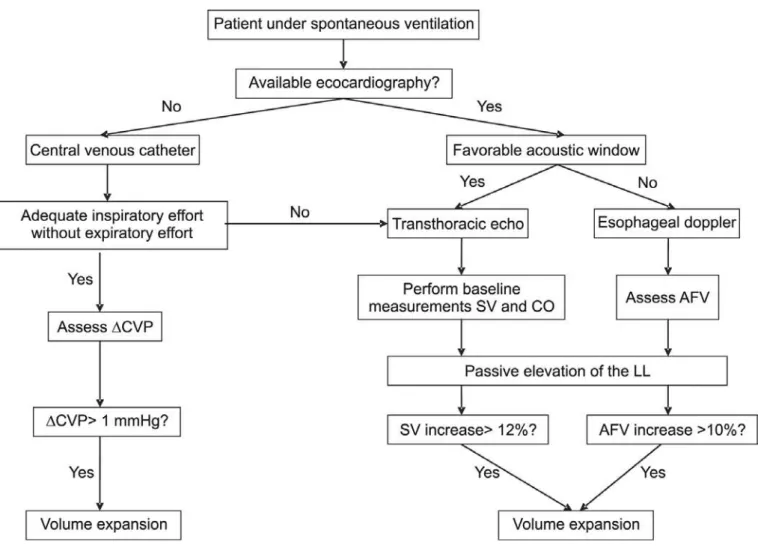
dy-namic variables found with echocardiography or esoph-ageal doppler. Based on these data, we suggest an al-gorithm for volume assessment in spontaneous breath-ing patients (Figure 1). However, before questionbreath-ing whether the patient is or not luid responsive, perhaps it would be more relevant to question if the patient ef-fectively needs volume. Sometimes less can be better.
Figure 1- Algorithm for volume responsiveness for the spontaneous breathing patient.
*he ∆CVP should be assessed in an adequate inspiration of the respiratory cycle, without respiratory efort. In patients with a signiicant expiratory efort, this method is less accurate and not recommended.
RESUMO
A avaliação da responsividade a volume no paciente em venti-lação espontânea representa um desaio para o intensivista. A maior parte dos conhecimentos adquiridos sobre interação coração-pulmão e o cálculo de índices dinâmicos de responsividade a luidos podem não ser adequados para essa avaliação. Historicamente, as variáveis mais frequentemente utilizadas para guiar a responsividade a volume têm sido as medidas estáticas de pré-carga. Mais recentemente, índi-ces dinâmicos obtidos por dispositivos menos invasivos têm sido mais usados, apesar de sua eicácia para esse im em pacientes em ventilação espontânea ainda não ter sido adequadamente estabelecida. O objeti-vo deste estudo foi revisar as principais evidências sobre a avaliação da responsividade a volume nos pacientes em ventilação espontânea. A pesquisa na literatura demonstrou escassez nas evidências para
utiliza-ção de medidas estáticas da volemia como as pressões de enchimento e o volume diastólico inal dos ventrículos. Medidas dinâmicas como variação da pressão de pulso e outros índices também não foram ade-quadamente testados durante a ventilação espontânea. Resultados favoráveis foram obtidos com a variação dinâmica da pressão venosa central e com parâmetros dinâmicos que utilizam o ecocardiograma transtorácico ou doppler esofágico associado à elevação passiva dos membros inferiores. Conclui-se que embora a variação da pressão ve-nosa central e variáveis obtidas com o ecocardiograma transtorácico ou doppler esofágico possam ser úteis na avaliação da responsividade a volume em pacientes sob ventilação espontânea, deinitivamente são necessários mais estudos neste grupo de pacientes.
Descritores: Deslocamentos de luídos; Hidratação/métodos; Determinação do volume sanguíneo/métodos; Volume sistólico; Volume de ventilação pulmonar;Hemodinâmica
REFERENCES
1. National Heart, Lung, and Blood Institute Acute Respira-tory Distress Syndrome (ARDS) Clinical Trials Network, Wiedmann HP, Wheller AP, Bernard GR, hompson BT, Hayden D, deBoisblanc B, Connors AF Jr, Hite RD, Ha-rabin AL. Comparison of two luid-management strategies in acute lung injury. New Engl J Med. 2006;354(24):2564-75. Comment in: ACP J Club. 2006;145(3):69. Can J Ana-esth. 2007;54(1):73-5. N Engl J Med. 2006;355(9):956-7; author reply 958. N Engl J Med. 2006;355(9):957-8; au-thor reply 958. N Engl J Med. 2006;354(24):2598-600. N Engl J Med. 2006;355(11):1175; author reply 1176. N Engl J Med. 2006;355(11):1175; author reply 1176. N Engl J Med. 2006;355(11):1175; author reply 1176. 2. Durairaj L, Schmidt GA. Fluid therapy in resuscitated
sep-sis: less is more. Chest. 2008;133(1):252-63.
3. Guyton AC, Hall JE. Cardiac output, venous return and their regulation. In: Guyton AC, Hall JE. Textbook of me-dical physiology. 11th ed. Philadelphia: Saunders; 2006. p 232-45.
4. Pinsky MR. Heart-lung interactions. Cur Opin Crit Care. 2007;13(5):528-31.
5. Feihl F, Broccard AF. Interactions between respiration and systemic hemodynamics. Part I: basic concepts. Intensive Care Med. 2009;35(1):45-54.
6. Feihl F, Broccard AF. Interactions between respiration and systemic hemodynamics. Part II: practical implications in critical care. Intensive Care Med. 2009;35(2):198-205. 7. Perel A, Pizov R, Cotev S. Systolic blood pressure
varia-tion is a sensitive indicator of hypovolemia in ventilated dogs subjected to graded hemorrhage. Anesthesiology. 1987;67(4):498-502.
8. Michard F, Boussat S, Chemla D, Anguel N, Mercat A, Lecarpentier Y, et al. Relation between respiratory changes in arterial pulse pressure and luid responsiveness in septic
patients with acute circulatory failure. Am J Respir Crit Care Med. 2000;162(1):134-8.
9. De Backer D. Stroke volume variations. Minerva Aneste-siol. 2003;69(4):285-8.
10. Charron C, Caille V, Jardin F, Vieillard-Baron A. Echocar-diographic masurement of fuid responsiveness. Curr Opin Crit Care. 2006;12(3):249-54.
11. Michard F, Teboul JL. Predicting fuid rsponsiveness in ICU patients: a critical analysis of the evidence. Chest. 2002;121(6):2000-8.
12. De Backer D, Heenen S, Piognerelli M, Koch M, Vin-cent JL. Pulse pressure variations to predict luid respon-siveness: inluence of tidal volume. Intensive Care Med. 2005;31(4):517-23. Comment in: Intensive Care Med. 2005;31(4):499-500.
13. Perner A, Faher T. Stroke volume variation does not pre-dict luid responsiveness in patients with septic shock on pressure support ventilation. Acta Anaesthesiol Scand. 2006;50(9):1068-73.
14. Oliveira RH, Azevedo LC, Park M, Schettino GP. Inluen-ce of ventilatory settings on static and functional hemody-namic parameters during experimental hypovolaemia. Eur J Anaesthesiol. 2009;26(1):66-72.
15. Coudray A, Romand JA, Treggiari M, Bendjelid K. Fluid responsiveness in spontaneously breathing patient: a re-view of indexes used in intensive care. Crit Care Med. 2005;33(12):2757-62. Comment in: Crit Care Med. 2006;34(8):2266-7; author reply 2267.
16. Teboul JL, Monnet X. Prediction of volume responsiveness in critically ill patients with spontaneous breathing activi-ty. Curr Opin Crit Care. 2008;14(3):334-9.
author reply 814-5. N Engl J Med. 2000;343(11):814; au-thor reply 814-5.
18. Dias FS, Rezende E, Mendes CL, Réa-Neto A, David CM, Schettino G, et al. Parte II: monitorização hemodinâmica básica e cateter de artéria pulmonar. Rev Bras Ter Intensi-va. 2006;18(1):63-77.
19. Dellinger RP, Levy MM, Carlet JM, Bion J, Parker MM, Jaeschke R, Reinhart K, Angus DC, Brun-Buisson C, Be-ale R, Calandra T, Dhainaut JF, Gerlach H, Harvey M, Marini JJ, Marshall J, Ranieri M, Ramsay G, Sevransky J, hompson BT, Townsend S, Vender JS, Zimmerman JL, Vincent JL; International Surviving Sepsis Campaign Gui-delines Committee; American Association of Critical-Care Nurses; American College of Chest Physicians; American College of Emergency Physicians; Canadian Critical Care Society; European Society of Clinical Microbiology and In-fectious Diseases; European Society of Intensive Care Me-dicine; European Respiratory Society; International Sepsis Forum; Japanese Association for Acute Medicine; Japane-se Society of Intensive Care Medicine; Society of Critical Care Medicine; Society of Hospital Medicine; Surgical In-fection Society; World Federation of Societies of Intensive and Critical Care Medicine. Surviving Sepsis Campaign: international guidelines for management of severe sepsis and septic shock: 2008. Crit Care Med. 2008;36(1):296-327. Erratum in: Crit Care Med. 2008;36(4):1394-6. 20. Rivers E, Nguyen B, Havstad S, Ressler J, Muzzin A,
Kno-blich B, Peterson E, Tomlanovich M; Early Goal-Directed herapy Collaborative Group. Early goal-directed therapy in treatment of severe sepsis and septic shock. N Engl J Med. 2001;345(19):1368-77.
21. Schneider AJ, Teule GJ, Groeneveld AB, Nauta J, Hei-dendal GA, hijs LG. Biventricular performance during volume loading in patients with early septic shock, with emphasis on the right ventricle: a combined hemodyna-mic and radionuclide study. Am Heart J. 1988;116(1 Pt 1):103-12.
22. Wagner JG, Leatherman JW. Right ventricular end-diasto-lic volume as a predictor of the hemodynamic response to a luid challenge. Chest. 1998;113(4):1048-54. Comment in: Chest. 1998;114(4):1226-7.
23. Marik PE, Baram M, Vahid B. Does central venous pressure predict luid responsiveness? A systematic review of the lite-rature and the tale of seven mares. Chest. 2008;134(1):172-8. Comment in: Chest. 2008;134(6):1351-2; author reply 1352-3. Chest. 2008;134(6):1352; author reply 1352-3. 24. Kumar A, Anel R, Bunnell E, Habet K, Zanotti S,
Mar-shall S, et al. Pulmonary artery occlusion and central ve-nous pressure fail to predict ventricular illing volume, car-diac performance, or the response to volume infusion in normal subjects. Crit Care Med. 2004;32(3):691-9. 25. Diebel LN, Wilson RF, Tagett MG, Kline RA.
End-diasto-lic volume. A better indicator of preload in the critically ill. Arch Surg. 1992;127(7):817-21; discussion 821-2.
Com-ment in: Arch Surg. 1993;128(3):358.
26. Diebel L, Wilson RF, Heins J, Larky H, Warsow K, Wilson S. End-diastolic volume versus pulmonary artery wedge pressure in evaluating cardiac preload in trauma patients. J Trauma. 1994;37(6):950-5.
27. Magder S, Georgiadis G, Cheong T. Respiratory variations in right atrial pressure predict the response to luid challen-ge. J Crit Care. 1992;7(2):76-85.
28. Magder S, Lagonidis D. Efectiveness of albumin versus normal saline as a test of volume responsiveness in post-cardiac surgery patients. J Crit Care. 1999;14(4):164-71. 29. Heenen S, De Backer D, Vincent JL. How can the
res-ponse to volume expansion in patients with sponta-neous respiratory movements be predicted? Crit Care. 2006;10(4):R102.
30. Magder S. Predicting volume responsiveness in sponta-neously breathing patients: still a challenging problem. Crit Care. 2006;10(5):165.
31. Monnet X, Rienzo M, Osman D, Anguel N, Richard C, Pinsky MR, Teboul JL. Passive leg raising predicts luid responsiveness in the critically ill. Crit Care Med. 2006;34(5):1402-7. Comment in: Crit Care Med. 2006;34(5):1559-60.
32. Soubrier S, Saulnier F, Hubert H, Delour P, Lenci H, Oni-mus T, et al. Can dynamic indicators help the prediction of luid responsiveness in spontaneously breathing critically ill patients? Intensive Care Med. 2007;33(7):1117-24. Comment in: Intensive Care Med. 2007;33(7):1111-3. 33. Slama M, Maizel J. Echocardiographic measurement of
ven-tricular function. Curr Opin Crit Care. 2006;12(3):241-8. Review.
34. Beaulieu Y. Bedside echocardiography in the assessment of the critically ill. Crit Care Med. 2007; 35(5 Suppl): S235-49.
35. Dark PM, Singer M. he validity of trans-esophageal Dop-pler ultrasonography as a measure of cardiac output in cri-tically ill adults. Intensive Care Med. 2004;30(11):2060-6 36. Lamia B, Ochagavia A, Monnet X, Chemla D, Richard C,
Teboul JL. Echocardiographic prediction of volume respon-siveness in critically ill patients with spontaneously breathing activity. Intensive Care Med. 2007;33(7):1125-32.
37. Maizel J, Airapetian N, Lorne E, Tribouilloy C, Massy Z, Slama M. Diagnosis of central hypovolemia by using pas-sive leg raising. Intenpas-sive Care Med. 2007;33(7):1133-8. 38. Rivers EP. Fluid-management strategies in acute lung
injury – liberal, conservative, or both? N Engl J Med. 2006;354(24): 2598-600. Comment on: N Engl J Med. 2006;354(24):2564-75.
39. Bagshaw SM, Bellomo R. he inluence of volume manage-ment on outcome. Curr Opin Crit Care. 2007;13(5):541-8. 40. De Backer D, Pinsky MR. Can one predict luid