Systematic Review
Revisão Sistemática
ISSN 2317-1782 (Online version)This is an Open Access article distributed under the terms of the Creative Commons Attribution License, which permits unrestricted use, distribution, and reproduction in any medium, provided the original work is properly cited.
Do gender and age influence hard palate
dimensions? A systematic review
O gênero e a idade influenciam as dimensões
do palato duro? Revisão sistemática da literatura
Luana Cristina Berwig
1Mariana Marquezan
1Jovana de Moura Milanesi
1Márlon Munhoz Montenegro
2Thiago Machado Ardenghi
1Ana Maria Toniolo da Silva
1Keywords
Palate, Hard Measures Evaluation Review Gender Age Groups
Descritores
Palato duro Medidas Avaliação Revisão Gênero Grupos Etários
Correspondence address: Luana Cristina Berwig Av. Roraima, 1000, Prédio 26, Sala 1418, Km 9, Camobi, Santa Maria (RS), Brasil. CEP: 97105-900. E-mail: [email protected]
Received: October 11, 2017
Accepted: February 14, 2018
Study conducted at Departamento de Fonoaudiologia, Universidade Federal de Santa Maria – UFSM - Santa Maria (RS), Brasil.
1 Universidade Federal de Santa Maria – UFSM - Santa Maria (RS), Brasil.
2 Faculdade Especializada na área de Saúde do Rio Grande do Sul – FASURGS - Passo Fundo (RS), Brasil. Financial support: nothing to declare.
Conflict of interests: nothing to declare.
ABSTRACT
Purpose: Analyze the influence of gender and age on hard palate dimensions and verify the reference parameters
available in the literature. Research strategies: Two reviewers independently performed a search at the Cochrane Library, PubMed-Medline and Web of Knowledge databases using descriptors according to the syntax rules of each database. Selection criteria: Observational or experimental human studies evaluating the dimensions of the hard palate or maxillary dental arch, with at least one transverse, vertical or sagittal plane measurement, in normal occlusions or class I malocclusions, and comparisons of the dimensions between genders and/or ages. Data analysis: Descriptive analysis with the following subdivisions: design, sample, evaluation instruments,
measurements in millimeters, and statistical analysis. Quality of the included studies was verified by the
Newcastle - Ottawa Quality scale. Results: Eighteen studies were selected and 11 presented results for hard palate or maxillary dental arch dimensions according to gender, six in age and gender and one in age only. Conclusion: The dimensions were larger in males and progressive increase in the measurements was observed from birth to the permanent dentition period.
RESUMO
Objetivo: Analisar a influência do gênero e da idade nas dimensões do palato duro, bem como verificar os
parâmetros de referência disponíveis na literatura. Estratégia de pesquisa: Dois examinadores realizaram a pesquisa de forma independente nas bases de dados Cochrane Library, PubMed-Medline e Web of Knowledge utilizando os descritores de acordo com as regras de sintaxe de cada banco de dados. Critérios de seleção: Estudos
em humanos observacionais ou experimentais, que avaliaram as dimensões do palato duro ou do arco dentário maxilar com pelo menos uma mensuração no plano transversal, vertical ou sagital em oclusões normais ou más oclusões classe I e que realizaram comparações das dimensões entre os gêneros e/ou idades. Análise de
dados: Análise descritiva, seguindo subdivisões: delineamento, amostra, instrumentos de avaliação, medidas em
milímetros e análise estatística. A qualidade dos estudos incluídos foi verificada através da escala “Newcastle -
Ottawa Quality”. Resultados: Foram selecionados 18 estudos. Destes, 11 apresentaram resultados das dimensões do palato duro ou do arco dentário maxilar conforme o gênero, seis em idade e gênero e um somente em idade.
Conclusão: As medidas foram maiores no gênero masculino e houve um aumento progressivo nas dimensões
INTRODUCTION
Orofacial myofunctional evaluation includes a visual and
subjective inspection of the hard palate through anthroposcopic
assessment. Current clinical assessment protocols include
evaluation of width and depth of the hard palate
(1,2), because
the morphology of the structures of the stomatognathic system
are crucial for the correct processing functions of this system
(3).
Although anthroposcopic assessment of the hard palate
is the most frequently used method among pathologists, it
has limitations because of the lack of clinical parameters to
classify width and depth of the hard palate as normal, reduced
or increased.
Current research on orofacial myology is aimed at studying
quantitative methods of evaluation that can complement orofacial
myofunctional clinical examination. In the literature, there are
some resources for quantitative assessment of the hard palate
in research whose objective is to compare the dimensions of
the hard palate between different clinical groups
(4-8)or compare
qualitative and quantitative evaluations of the hard palate
(3,9).
However, in order to make the use of such resources feasible in
clinical practice, knowledge is required of reference parameters
for quantitative analysis of the hard palate according to gender
and age.
Therefore, this systematic review of the literature is relevant
because it seeks to answer the following research questions:
Do age and gender influence hard palate dimensions? What are
the reference parameters of hard palate dimensions?
The objective of this study was to conduct a systematic
review of the literature to evaluate the influence of gender and
age on hard palate dimensions as well as check the reference
parameters available in the literature.
Research strategy
The aim of this systematic review of the literature was to
assess the association between gender and age and hard palate
dimensions. This is not a systematic review of intervention
as described in
the Cochrane Handbook and the PRISMA
statement
. However, the PRISMA guidelines were followed
whenever possible.
Two examiners with knowledge in the field conducted the
research independently (LCB AND MM). They searched for
articles published until June 2017 in the Cochrane Library,
PubMed-Medline and Web of Knowledge.
Only articles published in English were considered. Appropriate
adjustments were made to the keywords to follow the syntax
rules of each database (Table 1).
The two examiners evaluated the titles and abstracts of all
studies they had found. Abstracts with sufficient information
to allow inclusion or exclusion decisions were analyzed in full
prior to the final decision. Articles that had appeared in different
databases were considered only once. Different decisions by
the two researchers were resolved by consensus. The selected
articles were then carefully analyzed for quality assessment,
bias control and data extraction.
The search had to be broadened to include studies that had
performed measurements of the maxillary dental arch, as there
were few papers that had analyzed the hard palate. This inclusion
was made because the hard palate and the maxillary dental
arch are closely related, since they are on the same plane of the
maxilla and have a similar shape.
Selection criteria
This research included experimental or observational studies
conducted with humans which assessed the dimensions of the
hard palate or maxillary dental arch with at least a measurement
in the transverse, vertical or sagittal plane in normal occlusions
or Angle Class I malocclusions and compared such dimensions
with gender and/or age.
Studies were excluded when they had samples with craniofacial
deformities, cleft palate, syndromes, mouth breathing, crossbite,
open bite, and history of orthodontic treatment.
Data analysis
After the selected articles were read in full, the following
data were extracted: name of authors, year of publication,
country where the study was conducted, study design, objective
of the study, characteristics of the sample, instruments used for
measuring the hard palate or maxillary dental arch, description
of the measures undertaken, average of measures in accordance
with gender and/or age and significance value (p-value) when
available.
Table 1. Database and search strategies in use
Database Descriptors
Cochrane Library
http://cochrane.bvsalud.org/portal/php/index.php
(palat* or “dental arch”) and measure* or height or depth or width or dimension) and (Korkhaus or compass or caliper or cone-beam or cast) and (“age groups” or age or sex
or gender dimorphism or not (deformities or airway or cleft or implant).
PubMed-Medline
http://www.ncbi.nlm.nih.gov/pubmed
(palat* or “dental arch”) and measure* or height or depth or width or dimension) and (Korkhaus or compass or caliper or cone-beam or cast) and (“age groups” or age or sex
or gender dimorphism or not (deformities or airway or “Cleft Palate or cleft or implant” or “mini” or miniscrew implant or thickness). Filter was checked for studies using
HUMANS.
Web of Knowledge http://apps.webofknowledge.com
(palat* or “dental arch”) and (measure* or height or depth or width or dimension) and (Korkhaus or compass or caliper or cone-beam or cast) and (“age groups” or age or sex
Quality and risk of bias of the included studies were
assessed by means of the scale “
Newcastle - Ottawa Quality
”,
originally designed for cohort studies
(10), and subsequently
adapted for cross-sectional studies
(11). On the scale, the
score is given in number of stars comprising three domains:
selection, comparability and outcome/result. The maximum
score can be nine points for cohort studies and ten points
for cross-sectional studies. The higher the score achieved,
the greater the internal quality and the lower the risk of bias
in the study.
RESULTS
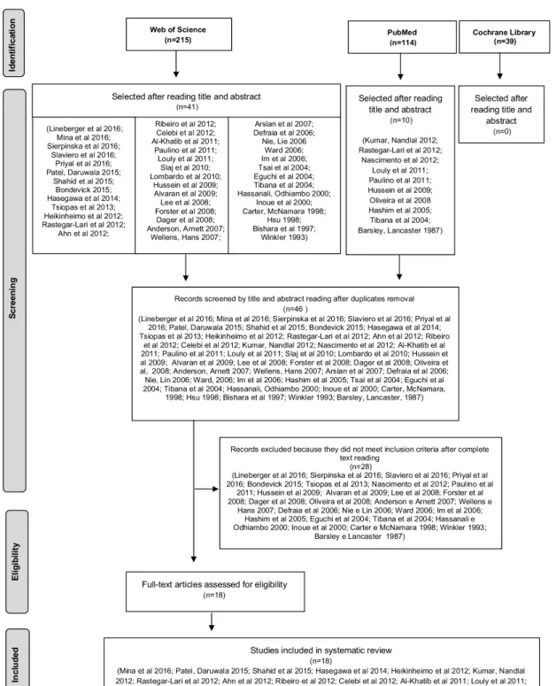
The flowchart shows the results of the searches (Figure 1):
215 studies were found in the database Web of Knowledge,
114 in Pubmed-Medline and 39 in the Cochrane Library.
According to the selection criteria, 46 studies were selected
in accordance with the title and abstract, five of which were
excluded for being duplicated. After the articles were read in
full, 28 were excluded because they did not fulfill the selection
criteria, while 18 studies were included.
In the phase of data extraction from the 18 studies included
in this systematic review, there was variability in the types of
measurements and reference points. Therefore, measurements
were made of the vertical transverse, and sagittal planes, which
are those of greatest interest to speech-language therapy.
To adequately present a summary of the results of the
18 studies in Table 2, standardization was applied to the names
of measures and their appropriate abbreviations with mention
to a point of reference in use (Chart 1). When the reference
points used for measurement were marked on the gums, they
were considered as hard palate dimensions. When the reference
points were marked on the teeth (cusps, grooves or pits), they
were considered as maxillary dental arch dimensions.
The designations of hard palate measures (Chart 1) were
standardized because of the lack of standardization in the use of
nomenclatures for the measurements performed in the 18 studies.
It was found that five studies had termed the measures of the
sagittal plane of the arch as depth
(12,13,24-26), while the other four
had called them length
(15,19,20,23). In four studies, the measure
called “depth” was related to the vertical plane
(19,23,27,28). Thus, we
chose to standardize the measurements of the sagittal plane as
“length” and tjose of the vertical plane as “depth”. The measures
of the transverse plane were called “width” (Chart 1).
All of the included studies performed the measurements based
on plaster casts of the maxillary dental arch. The measurements
were performed directly on the models with a dial or a digital
caliper
(14,15,17-19,20,23,26,29), a dial caliper with a gauge to measure
palate height
(19)or with a three-dimensional Korkhaus compass
(23);
in scanned models, in pictures and copiers, they were measured
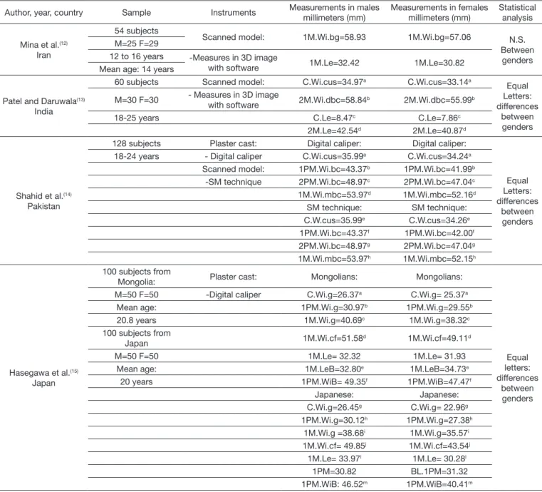
Table 2. Studies comparing hard palate or maxillary dental arch dimensions between genders and/or ages
Author, year, country Sample Instruments Measurements in males millimeters (mm)
Measurements in females millimeters (mm)
Statistical analysis
Mina et al.(12)
Iran
54 subjects
Scanned model: 1M.Wi.bg=58.93 1M.Wi.bg=57.06 N.S. Between
genders M=25 F=29
12 to 16 years -Measures in 3D image
with software 1M.Le=32.42 1M.Le=30.82 Mean age: 14 years
Patel and Daruwala(13)
India
60 subjects Scanned model: C.Wi.cus=34.97a C.Wi.cus=33.14a
Equal Letters: differences
between genders M=30 F=30 - Measures in 3D image
with software 2M.Wi.dbc=58.84
b 2M.Wi.dbc=55.99b
18-25 years C.Le=8.47c C.Le=7.86c
2M.Le=42.54d 2M.Le=40.87d
Shahid et al.(14)
Pakistan
128 subjects Plaster cast: Digital caliper: Digital caliper:
Equal Letters: differences
between genders 18-24 years - Digital caliper C.Wi.cus=35.99a C.Wi.cus=34.24a
Scanned model: 1PM.Wi.bc=43.37b 1PM.Wi.bc=41.99b
-SM technique 2PM.Wi.bc=48.97c 2PM.Wi.bc=47.04c
1M.Wi.mbc=53.97d 1M.Wi.mbc=52.16d
SM technique: SM technique: C.W.cus=35.99e C.W.cus=34.26e
1PM.Wi.bc=43.37f 1PM.Wi.bc=42.00f
2PM.Wi.bc=48.97g 2PM.Wi.bc=47.04g
1M.Wi.mbc=53.97h 1M.Wi.mbc=52.15h
Hasegawa et al.(15)
Japan
100 subjects from
Mongolia: Plaster cast: Mongolians: Mongolians:
Equal letters: differences
between genders M=50 F=50 -Digital caliper C.Wi.g=26.37a C.Wi.g= 25.37a
Mean age: 1PM.Wi.g=30.97b 1PM.Wi.g=29.55b
20.8 years 1M.Wi.g=40.69c 1M.Wi.g=38.32c
100 subjects from
Japan 1M.Wi.cf=51.58d 1M.Wi.cf=49.11d M=50 F=50 1M.Le= 32.32 1M.Le= 31.93
Mean age: 1M.LeB=32.80e 1M.LeB=34.73e
20 years 1PM.WiB= 49.35f 1PM.WiB=47.47f
Japanese: Japanese: C.Wi.g=26.45g C.Wi.g= 22.96g
1PM.Wi.g=30.12h 1PM.Wi.g=27.38h
1M.Wi.g =38.68i 1M.Wi.g=35.57i
1M.Wi.cf= 49.85j 1M.Wi.cf=43.54j
1M.Le= 33.97l 1M.Le= 30.28l
1PM=30.82 BL.1PM=31.32 1PM.WiB: 46.52m 1PM.WiB=40.41m
Author, year, country Sample Instruments Measurements in males millimeters (mm)
Measurements in females millimeters (mm)
Statistical analysis
Ahn et al.(16) South Korea
66 subjects Scanned model: C.Wi.cus: C.Wi.cus:
Equal letters: differences
between genders
M=16 F=50 - Measures in 3D image
with software 6 years =32.33 6 years =32.11
6-14 years 8 years =34.33 8 years =33.97
9 years =32.92 9 years =33.69
10 years =33.77 10 years =35.14
11 years =33.53
C.Wi.cus: C.Wi.cus:
8 years =34.30
9 years =39.58 9 years =35.14
10 years =38.36a 10 years =35.78a
11 years =37.45 11 years =35.85
12 years =37.30b 12 years =35.75b
13 years =37.24c 13 years =35.70c
14 years =37.15d 14 years =35.58d
Celebi et al.(17) Turkey
142 subjects: Plaster cast: 1PM.Wi.cg=35.95 1PM.Wi.cg=35.56 N.S.
Between genders
M=64 F=78 -Digital caliper 1M.Wi.cf=46.25 1M.Wi.cf=45.18
14-15 years
Heikinhei-mo et al.(18) Finland
33 subjects: Plaster cast: C.Wi.cus: C.Wi.cus:
Descriptive analysis
M=15 F=18 -Digital caliper 7 years =32.49 7 years =32.51
Assessed at 7, 10, 12,
15 and 32 years 10 years =33.89 10 years =33.52
12 years =35.76 12 years =33.73
15 years =35.85 15 years =34.16
32 years =35.41 32 years =33.88
C.Wi.g: C.Wi.g:
7 years =25.70 7 years =25.63
10 years =26.26 10 years =26.32
12 years =26.02 12 years =24.96
15 years =25.66 15 years =24.95
32 years =24.91 32 years =24.54
1PM.Wi.lc: 1PM.Wi.lc:
7 years =31.42 7 years =31.25
10 years =31.67 10 years =31.83
12 years =31.44 12 years =31.04
15 years =31.68 15 years =30.99
32 years =31.04 32 years =30.81
2PM.Wi.lc: 2PM.Wi.lc:
7 years =35.93 7 years =35.34
10 years =36.22 10 years =35.95
12 years =36.67 12 years =36.32
15 years =36.70 15 years =35.92
32 years =35.91 32 years =35.67
1M.Wi.mlc: 1M.Wi.mlc:
7 years =40.26 7 years =39.87
10 years =41.10 10 years =40.72
12 years =41.71 12 years =41.14
15 years =41.89 15 years =40.94
32 years =41.03 32 years =40.24
1M.Wi.dlc 1M.Wi.dlc
7 years =41.80 7 years =40.97
10 years =42.94 10 years =42.29
12 years =43.09 12 years =42.79
15 years =43.41 15 years =42.41
32 years =42.99 32 years =41.98
1M.Wi.g: 1M.Wi.g:
7 years =32.96 7 years =32.96
10 years =33.76 10 years =33.95
12 years =34.72 12 years =34.70
15 years =35.37 15 years =34.78
32 years =35.31 32 years =34.72
Author, year, country Sample Instruments Measurements in males millimeters (mm)
Measurements in females millimeters (mm)
Statistical analysis
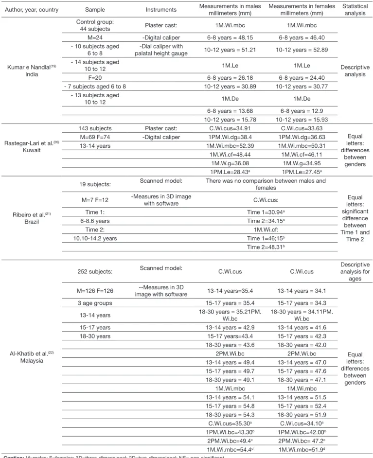
Kumar e Nandlal(19)
India
Control group:
44 subjects Plaster cast: 1M.Wi.mbc 1M.Wi.mbc
Descriptive analysis M=24 -Digital caliper 6-8 years = 48.15 6-8 years = 46.40
- 10 subjects aged 6 to 8
-Dial caliper with
palatal height gauge 10-12 years = 51.21 10-12 years = 52.89 - 14 subjects aged
10 to 12 1M.Le 1M.Le
F=20 6-8 years = 26.18 6-8 years = 24.40 - 7 subjects aged 6 to 8 10-12 years = 30.89 10-12 years = 30.77
- 13 subjects aged
10 to 12 1M.De 1M.De
6-8 years = 13.68 6-8 years = 12.9 10-12 years = 15.78 10-12 years = 15.93
Rastegar-Lari et al.(20)
Kuwait
143 subjects Plaster cast: C.Wi.cus=34.91 C.Wi.cus=33.63
Equal letters: differences
between genders M=69 F=74 -Digital caliper 1PM.Wi.dg=38.4 1PM.Wi.dg=36.63
13-14 years 1M.Wi.mbc=52.39 1M.Wi.mbc=50.31 1M.Wi.cf=48.44 1M.Wi.cf=46.11
1M.W.g=36.08 1M.W.g=34.95 1PM.Le=28.43a 1PM.Le=27.45a
Ribeiro et al.(21)
Brazil
19 subjects: Scanned model: There was no comparison between males and females
Equal letters: significant difference between Time 1 and
Time 2 M=7 F=12 -Measures in 3D image
with software C.Wi.cus:
Time 1: Time 1=30.94a
6-8.6 years Time 2=34.15a
Time 2: 1M.Wi.cf:
10.10-14.2 years Time 1=46;15b
Time 2=48.31b
Al-Khatib et al.(22)
Malaysia
252 subjects: Scanned model: C.Wi.cus C.Wi.cus
Descriptive analysis for
ages
M=126 F=126 --Measures in 3D
image with software 13-14 years=35.4 13-14 years = 34.1
Equal letters: differences
between genders 3 age groups 15-17 years = 35.4 15-17 years = 34.3
13-14 years 18-30 years = 35.21PM. Wi.bc
18-30 years = 34.11PM. Wi.bc 15-17 years 13-14 years = 42.9 13-14 years = 41.6 18-30 years 15-17 years=43.4 15-17 years = 42.3 18-30 years = 43.6 18-30 years = 42.0
2PM.Wi.bc 2PM.Wi.bc 13-14 years = 49.4 13-14 years = 47.0 15-17 years = 49.7 15-17 years = 47.6 18-30 years = 49.1 18-30 years = 47.1
1M.Wi.mbc 1M.Wi.mbc 13-14 years = 54.1 13-14 years = 51.5 15-17 years = 54.8 15-17 years = 52.4 18-30 years = 54.3 18-30 years = 51.9 C.Wi.cus=35.30a C.Wi.cus=34.10a
1PM.Wi.bc=43.30b 1PM.Wi.bc=42.00b
2PM.Wi.bc=49.4c 2PM.Wi.bc= 47.2c
1M.Wi.mbc=54.4d 1M.Wi.mbc=51.9d
Author, year, country Sample Instruments Measurements in males millimeters (mm)
Measurements in females millimeters (mm)
Statistical analysis
Louly et al.(23)
Brazil
66 subjects Plaster cast: C.Wi.cus: C.Wi.cus:
Equal letters: differences
between genders M=29 F=37 -Digital caliper 9 years =27.99 9 years =27.04
9-12 years -Three-dimensional
Korkhaus compass 10 years =26.72 10 years =26.73 11 years =26.56 11 years =26.91 12 years =26.51 12 years =25.78
1PM.Wi.cg: 1PM.Wi.cg 9 years =36.89 9 years =35.89 10 years =35.28 10 years =36.06 11 years =35.57 11 years =36.14 12 years =36.48 12 years =36.47
1M.Wi.cg: 1M.Wi.cg: 9 years =48.06 9 years = 45.74 10 years =46.69 10 years =48.05 11 years =47.14 11 years =48.47 12 years =48.93 12 years =47.61
2M.Wi.cf: 2M.Wi.cf: 10 years =50.70 10 years =52.16 11 years =52.02 11 years =52.98 12 years =53.44 12 years =53.56 Max.D.o:9 years =11.0 Max.D.o:9 years: 9.40
10 years =11.71a 10 years: 9.72a
11 years =11.0 11 years: 10.84 12 years =12.20 12 years: 10.87
1M.Le: 1M.Le: 9 years =40.00 9 years =38.40 10 years =39.00 10 years =39.36 11 years =39.05 11 years =39.76 12 years: 40.45 12 years =39.87
C.Le: C.Le: 9 years =14.50 9 years =13.30 10 years =13.57 10 years =14.09 11 years =14.27 11 years =14.63 12 years =15.37 12 years =15.12
1M.Le-C.Le: 1M.L-C.Le: 9 years =25.5 9 years =25.10 10 years =24.42 10 years =25.27 11 years =24.77 11 years =25.13 12 years =25.08 12 years =24.75
Lombardo et al.(24)
Italian and Spain
58 southern European
subjects: Scanned model: C.Wi.g=27.1 C.W.g=26.6
N.S. Between
genders M=21 F=37 -Measures in 2D image
with software 1M.Wi.g=36.7 1M.W.g=36.9 19-70 years 2M.Wi.g=42.2 2M.W.g=42.4 C.Le.g= 6.9 C.Le.g=6.6 1M.Le.g=29.3 1M.L.g=28.9 2M.Le.g=38.5 2M.Le.g=42.1
Slaj et al.(25)
Croatia
43 subjects Scanned model: C.Wi.buc= 37.51 C.Wi.cus=36.38
Descriptive analysis Angle Class I -Measures in 3D image
with software 1M.Wi.buc=56.22 1M.Wi.buc=54.37 M=19 F=24 1M.Le=9.50 1M.Le=8.59 15-18 years 1M.L =32.48 1M.L =30.96
Arslan et al.(26)
Turkey
65 subjects Plaster cast: C.Wi.cus=31.97 C.Wi.cus=31.29 Equal
letters: differences
between genders M=29 F=36 - Digital caliper 1PM.Wi.cg=35.55a 1PM.Wi.cg=34.43a
Mean age: 1M.Wi.cf=45.84b 1M.Wi.cf=44.15b
M= 9.44 years 1M.Le=28.16 1M.Le=26.88 F=9.74 years
Author, year, country Sample Instruments Measurements in males millimeters (mm)
Measurements in females millimeters (mm)
Statistical analysis
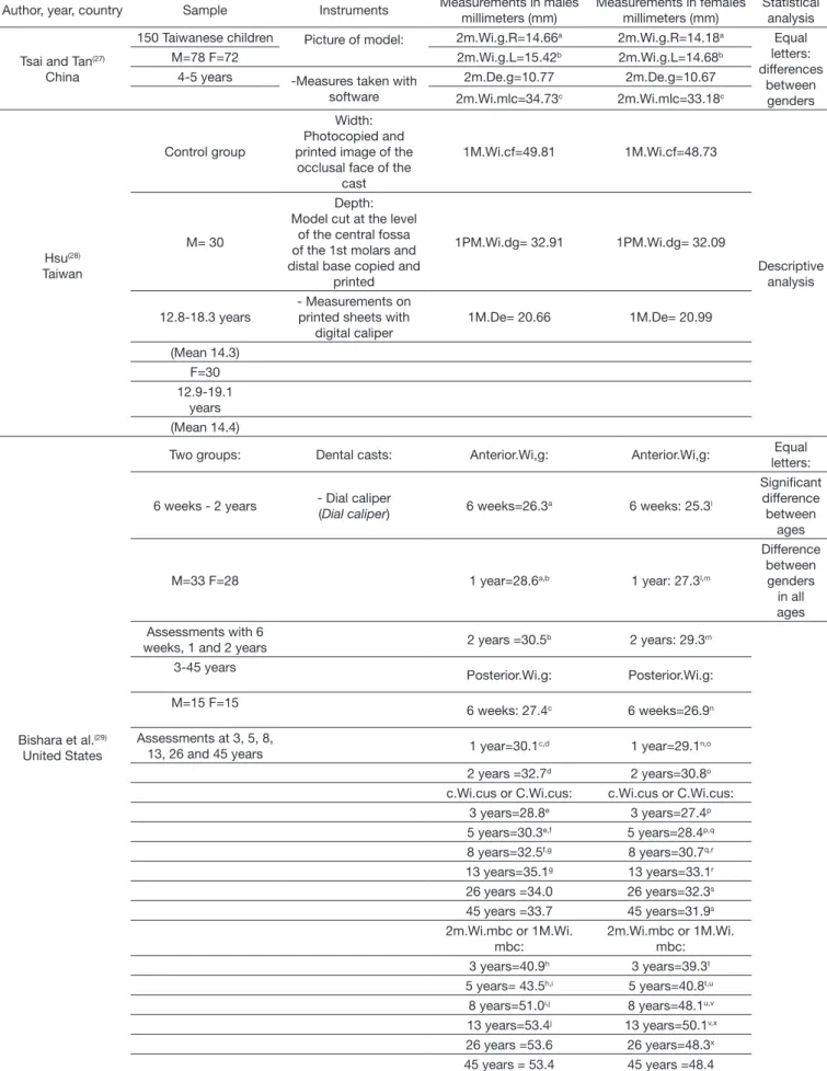
Tsai and Tan(27)
China
150 Taiwanese children Picture of model: 2m.Wi.g.R=14.66a 2m.Wi.g.R=14.18a Equal
letters: differences
between genders M=78 F=72 2m.Wi.g.L=15.42b 2m.Wi.g.L=14.68b
4-5 years -Measures taken with software
2m.De.g=10.77 2m.De.g=10.67
2m.Wi.mlc=34.73c 2m.Wi.mlc=33.18c
Hsu(28)
Taiwan
Control group
Width: Photocopied and printed image of the
occlusal face of the cast
1M.Wi.cf=49.81 1M.Wi.cf=48.73
Descriptive analysis M= 30
Depth: Model cut at the level
of the central fossa of the 1st molars and distal base copied and
printed
1PM.Wi.dg= 32.91 1PM.Wi.dg= 32.09
12.8-18.3 years
- Measurements on printed sheets with
digital caliper
1M.De= 20.66 1M.De= 20.99
(Mean 14.3) F=30 12.9-19.1
years (Mean 14.4)
Bishara et al.(29)
United States
Two groups: Dental casts: Anterior.Wi,g: Anterior.Wi,g: Equal letters:
6 weeks - 2 years - Dial caliper
(Dial caliper) 6 weeks=26.3
a 6 weeks: 25.3l
Significant difference between
ages
M=33 F=28 1 year=28.6a,b 1 year: 27.3l,m
Difference between genders
in all ages Assessments with 6
weeks, 1 and 2 years 2 years =30.5b 2 years: 29.3m 3-45 years
Posterior.Wi.g: Posterior.Wi.g:
M=15 F=15
6 weeks: 27.4c 6 weeks=26.9n
Assessments at 3, 5, 8,
13, 26 and 45 years 1 year=30.1c,d 1 year=29.1n,o 2 years =32.7d 2 years=30.8o
c.Wi.cus or C.Wi.cus: c.Wi.cus or C.Wi.cus: 3 years=28.8e 3 years=27.4p
5 years=30.3e,f 5 years=28.4p,q
8 years=32.5f,g 8 years=30.7q,r
13 years=35.1g 13 years=33.1r
26 years =34.0 26 years=32.3s
45 years =33.7 45 years=31.9s
2m.Wi.mbc or 1M.Wi. mbc:
2m.Wi.mbc or 1M.Wi. mbc: 3 years=40.9h 3 years=39.3t
5 years= 43.5h,i 5 years=40.8t,u
8 years=51.0i,j 8 years=48.1u,v
13 years=53.4j 13 years=50.1v,x
26 years =53.6 26 years=48.3x
45 years = 53.4 45 years =48.4
Chart 1. Description of the measurements made in the studies, standardization of the nomenclature and abbreviation for data extraction
Author year
Description of measurements in the maxillary dental arch according to the selected papers
Standardized nomenclature and abbreviations used in this Review
Mina et al.
(12)
1. Intermolar width: distance between the buccal grooves of the 1st permanent molars.
2. Molar depth: distance between the contact point of the central incisors and the line connecting the 1st permanent molars.
1. Width of the maxillary 1st molar between the buccal grooves (1M.Wi.bg).
2. 1st molar length: (1M.Le)
Patel and Daruwala
(13)
1. Intercanine width: distance between canine cusps.
2. Width between 2nd molars: distance between the distobuccal cusps of the 2nd molars.
3. Canine depth: distance between the contact point of the central incisors and the line connecting the canine cusps.
4. 2nd molar depth: distance between the contact point of the central incisors and the line connecting the distobuccal cusps of the 2nd molars.
1. Maxillary canine width between cusps (C.Wi.cus). 2. Maxillary 2nd molar width between the distobuccal cusps (2M.Wi.dbc).
3. Maxillary canine length (C.Le). 4. Maxillary 2nd molar length (2M.Le).
Shahid et al.
(14) 1. Intercanine width: distance between canine cusps.
2. Length between 1st premolars: distance between the buccal cusps of the 1st premolars.
3. Length between 2nd premolars: distance between the buccal cusps of the 2nd premolars.
4. Width between 1st molars: distance between the mesiobuccal cusps of the 1st molars.
1. Maxillary canine width between cusps (C.Wi.cus). 2. Width of the 1st maxillary premolar between the buccal cusps (1PM.Wi.bc).
3. Width of the 2nd maxillary premolar between the buccal cusps (2PM.Wi.bc).
4. Width of the 1st molar maxillary between the mesiobuccal cusps (1M.Wi.mbc).
Hasegawa et al.
(15)
1. Intercanine lingual: distance between the canines at the intersection of the gingival rim and the long axis of the tooth.
2. Interpremolar lingual: distance between the 1st premolars at the intersection of the gingival rim and the long axis of the tooth.
3. Intermolar lingual: distance between 1st molars at the gingival level of the lingual groove.
4. Intermolar central: distance between the central fossa of the 1st molars. 5. Coronal arch length: measured between the most anterior point of the gingiva in the mesial contact area of the central incisors and the most distal point of the 1st molars.
6. Basal arch length: measured between the distal point of the 1st molars and the most anterior point of the basal arch.
7. Basal arch width: measured between the most concave point of the basal bone in the area of the 1st premolars.
1. Canine width between points at the gingival level (C.Wi.g).
2. Width of the 1st premolar between points at the gingival level (1PM.Wi.g).
3. Width of the 1st molar between points at the gingival level (1M.Wi.g).
4. Width of the maxillary 1st molar between the central fossa (1M.Wi.cf).
5. 1st molar length (1M.Le). 6. Basal arch length (1M.LeB). 7. Basal length (1PM.WiB).
Ahn et al.
(16)
1. Intercanine width: distance between crown cusps of deciduous or permanent canines.
1. Maxillary canine width between the cusps of deciduous canines (c.Wi.cus).
2. Maxillary canine width between permanent canine cusps (C.Wi.cus).
Celebi et al.
(17)
1. Interpremolar Width: distance between the central grooves of the 1st premolars.
2. Intermolar Width: distance between the central fossa of the 1st molars.
1. Width of the 1st maxillary premolar between the central grooves (1PM.Wi.cg).
2. Width of the maxillary 1st molar between the central fossae (1M.Wi.cf).
Heikinheimo et al.
(18)
1. Intercanine width:
a) distance between the canine cusps;
b) distance between the canines measured through the intersection of the gingival rim and the long axis of the tooth.
2. Inter-bicuspid width of the 1st premolar: distance between the lingual cusps of the 1st premolars;
3. Inter-bicuspid width of the 2nd premolar: distance between the lingual cusps of the 2nd premolars;
4. Intermolar Width:
a) distance between the mesiolingual cusps; b) distance between the distolingual cusps;
c) distance from the gingival rim to the level of the mesiolingual cusps.
1. Maxillary canine width: a) between the cusps (C.Wi.cus). b) at the gingival level (C.Wi.g).
2. Width of the 1st maxillary premolar between the lingual cusps (1PM.Wi.lc).
3. Width of the 2nd maxillary premolar between the lingual cusps (2PM.Wi.lc).
Author year
Description of measurements in the maxillary dental arch according to the selected papers
Standardized nomenclature and abbreviations used in this Review
Kumar and Nandlal
(19)
1. Intermolar width: maximum rectilinear distance between the tips of the mesiobuccal cusps of the 1st molars.
2. Maxillary arch length: distance from the line connecting the 1st molars to the labial surface of the central incisors.
3. Palate depth: from the line corresponding to the intermolar distance to the palate.
1. Width of the maxillary 1st molar between the mesiobuccal cusps (1M.Wi.mbc).
2. 1st molar length (1M.Le).
3. Depth of the maxillary 1st molar (1M.De).
Rastegar
-Lari et al.
(20) 1. Width between canines: distance between canine cusps.
2. Width between the 1st premolars: distance between the distal groove of the 1st premolar.
3. Width between the 1st molars:
a) distance between the mesiobuccal cusps of the 1st molars; b) distance between the central fossa of the 1st molars;
c) distance between the midpoint of the lingual faces of the 1st molars. 4. Arch length: from the point of contact between the central incisors to the line connecting the mesial point of the 1st premolars.
1. Maxillary canine width between cusps (C.Wi.cus). 2. Width of the maxillary 1st premolar between the distal grooves (1PM.Wi.dg).
3. Width of the maxillary 1st molar (1M.Wi): a) between the mesiobuccal cusps (1M.Wi.mbc); b) between the central fossa (1M.Wi.cf); c) at the gingival level (1M.Wi.g).
4. Length of the maxillary 1st premolar (1PM.Le).
Ribeir
o et al.
(21)
1. Intercanine width: distance between canine cusps.
2. Intermolar width: distance between the midpoint of the mesiopalatal, distopalatal, mesiobuccal and distobuccal cusps.
1. Maxillary canine width between cusps (C.Wi.cus). 2. Maxillary 1st molar width between the central fossa (1M.Wi.cf).
Al-Khatib et
al.
(22) 1. Intercanine distance: distance between canine cusps.
2. Inter-1st premolar distance: distance between the buccal cusps of the 1st premolars.
3. Inter-2nd premolar distance: distance between the buccal cusps of the 2nd premolars.
4. Intermolar distance: distance between the mesiobuccal cusps of the 1st molars.
1. Maxillary canine width between cusps (Wi.Ca.cus). 2. Width of the maxillary 1st premolar between buccal cusps (1PM.Wi.bc).
3. Width of the maxillary 2nd premolar between buccal cusps (2PM.Wi.bc).
4. Width of the maxillary 1st molar between the mesiobuccal cusps (1M.Wi.mbc).
Louly et al.
(23)
1. Intercanine width: distance between canine cusps.
2. Inter 1st premolar width: distance between the central grooves of the 1st premolars.
3. Inter 1st molar width: distance between the central grooves of the 1st molars. 4. Inter-2nd molar width: distance between the central grooves of the 2nd molars.
5. Maxillary depth: from the line that connects the occlusal plane to the greater depth of the palate.
6. Total arch length: perpendicular distance from the line connecting the central incisors and the upper point of the palatine raphe to the line that measures the depth at the level of the 1st molars.
7. Length of the anterior arch segment: perpendicular distance from the line connecting the central incisors to the line connecting the distal surfaces of the canines.
8. Length of the posterior arch segment: difference between the total length of the arch and the length of the anterior segment.
1. Maxillary canine width between cusps (C.Wi.cus). 2. Width of the maxillary 1st premolar between the central grooves (1PM.Wi.cg).
3. Width of the maxillary 1st molar between the central grooves (1M.Wi.cg).
4. Width of the maxillary 2nd molar between the central grooves (2M.Wi.cg).
5. Maximum maxillary depth at the level of the occlusal plane (Max.De.o).
6. Maxillary 1st molar length (1M.Le). 7. Maxillary canine length (C.Le).
8. Difference between maxillary 1st molar length and maxillary canine length (1M.Le- C.Le).
Lombar
do et al.
(24)
1. Intercanine diameter: distance between the most prominent points in the central axis of the lingual surface of the canine crown.
2. Intermolar diameter at the 1st molar level: distance between the most prominent points of the lingual surface of the 1st molars in the center of the clinical crown.
3. Intermolar diameter at the level of the 2nd molars: distance between the most prominent points of the lingual surface of the 2nd molars in the center of the clinical crown.
4. Canine depth: is the distance between the point between the central incisors and the line that connects the most prominent points in the central axis of the lingual surface of the canine crown.
5. 1st molar depth: the distance of the point between the central incisors and the line that connects the 1st molars.
6. 2nd molar depth: the distance of the point between the central incisors and the line that connects the 2nd molars.
1. Canine width between points at the gingival level (C.Wi.g).
2. Width of the 1st molar between points at the gingival level (1M.Wi.g).
3. Width of the 2nd molar between points at the gingival level (2M.Wi.g).
Author year
Description of measurements in the maxillary dental arch according to the selected papers
Standardized nomenclature and abbreviations used in this Review
Slaj et al.
(25)
1. Canine width: distance between the clinical points of the canine brackets.
2. Intermolar width: distance between the clinical points of the 1st molar brackets.
3. Canine depth: distance from the line connecting the clinical points of the canine brackets to the point between the central incisors.
4. Molar depth: distance from the line connecting the clinical points of the 1st molar brackets to the point between the central incisors.
1. Maxillary canine width between the buccal (C.Wi.buc). 2. Maxillary 1st molar width between the buccal (1M.Wi.buc).
3. Maxillary canine length (C.Le). 4. Maxillary 1st molar length (1M.Le).
Arslan et al.
(26)
1. Maxillary canine width: distance between canine cusps.
2. Maxillary pre-molar width: distance between the central grooves of the 1st premolars.
3. Maxillary molar width: distance between the points of the central fossa of the 1st molars.
4. Maxillary arch depth: perpendicular distance from the labial surface of the central incisors to the line between the central fossae of the 1st molars.
1. Maxillary canine width between cusps (Ca.Wi.cus). 2. 1st premolar width between the central grooves (1PM.Wi.cg).
3. Maxillary 1st molar width between the central fossa (1M.Wi.cf).
4. Maxillary 1st molar length (1M.Le).
T
sai and T
an
(27)
1. Right-side palatal width: distance between the cervical point of the right deciduous 2nd molar to the point of the perpendicular line in the palatine raphe.
2. Left-side palatal width: distance between the cervical point of the left deciduous 2nd molar to the point of the perpendicular line in the palatine raphe.
3. Palate depth: distance between the point of the palatine raphe to the line connecting the 2nd deciduous molars to the gingival level.
4. Dental arch width: Distance between the mesiolingual cusps of the upper deciduous 2nd molars.
1. Width of the deciduous 2nd molar from the point to the gingival level on the right side (2m.Wi.g.R) 2. Width of the deciduous 2nd molar from the point to the gingival level on the left side (2m.Wi.g.L)
3. Gingival palatine depth (2m.De.g).
4. Deciduous 2nd molar width between mesiolingual cusps (2m.Wi.mlc).
Hsut
(28)
1. Upper intermolar width: distance between the central fossa of the upper 1st molars.
2. Upper interpremolar width: distance between the distal grooves of the upper 1st premolars.
3. Palate depth: distance from the deepest part of the palate to the line connecting the central fossae of the upper 1st molars.
1. Maxillary 1st molar width between the central fossa (1M.Wi.cf).
2. Maxillary 1st premolar width between the distal grooves (1PM.Wi.dg).
3. Maxillary 1st molar depth (1M.De).
Bishara et al.
(29)
6 weeks - 2 years
1. Anterior maxillary arch width: distance between lateral groove points at the alveolar ridge level.
2. Posterior maxillary arch width: distance between the points of the posterior margin of the gingiva at the alveolar ridge level.
3 - 45 years
1. Intercanine width: distance between the tips of the canine cusps. 2. Deciduous intermolar width: distance between the tips of the mesiobuccal cusps of the 2nd deciduous molars (3 to 5 years). 3. Permanent intermolar width: distance between the tips of the mesiobuccal cusps of the 1st permanent molars for all subsequent ages.
1. Anterior maxillary width between the points at the gingival level (Anterior.Wi.g).
2. Posterior maxillary width between the points at the gingival level (Posterior.Wi.g).
1. Maxillary canine width between cusps (C.Wi.cus). 2. Deciduous 2nd molar width between the mesiobuccal cusps (2m.Wimbc).
3. Permanent 2nd molar width between the mesiobuccal cusps (2M.Wi.mbc).
with software
(12-14,16,21,22,24,25,27); and in printed copies of the
models, measurements were performed with a digital caliper
(28).
Most of the reference points used for measurements used some
anatomical point of the teeth (maxillary dental arch dimensions)
while in only six studies
(15,18,20,24,27,29)at least one measurement
was based on some point in the gingival edge, which allowed
hard palate dimensions to be determined. All studies considered
the transverse plane while making the measurements, while nine
also made measurements on the sagittal plane
(12,13,15,19,20,23-26)and
four, on the vertical plane
(19,23,27,28).
Analysis of hard palate or maxillary dental arch
dimensions according to gender and age
The 18 studies included in this review were published between
1997 and 2016. Thirteen had a cross-sectional design
(12-15,17,19,20,22-25,27,28),
while five had a longitudinal design
(16,18,21,26,29). Eleven studies
reported hard palate or maxillary dental arch dimensions according
to gender
(12-15,17,20,24-28), six according to age and gender
(16,18,19,22,23,29)and only one study showed hard palate dimensions according
to age range
(21)(Table 2).
There were some findings after analysis of information
intended for comparison of hard palate and maxillary dental
arch dimensions as far as gender is concerned (Table 2). A study
made a comparison between boys and girls with deciduous teeth
(children aged 4 to 5 years)
(27)and found that Chinese boys
showed greater maxillary width, as well as higher left and right
maxillary width at the level of the second deciduous molars than
girls, but there was no difference in hard palate depth.
Another study was conducted with a sample with mean age
of nine years for both boys and girls
(26). Boys had significantly
higher maxillary width at the level of the first premolars and
molars, but there was no difference between boys and girls in
maxillary width between the canines and in anteroposterior
maxillary length until the first molars. In the mixed dentition
phase, one of the studies found significantly higher hard palate
in boys only at ten years of age
(23).
The analysis of the studies described in Table 2 showed that
virtually all the means of the measurements performed in the
transverse plane (width), regardless of the point of reference
in use, were higher in males. As regards studies that made a
statistical analysis to compare the values found in the transverse
plane (width) between males and females in the permanent
dentition phase, it was found that five
(13-15,22,29)found a significant
difference between males and females, while four others
(12,17,20,24)showed no difference.
Of the 12 studies performed with subjects in the
permanent dentition phase addressing a comparison between
genders
(12-15,17,18,20,22,24,25,28,29), six made at least one measurement of
the maxillary dental arch on the sagittal plane (length)
(12,13,15,20,24,25).
Five of such studies compared differences in maxillary length
between genders
(12,13,15,20,24).
As regards the results found in these five studies,
Hasegawa et al.
(15)found that hard palate length until the level
of the first molars was significantly greater in Mongolian women
while anteroposterior maxillary length was significantly greater
in Japanese males. Two studies found significantly higher
maxillary length in males
(13,20), while the other two
(12,24)found
no difference.
During measurements on the vertical plane (depth) in
the permanent dentition phase, only one study had measured
maxillary depth of the first molars, and the means were very
similar between genders
(28).
With respect to hard palate or maxillary dental arch dimensions
according to age, most of the studies made only a descriptive
analysis of the results
(18,19,22)or an analytical statistical analysis for
comparison between genders at different ages
(16,23). A statistical
comparison between measurements at different ages was only
made in two studies
(21,29).
The longitudinal study of Ribeiro et al.
(21)found that the
width values of the maxillary dental arch between the canines
and first premolars in the age range of 10 to 14 years were
significantly higher than those of children aged six to eight
years. The difference between the two periods was equal to
3.21 mm in width between the canines and 2.16 mm in width
between the first molars
(21).
Finally, Bishara et al.
(29)made a longitudinal evaluation of
subjects from birth to 45 years of age through measurements
on the transverse plane (width). From six weeks to two years
of age, i.e., from the period of gingival rims until deciduous
dentition, there was an increase of 4.2 mm of the average width
of the anterior hard palate in boys and 4.0 mm in girls, as well as
an increase of 5.3 mm in the width of the posterior hard palate
in boys and 3.9 mm in girls. From three up to 13 years of age,
width of the anterior and posterior maxillary arch has gradually
increased, and there was statistical significance between ages.
From three to five years, average width between the canines
increased by 1.5 mm in boys and 1.0 in girls, and from eight to
13 years, it increased by 2.6 mm and 2.4 mm in boys and girls,
respectively. Average width between the second deciduous molars
from three to five years increased by 2.6 mm in boys and 1.5 mm
in girls, while the average between the first permanent molars
increased by 2.4 mm in boys and 2.0 mm in girls from eight
to 13 years of age. Moreover, in women from 26 to 45 years,
there was a decrease in width between the canines, and from
13 to 26 years, width decreased between the first molars. In the
present study, all the averages of the measurements made at
different ages were significantly higher in males
(29).
Table 3. Internal quality and risk control of bias according to the “Newcastle - Ottawa Quality” Scale
Author Design Selection Comparability Result Total Mina et al.(12) Cross-sectional 4 (10)
Patel and Daruwala(13) Cross-sectional 3 (10)
Shahid et al.(14) Cross-sectional 4 (10)
Hasegawa et al.(15) Cross-sectional 5 (10)
Ahn et al.(16) Longitudinal 5 (9)
Celebi et al.(17) Cross-sectional 5 (10)
Heikinheimo et al.(18) Longitudinal 5 (9)
Kumar and Nandlal(19) Cross-sectional 4 (10)
Rastegar-Lari et al.(20) Cross-sectional 5 (10)
Ribeiro et al.(21) Longitudinal 4 (9)
Al-Khatib et al.(22) Cross-sectional 5 (10)
Louly et al.(23) Cross-sectional 5 (10)
Lombardo et al.(24) Cross-sectional 4 (10)
Slaj et al.(25) Cross-sectional 3 (10)
Arslan et al.(26) Longitudinal 4 (9)
Tsai and Tan(27) Cross-sectional 3 (10)
Hsu(28) Cross-sectional 3 (10)
Bishara et al.(29) Longitudinal 5 (9)
Evaluation of quality and risk of bias
The score for the analysis of internal quality and control
of bias is based on the “
Newcastle - Ottawa Quality
” scale
(Table 3). It ranged between three and five for studies with a
cross-sectional design (maximum of 10 points) and between
four and five in studies with a longitudinal design (maximum
of 9 points). Whereas a higher score represents a better quality
and lower risk of bias, the studies analyzed in this systematic
review had low to intermediate quality.
CONCLUSION
The averages of width measurements of the hard palate and
the dental maxillary arch were higher in males in the majority of
studies, and most of the selected articles found some significant
difference between genders.
In two of the studies that investigated the influence of age
on hard palate or maxillary dental arch dimensions, there was
statistical difference between the measurements according to
age or age range. This suggests the influence of age on the
transverse dimensions of the maxillary dental arch.
A comprehensive analysis of the results showed that, as
expected, the average reference values gradually increased since
birth until approximately the ages between 12 and 15 years, a
period which corresponds to the permanent dentition. There are
some reference values (expressed as average) from convenience
a sample, which limits the extrapolation of these results to
other populations. In addition, values of the measurements
on the transverse plane outnumber those on the vertical and
sagittal planes.
All studies were based on plaster casts, and the measurements
were performed directly on the models, on scanned models
or on printed images. The instruments used for making such
measurements were calipers, a three-dimensional compass
and software.
Reference parameters found for quantitative analysis of
the hard palate according to gender and age, especially in the
Brazilian population, are still scarce. In addition, few studies
so far have used reference points based on the gingival edge, as
well as the vertical and sagittal planes, to make measurements of
the hard palate. As a result, further research should address the
quantitative assessment of the hard palate according to gender and
age on representative samples of Brazilian population. This way,
quantitative evaluation of the hard palate can be possibly used
to support anthroposcopic assessment in clinical practice.
REFERENCES
1. Felício CM, Folha GA, Ferreira CL, Medeiros AP. Expanded protocol of orofacial myofunctional evaluation with scores: validity and reliability. Int J Pediatr Otorhinolaryngol. 2010;74(11):1230-9. http://dx.doi.org/10.1016/j. ijporl.2010.07.021. PMid:20800294.
2. Marchesan IQ, Berretin-Félix G, Genaro KF. MBGR protocol of orofacial myofunctional evaluation with scores. Int J Orofacial Myology. 2012;38:38-77. PMid:23362752.
3. Maria CM. Silva AMTd, Busanello-Stella AR, Bolzan GdP, Berwig LC. Avaliação da profundidade do palato duro: correlação entre o método
quantitativo e qualitativo. Rev CEFAC. 2013;15(5):1292-9. http://dx.doi. org/10.1590/S1516-18462013005000029.
4. Freitas F, Bastos E, Primo L, Freitas V. Evaluation of the palate dimensions of patients with perennial allergic rhinitis. Int J Paediatr Dent. 2001;11(5):365-71. http://dx.doi.org/10.1046/j.0960-7439.2001.00292.x. PMid:11572268. 5. Ghasempour M, Mohammadzadeh I, Garakani S. Palatal arch diameters of
patients with allergic rhinitis. Iran J Allergy Asthma Immunol. 2009;8(1):63-4. PMid:19279362.
6. Berwig LC, Silva AM, Côrrea EC, Moraes AB, Montenegro MM, Ritzel RA. Hard palate dimensions in nasal and mouth breathers from different etiologies. J Soc Bras Fonoaudiol. 2011;23(4):308-14. http://dx.doi. org/10.1590/S2179-64912011000400004. PMid:22231050.
7. Berwig LC, Montenegro MM, Ritzel RA, Silva AMT, Corrêa ECR, Mezzomo CL. Influence of the respiratory mode and nonnutritive sucking habits in the palate dimensions. Braz J Oral Sci. 2011;10(1):42-9. 8. Berwig LC, Silva AMT, Côrrea ECR, Moraes AB, Montenegro MM,
Ritzel RA. Análise quantitativa do palato duro em diferentes tipologias faciais de respiradores nasais e orais. Rev CEFAC. 2012;14(4):616-25. http://dx.doi.org/10.1590/S1516-18462011005000134.
9. Costa TLS, Silva HJ, Cunha DA. Análise qualitativa inter-observadores e avaliação morfométrica do palato duro. Rev CEFAC. 2005;7(3):326-35. 10. Wells G, Shea B, O’Connell D, Peterson J, Welch V, Losos M, et al. The
Newcastle-Ottawa Scale (NOS) for assessing the quality of nonrandomised studies in meta analyses [Internet]. Ottawa: University of Ottawa, 2001 [citado em 2006 Nov 2]. Disponível em: http://www.ohri.ca/programs/ clinical_epidemiology/oxford.htm
11. Herzog R, Álvarez-Pasquin MJ, Díaz C, Del Barrio JL, Estrada JM, Gil Á. Are healthcare workers’ intentions to vaccinate related to their
knowledge, beliefs and attitudes? A systematic review. BMC Public
Health. 2013;13(1):154. http://dx.doi.org/10.1186/1471-2458-13-154. PMid:23421987.
12. Mina M, Borzabadi-Farahani A, Tehranchi A, Nouri M, Younessian F. Mathematical beta function formulation for maxillary arch form prediction in normal occlusion population. Odontology. 2016;105(2):229-36. http:// dx.doi.org/10.1007/s10266-016-0244-7. PMid:27167385.
13. Patel MN, Daruwala NR. Appraisal of dental arch dimension in Gujarati males and females. Adv Hum Biol. 2015;5(3):61-7.
14. Shahid F, Alam MK, Khamis MF, Honda Y, Sugita Y, Maeda H. Geomorphometrics of tooth size and arch dimension analysis by conventional digital caliper and digital stereomicroscope to establish standard norms for the pakistani population. J Hard Tissue Biol. 2015;24(2):155-68. http:// dx.doi.org/10.2485/jhtb.24.155.
15. Hasegawa Y, Amarsaikhan B, Chinvipas N, Tsukada SI, Terada K, Uzuka S, et al. Comparison of mesiodistal tooth crown diameters and arch dimensions between modern Mongolians and Japanese. Odontology. 2014;102(2):167-75. http://dx.doi.org/10.1007/s10266-013-0130-5. PMid:24026430. 16. Ahn JS, Park MS, Cha HS, Song HC, Park YS. Three-dimensional
interpretation of intercanine width change in children: a 9-year longitudinal study. Am J Orthod Dentofacial Orthop. 2012;142(3):323-32. http://dx.doi. org/10.1016/j.ajodo.2012.04.012. PMid:22920698.
17. Celebi AA, Tan E, Gelgor IE. Determination and application of Pont’s Index in Turkish Population. Sci World J. 2012;2012:1-5. http://dx.doi. org/10.1100/2012/494623. PMid:22654616.
18. Heikinheimo K, Nystrom M, Heikinheimo T, Pirttiniemi P, Pirinen S. Dental arch width, overbite, and overjet in a Finnish population with normal occlusion between the ages of 7 and 32 years. Eur J Orthod. 2012;34(4):418-26. http://dx.doi.org/10.1093/ejo/cjr025. PMid:21357654. 19. Kumar SS, Nandlal B. Effects of asthma and inhalation corticosteroids
on the dental arch morphology in children. J Indian Soc Pedod Prev Dent. 2012;30(3):242-9. http://dx.doi.org/10.4103/0970-4388.105018. PMid:23263429.
21. Ribeiro JS, Ambrosio AR, Santos-Pinto A, Shimizu IA, Shimizu RH. Evaluation of transverse changes in the dental arches according to growth pattern: a longitudinal study. Dental Press J Orthod. 2012;17(1):66-73. http://dx.doi.org/10.1590/S2176-94512012000100010.
22. Al-Khatib AR, Rajion ZA, Masudi SM, Hassan R, Anderson PJ, Townsend GC. Tooth size and dental arch dimensions: a stereophotogrammetric study in Southeast Asian Malays. Orthod Craniofac Res. 2011;14(4):243-53. http://dx.doi.org/10.1111/j.1601-6343.2011.01529.x. PMid:22008304. 23. Louly F, Nouer PRA, Janson G, Pinzan A. Dental arch dimensions in the
mixed dentition: a study of Brazilian children from 9 to 12 years of age. J Appl Oral Sci. 2011;19(2):169-74. http://dx.doi.org/10.1590/S1678-77572011000200014. PMid:21552719.
24. Lombardo L, Saba L, Scuzzo G, Takemoto K, Oteo L, Palma JC, et al. A new concept of anatomic lingual arch form. Am J Orthod Dentofacial Orthop. 2010;138(3):260.e1-13, discussion 260-1. http://dx.doi.org/10.1016/j. ajodo.2010.04.022. PMid:20816292.
25. Slaj M, Spalj S, Pavlin D, Illes D, Slaj M. Dental archforms in dentoalveolar Class I, II and III. Angle Orthod. 2010;80(5):919-24. http://dx.doi. org/10.2319/112609-672.1. PMid:20578864.
26. Arslan SG, Kama JD, Sahin S, Hamamci O. Longitudinal changes in dental arches from mixed to permanent dentition in a Turkish population. Am J Orthod Dentofacial Orthop. 2007;132(5):15-21. http://dx.doi.org/10.1016/j. ajodo.2007.06.009. PMid:18005825.
27. Tsai HH, Tan CT. Morphology of the palatal vault of primary dentition in transverse view. Angle Orthod. 2004;74(6):774-9. PMid:15673140. 28. Hsu BS. The nature of arch width difference and palatal depth of the anterior
open bite. Am J Orthod Dentofacial Orthop. 1998;113(3):344-50. http:// dx.doi.org/10.1016/S0889-5406(98)70307-5. PMid:9517728.
29. Bishara SE, Jakobsen JR, Treder J, Nowak A. Arch width changes from 6 weeks to 45 years of age. Am J Orthod Dentofacial Orthop. 1997;111(4):401-9. http://dx.doi.org/10.1016/S0889-5406(97)80022-4. PMid:9109585.
Author contributions
LCB was responsible for data collection, tabulation and analysis and drafting the article; MM was responsible for data collection and analysis and drafting the article; JMM and MMM were responsible for data analysis and drafting the article;
TMA and AMTS gave general advice on the stages of drafting the article and