Anatomical evaluation of the cervical vertebrae of Wistar rats
by means of digital radiographs and its correlation with the
maturation stages of human cervical vertebrae
Roberto Hiroshi Matsui1, Julio Cezar de Melo Castilho2, Luiz César de Moraes3, Mônica Fernandes Gomes4, Kurt Faltin Júnior5, Miriam Yumi Matsui6
Introduction:Biological age is an important parameter for growth and development assessment. It can be evaluated through the observation of radiographic changes in skeletal maturation of cervical vertebrae. Objective:This study aims to: a) verify if there is correlation between growth curve and the stages of bone age of animals used in laborato-ries, by evaluating radiographs of the cervical vertebrae; b) correlate these stages with their correspondents in humans.
Methods: 35 Wistar rats were evaluated for a period of 160 days, starting at day 22nd (weaning), with cross sections for periodic weighing, length measurement and digital radiography. Radiographs of the cervical vertebrae (C2 and C3) were measured by means of a computer program (Radio IMP). Data were submitted to statistical analysis (ANOVA) and Pear-son correlation. Results: Growth spurt was characterized by fast increasing in weight and length. Through ANOVA, differences were observed in the cervical measurements between days 22, 97, 127, 157, 187 and 217 (p <0.001). A high correlation was found between increasing in body length and weight, as well as in cervical vertebrae height (r = 0.86). In-crements in concavities of vertebrae were also observed, similar to humans. Conclusions: There is correlation between body growth and maturation of cervical vertebrae in rats. Despite the continuous development of concavities, it was not possible to clearly identify the 5/6 stages as in studies of cervical vertebrae maturation in humans.
Keywords:Growth and development. Cervical vertebrae. Radiography.
How to cite this article: Matsui RH, Castilho JCM, Moraes LC, Gomes MF, Faltin Júnior K, Matsui MY. Anatomical evaluation of the cervical vertebrae of Wistar rats by means of digital radiographs and its correlation with the matu-ration stages of human cervical vertebrae. Dental Press J Orthod. 2013 July-Aug;18(4):82-8.
Submitted: January 13, 2011 - Revised and accepted: December 03, 2011
Contact address: Roberto Hiroshi Matsui
Av. Santa Catarina, 1825 – CEP: 04378-300 – São Paulo / SP, Brazil E-mail : [email protected]
» The authors report no commercial, proprietary or financial interest in the prod-ucts or companies described in this article.
1 Assistant Professor, Orthodontics and Dentofacial Orthopedics, UNIP. 2 Associate Professor, Diagnosis and Surgery, UNESP.
3 Professor, Diagnosis and Surgery, UNESP.
4 Associate Professor, Oral Pathology, Biosciences and Oral Diagnosis, UNESP. 5 Full Professor, Orthodontics and Dentofacial Orthopedics, UNIP.
6 Master student, Oral Pathology, UNESP.
Introdução:a idade biológica é um parâmetro importante na avaliação do crescimento e desenvolvimento, podendo ser avaliada por meio da observação de alterações na maturação óssea das vértebras. Objetivo: o presente estudo visa descrever e relacionar a curva de crescimento de ratos utilizados em pesquisas laboratoriais com os estágios de idade óssea, avaliados por radiografias de vértebras cervicais, e correlacionar esses estágios com estudos correspondentes em humanos. Métodos: foram avaliados 35 ratos Wistar em um período de 160 dias, iniciando no 22° dia de vida (desma-me), com cortes transversais periódicos para pesagem, medição do comprimento e radiografias digitais. As radiografias das vértebras cervicais (C2 e C3) foram mensuradas por meio de um programa de computador (Radio IMP). Os dados foram submetidos à análise estatística de variância (ANOVA). Resultados: o surto de crescimento caracterizou-se por aumento rápido de peso e comprimento, seguido por um período de crescimento lento e de estabilidade. Uma alta correlação (r = 0,86) foi verificada entre o aumento de peso e o comprimento do corpo, bem como o comprimento das vértebras cervicais. Incrementos nas concavidades das vértebras dos ratos foram observados, semelhantemente aos resultados obtidos em estudos em humanos. Conclusões: existe correlação entre o crescimento corporal de ratos e a maturação das vértebras cervicais. Apesar da detecção de desenvolvimento contínuo de concavidades das vértebras, não foi possível identificar claramente os 5 ou 6 estágios de maturação óssea descritos em seres humanos.
INTRODUCTION
The understanding of facial growth is indispensable to the orthodontic and orthopedic treatment planning. Treatment of skeletal, dental and muscular discrepan-cies should focus on mixed dentition, preferably before completion of second dentition1. Since the development
of structures of stomatognathic system occurs simulta-neously with the body growth,2 the comprehension of
the process of growth as a whole, by studying its mo-ments of peaks and stability, is essential.
Obtaining information concerning growth in-cludes the use of laboratory animals. It would not be the ideal though, to compare the effects of proposed therapies for patients with mixed dentition using adult rats. Ideally, they should be studied in the cor-responding phases of human growth, so that treat-ment effects can be evaluated and compared.
This concern about correlating humans and ani-mals according to age is present in recent studies.3
In order to understand the influence of age on the efficacy of therapies with stem cells, the authors com-pared stem cell markers in younger (4 months) and older (15 months) rats, suggesting correlation be-tween their findings and therapies in elderly patients. Besides the use of chronological age, evaluating biological age of humans and animals undergoing growth is also important to establish reliable correla-tions between them.
The growth process can be assessed by changes in the morphology of different bones during its maturation.4,5
The comparison between chronological age and bone age (by means of lateral cephalometric and car-pal radiographs) shows that chronological age of a person does not always precisely correlate with the bone age. The latter may present himself advanced or delayed in relation to the first.6,7,8
Besides the assessment of cervical vertebrae mat-uration9,10,11 and skeletal maturation of the hand and
wrist,12,13 several other methods for measurement
of age are studied and compared, such as dental age,14 variations in height and weight,4 mandibular
growth15,16 and expression of secondary sexual
char-acteristics,17,18 each of them used according to the
interests of diagnosis.
The cervical vertebrae maturation (CVM) in humans can be classified in five stages (CVM I to V)19 or six
stag-es (initiation, acceleration, transition, deceleration, and
final maturation).20 This evaluation method is
sta-tistically valid and reliable,19,20,21 presenting the same
clinical value to the evaluation of hand and wrist. The inherent advantage of this method would be elimi-nating the need for radiographs other than those that comprise regular orthodontic exams.
Often the stages of growth and development are studied in laboratory animals based on chronologi-cal age.22 There are, however, no studies evaluating
radiographic stages of bone development and its cor-relation with the development of cervical vertebrae during the pubertal growth in humans.
Thus, this study aimed: 1) to observe, by means of digital radiographs, the anatomical stages of maturation of cervical vertebrae 2 and 3 (C2 and C3) of animals (Wistar rats) during growth, correlating bone age with chrono-logical age, weight and length; and 2) compare cervical vertebrae maturation stages in rats and in humans.
MATERIAL AND METHODS Ethical aspects
Approval was obtained from the Ethics Com-mittee of Research Involving Animals, School of Dentistry, UNESP-São José dos Campos, protocol 046/2007-PA/CEP.
Characteristics of the animals
Thirty five Wistar rats (Rattus norvegicus albinus), 20 males and 15 females, were supplied by the vivari-um of the School of Dentistry, Universidade Estadual Paulista Julio de Mesquita Filho, Campus de São José dos Campos - SP.
The study started after weaning of the animals and with the introduction of solid food diet at 22 days after birth. At this phase, their weight was approxi-mately 80-100 grams. The animals were kept in cag-es, each with five rats in light-dark cycle of 12 hours with controlled temperature and humidity and fed with a balanced diet (Labina®, Guabi -
Guabinutrila-bor for rats and mice) and water ad libitum.
The animals were treated with single doses of polyvalent anthelmintic (Zentel®, São Paulo, Brasil)
and multivitamin (Vita Gold Potenciado®, Tortuga
Companhia Zootécnica Agrária, São Paulo, Brasil), 40 drops per liter of solution for 5 days.
Periods of observation
Considering that within 1.5 months, rats reach their puberty, and after 6 months, they reach matu-rity,23 a wide age range was included taking in
con-sideration the need of observation from the initial to the final phase of growing.
All animals were examined 22 days after birth and thereafter, every 14 days until the observation of growth peak. After that, exams were done every 29 days, until the completion of maturity phase and adulthood (160 days), totaling nine acquisitions of ra-diographic images per animal.
Since rats were observed for more than 7 months, the observation period of this studied included: wean-ing, puberty and maturity.
In adulthood, each rat month is approximately equivalent to 2.5 human years.24 It was considered
that main changes are expected to happen during the growth peak, explaining the higher frequency of ex-amination during this period.
Preparation of the animals for X-ray analysis After weighing, the animals were anesthetized with a combination of ketamine chloride (Vetaset®,
Fort Dodge Animal Health Fort Dodge, Iowa, USA) dose of 5 mg/kg, associated with xylazine (Rom-pum®, Bayer S.A. - Saúde Animal, São Paulo, Brazil),
sedative, analgesic and muscle relaxant at a dose of 10 mg/kg, administered intramuscularly.
RADIOGRAPHIC ANALYSIS Protocol of image acquisition of the cervical vertebrae
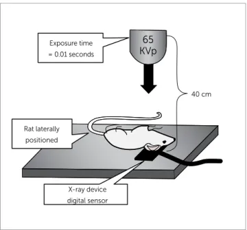
A wooden table, especially made for this purpose, was adapted for the positioning of the animal in a niche excavated to accommodate the sensor (Fig 1). After immobilization and positioning of the rat through this positioner, a digital radiography was obtained using the system Visualix Gx-S-HDI (Gendex Dental Sys-tem, Dentsply International, Chicago, IL, USA). This system captures digital radiographic image by means of a CCD sensor (Charge–Coupled Device), with active area of 20.0 mm (width) x 30.0 mm (length). The X-ray machine used was Gendex 765 DC (Gendex Den-tal X-Ray Division, Dentsply International Inc., IL, USA), set to 65KVp and 7mA. The rats were placed on the sensor positioned parallel to the ground with
the central beam of X-rays directed perpendicular to the table. The focus-object distance was 40 cm, 90 de-gree angle of incidence and duration of exposure to X-rays of 0.01 seconds. The digital radiographic images were stored in JPEG file format.
Anatomical analysis of cervical vertebrae in rats The animals were followed-up until the final phase of growth (160 days). Each animal had a chart and spreadsheet for analysis of the maturation of cer-vical vertebrae (C2 and C3) and other growth indices such as body length, weight, chronological age, for comparison of stages of development and growth.
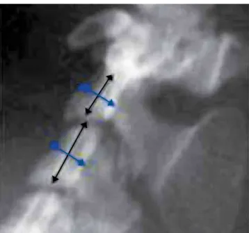
In order to evaluate the growth and anatomical changes of the vertebrae, the diameter, height, width and area were measured and calculated (Fig 2).
The analysis of maturation of cervical vertebrae (C2 and C3) of animals was performed based on the method of Hassel and Farman20 summarized in Table 1
and schematically shown in Figure 3. The following changes were made to this method: it was used digital radiography and the cervical vertebrae was measured with the Radio IMP software (Radiocef).
Analysis of body length and weight of rats The body length was measured by a metal rule (Tramontina, RS, Brazil) in millimeters, from the
Figure 1 - Schematic representation of radiographs taken using the Visualix Gx-S-HDI system.
40 cm Exposure time
= 0.01 seconds
65 KVp
Rat laterally positioned
RESULTS
Sequences of measurements and analysis of gradual length and weight
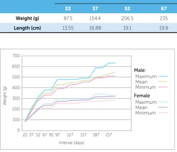
An increase in weight and length was observed for all animals (Tables 2 and 3).
Statistically significant differences were found be-tween males and females in relation to weight. As ex-pected, greater weight was found in males, when compared to females.
Three distinct phases could be observed:
1) Growth spurt from the weaning day to their 10-13th week, when females and males respectively
reached 250% and 350% of their initial weight. 2) Slow growth period: from 10-13th to 27th week.
3) Final growth phase: when males continued to gain weight, while females stabilized (Fig 4).
In relation to length, the first and second phases were very similar to the weight curve. In the growth spurt phase, the animals increased approximately 70% of their initial length. After that, increasing in length became slow and constant.
The final phase of growth was observed when losses or gains in weight were observed and length (Fig 5) was constant. The breeding phase began be-fore the end of the growth in length and weight, cor-responding to a month old.
outer portion of the nose to the beginning of the tail. The animals were weighed on a digital scale (Filizola BP15, Campo Grande, MS) in grams.
Statistical analysis
Normality test (Anderson-Darling, through Minitab 16 Software) was done and it was verified that the variables were normally distributed. Data were then tested for analysis of variance (ANOVA) and Pearson correlation.
Figure 2 - Digital radiography in lateral view of cervical vertebrae (C2 and C3) of adult rat, for linear measurement.
Table 1 - Maturation stage of cervical vertebrae.
Stage Growing
activity Inferior margin
Vertebrae morphology
Initiation High Flat (C2, C3, C4) Triangular
Acceleration High Concavity (C2 and C3) Flat (C4)
Rectangular (C3)
(width> height)
Transition Moderate
Apparent concavity
(C2 and C3) Initial concavity (C4)
Rectangular
(width> height) (C3, C4)
Deceleration Low Apparent concavity (C2, C3 and C4)
Squared (C3, C4)
Maturation Insignificant Accentuated concavity (C2, C3 and C4)
Squared
(C3, C4)
Final Completed Deep concavity (C2, C3 and C4)
Rectangular
(height > width)
4 5 6
Figure 3 - Six stages of maturation of cervical vertebrae according to Hassel and Farman20.
1 2
C2
3 C3
C4
C5
Table 2 - Mean body length and weight of females.
Days of life
22 37 52 67 97 127 157 187 217
Weight (g) 87 125.03 190 231.33 357 405.66 454.33 500.66 511.66
Length (cm) 13.85 16.13 18.82 20.73 23.16 24.23 24.86 25.36 26
Table 3 - Mean body length and weight of males.
Days of life
22 37 52 67 97 127 157 187 217
Weight (g) 87.5 154.4 206.5 235 273.2 279.8 285.5 317.5 317
Length (cm) 13.55 16.88 19.1 19.9 21.25 21.3 21.75 22 22.05
Results of changes in cervical vertebrae of rats according to the Radio IMP software
Repeated Measures Analysis of Variance showed that males and females presented significant differ-ences in height of the cervical vertebrae along the as-sessments (p< 0.001).
Cervical vertebrae showed statistically different values between days 22, 97, 127, 157, 187 and 217 (p <0.001) but presented similar values on days 37 (p = 0.5489), 52 (p = 0.9852) and 67 (p = 0.7560) (Fig 6).
Increasing of the cervical vertebrae was relatively constant from weaning day to 22nd week. After that, a
stable phase was observed.
In relation to morphological changes, increasing in concavities on the bottom edge of the vertebrae of the animals was observed. From the radiographic exams, these changes allowed the differentiation of stages: Initial, growth spurt and final.
According to Pearson correlation, a high correla-tion (r = 0.86) was found between changes of cervi-cal vertebrae and growth (increase in body length). The height and width of C2 and C3 vertebrae of rats increased along with growth.
DISCUSSION
In the present study, increasing in concavities on the bottom edge of the vertebrae of the animals was observed according to chronological age, consistent with studies about bone maturation in humans.4
While in humans, increasing of height of the ver-tebrae is observed during the entire period of growth regardless of sex,4 the present study presented higher
values for males than females.
In rats, it was not possible to clearly identify the five or six stages as in studies of human cervical vertebrae.18,19
Figure 4 - Graphical representation of mean, minimum and maximum weight for females and males.
Maximum Male:
Female Maximum Mean
Mean Minimum
Minimum
22 37 52 67 81 97 127
0 100 200 300
W
eight (g)
400 500 600 700
Interval (days)
157 187 217
Figure 5 - Graphical representation of the mean length of females and males.
22 37 52 67
0 5 10 15 20 25 30
97 127
Interval (days)
157 187 217
Females
L
ength (cm)
Males
Figure 6 - Graphical representation of the growth of vertebra (C2).
22 0 1 2 3 4 5 6 7 8 9 0
37
C2 vertebra length (mm)
52 67 97 127
Interval (days)
157 187 217
Even in humans, however, there are still some diffi-culties regarding the identification of the maturation stage. The comparison of three mostly used meth-ods for cervical vertebrae in orthodontic patients shows that the determination of skeletal maturation stage via cervical vertebrae has satisfactory clinical applicability.25 However, it is also suggested
modifi-cations in order to facilitate the classification of the maturation stage.
Width measurements showed considerable oscil-lations, probably depending on the difficulty to posi-tion the rats during radiograph exam.
Even with the standardization of the position of the rats, a minimum of rotating or tilting the animal’s head in relation to the body may have caused differ-ences in measuring the widths of the cervical verte-brae, causing differences in the results.
It is suggested that among the cervical vertebrae, one should also be studied is the axis (first cervical ver-tebra) due to its relationship with the articular func-tion and posifunc-tioning of the head of the animal / patient. Height measurements for vertebrae C2 and C3 in-creased according to body growth (increase in length and weight) in animals.
The existence of good correlation between the changes in the concavity of the lower border, height, and shape of cervical vertebrae in humans,26 in
accor-dance to this study, suggests that these parameters are useful to evaluate maturation of cervical vertebrae.
Unlike what is expected in humans, however, there were no differences between rats with regard to body length and height of the vertebrae, which should be explained due to the environmental stan-dardization and parity in the growth of animals.
The same strain, same food and space are factors that may have influenced the growth pattern of the animals, causing them to reach the same stability in the final period of growth.
In this context, we suggest a new study with differ-ent food and environmdiffer-ent for observation of differenc-es in growth and development between the animals.
The weight, especially in males, showed significant differences. Part of this difference in weight is due to fat accumulation. The length and weight of females was sig-nificantly smaller than males, even with the same food.
Comparisons between chronological and bone age show that in humans, there is a significant correlation between them. Besides that, the correlation is higher between radiographs of cervical vertebra and hand and wrist, than chronological age.27
Also regarding the relation between growth and the maturation of the vertebrae, as well as develop-ment of mandibular length and height, significant increments during growth are present for all param-eters, demonstrating that the analysis of cervical ver-tebrae maturation is an appropriate method for assess-ing the mandibular bone maturity by the analysis of lateral cephalometric radiography.28
CONCLUSIONS
A high correlation was found between increasing in body length and weight, as well as in cervical ver-tebrae height.
1. McNamara JA Jr, Brudon WL. Tratamiento ortodóncico y ortopédico en la dentición mixta. 2ª ed. Michigan: Needham; 1995.
2. Armond MC. Estimativa do surto de crescimento puberal pela
avaliação das vértebras cervicais em radiografias cefalométricas laterais [dissertação]. São José dos Campos (SP): Universidade Estadual Paulista Júlio de Mesquita Filho; 2000.
3. Asumda FZ, Chase PB. Age-related changes in rat bone-marrow
mesenchymal stem cell plasticity. BMC Cell Biol. 2011;12(1):44. Epub 2011 Oct 12.
4. Tavano O, Freitas JAS, Lopes ES. Greulich & Pyle e Tanner & Whitehouse: comparação entre duas tabelas de avaliação de idade biológica através do desenvolvimento ósseo. Clin Pediatr. 1982;5(6):7-21.
5. Van der Linden FPG. Crescimento e Ortopedia Facial. 2ª ed. Rio de
Janeiro: Quintessence; 1990.
6. Camargo GTL, Cunha TGE. Estudo do sincronismo entre o índice de
maturação das vértebras cervicais, idade dentária e idade carpal com a idade cronológica. Sotau Rev Virtual Odontol. 2007;2(1):2-7.
7. Fishman LS. Radiographic evaluation of skeletal maturation: a clinically oriented method based on hand-wrist films. Angle Orthod. 1982 Abr;52(2):88-112.
8. Schusterchitz T, Haiter NF. Estudo comparativo entre maturação das
vértebras cervicais e a região carpal. Ortod. 2002;35(3):33-42.
9. Horliana RF. Estudo da relação entre os estágios de maturidade óssea
avaliados em radiografias de mão e punho e das vértebras cervicais em telerradiografias em norma lateral [dissertação]. São Paulo (SP): Universidade de São Paulo; 2005.
10. Baccetti T, Franchi L, McNamara Jr. JA. The cervical vertebral maturation (CVM) method for the assessment of optimal treatment timing in dentofacial orthopedics. Semin Orthod. 2005;11(3):119-29.
11. Paiva GAN, Barbosa RS, Ferreira EEM, Carvalho PEG, Ferreira RI. Avaliação radiográfica das vértebras cervicais como método para estimativa da maturidade esquelética. Ciênc Odontol Bras. 2007;10(1):54-63. 12. Grave B, Brown T, Townsend G. Comparison of cervicovertebral
dimensions in Australian Aborigines and Caucasians. Eur J Orthod. 1999;21(2):127-35.
13. Moraes MEL, Médici Filho E, Moraes LC. Surto de crescimento puberal: relação entre mineralização dentária, idade cronológica, idade dentária e idade óssea: método radiográfico. Rev Odontol UNESP. 1998;27(1):111-29. 14. Aguila JF, Berdasco A. Puberdade e maturação biológica. In: Aguila JF.
Crescimento craniofacial. São Paulo: Pancast; 1997. p. 46-84.
REFERENCES
15. O’Reilly MT, Yanniello GJ. Mandibular growth changes and maturation of cervical vertebrae: a longitudinal cephalometric study. Angle Orthod. 1988;58(1-2):179-84.
16. Ursi WJS. Crescimento das alterações mandibulares dos 6 aos 18 anos de idade. Rev Ortod. 1999;29(1):4-12.
17. Greulich WW, Pyle SI. Radiographic atlas of skeletal development of hand and wrist. 2nd ed. California: Stanford University; 1971.
18. Hägg U, Taranger J. Menarche and voice change as indicators of the pubertal growth spurt. Acta Odontol Scand. 1980;38(3):179-86. 19. Franchi L, Baccetti T, McNamara JA Jr. Mandibular growth as related to
cervical vertebral maturation and body height. Am J Orthod Dentofacial Orthop. 2000;118(3):335-40.
20. Hassel B, Farman AG. Skeletal maturation evaluation using cervical vertebrae. Am J Orthod Dentofacial Orthop. 1995;107(1):58-66. 21. Santos CBN, Almeida RR, Henriques JFC, Bertoz FA, Almeida RR.
Avaliação de um método de determinação do estágio de maturação esquelética utilizando as vértebras cervicais presentes nas
telerradiografias em norma lateral. Rev Dental Press Ortod Ortop Facial. 1998;3(3):76-77.
22. Santos CBN, Almeida RR. Estudo comparativo de dois métodos de avaliação da idade esquelética utilizando telerradiografias em norma lateral e radiografias carpais. Ortodontia. 1999;32(2):33-45. 23. Adams N, Boice R. A longitudinal study of dominance in an outdoor
colony of domestic rats. J Comp Psychol. 1983;97(1):24-33. 24. Ruth EB. Metamorphosis of the pubic symphysis. I. The white rat
(Mus norvegicus albinus). Anat Rec. 1935;64:1-7.
25. Jaqueira LM, Armond MC, Pereira LJ, Alcântara CE, Marques LS. Determining skeletal maturation stage using cervical vertebrae: evaluation of three diagnostic methods. Braz Oral Res. 2010;24(4):433-7. 26. San Román P, Palma JC, Oteo MD, Nevado E. Skeletal maturation
determined by cervical vertebrae development. Eur J Orthod. 2002;24(3):303-11.
27. Litsas G, Ari-Demirkaya A. Growth indicators in orthodontic patients. Part 2: comparison of cervical bone age to hand-wrist skeletal age. Relationship with chronological age. Eur J Paediatr Dent. 2010;11(4):176-80.