RBCCV 44205-1459 DOI: 10.5935/1678-9741.20130030
Initial and pioneer experience of transcatheter
aortic valve implantation (Inovare) through
femoral or iliac artery
Experiência inicial e pioneira do implante de valva aórtica transcateter (Inovare) por via femoral ou ilíaca
José Carlos Dorsa Vieira Pontes
1, João Jackson Duarte
2, Augusto Daige da Silva
3, Neimar Gardenal
4,
Amaury Mont’Serrat Ávila Souza Dias
2, Ricardo Adala Benfatti
2, Guilherme Viotto Rodrigues da
Silva
5, Amanda Ferreira Carli Benfatti
41. Ph.D., General Director of the University Hospital of the Federal University of Mato Grosso do Sul (UFMS), Campo Grande, MS, Brazil.
2. Cardiovascular Surgeon at UFMS University Hospital, Campo Grande, MS, Brazil.
3. Interventional Cardiologist at UFMS University Hospital, Campo Grande, MS, Brazil.
4. Cardiologist at UFMS University Hospital, Campo Grande, MS, Brazil. 5. Resident Physician in Cardiovascular Surgery at UFMS University
Hospital, Campo Grande, MS, Brazil.
Work carried out at University Hospital of the Federal University of Mato Grosso do Sul Medical School, Campo Grande, MS, Brazil.
Correspondence Address: José Carlos Dorsa Vieira Pontes
Universidade Federal de Mato Grosso do Sul – Hospital Universitário Av. Filinto Müller, s/n – Bairro Universitário – Campo Grande, MS, Brazil – Zip code: 79080-190.
E-mail: [email protected]
Article received on November 17th, 2012 Article accepted on February 7th, 2013 Abstract
Objective: This paper demonstrates the initial and pioneering experience implant of the Inovare prosthesis implant through transfemoral or iliac artery route.
Methods: Six patients underwent transcatheter aortic valve implantation. The access was femoral or iliac through which the delivery device, a latex balloon catheter with the crimped prosthesis, was inserted. Through the femoral introducer 24 Fr Gore® DrySeal sheath, an extra stiff guide
wire with non-traumatic tip was positioned in the left ventricle by passing through the valve ring. After balloon valvuloplasty, in cases of native valve stenosis, the prosthesis implantation was performed after hypotension induced by tachycardia and controlled by temporary pacemaker. The valve positioning was guided by TEE (transesophageal ecocardiography) and fluoroscopy, aiming to position a
third of the length of the prosthesis into the left ventricle cavity.
Results: The successful valve implantation was possible in six cases. There was no need of conversion to open surgery due to inability to access or graft migration. There were no intraoperative or hospital deaths. We observed a signiicant reduction in the mean gradient of 66.84±15.46 mmHg to 19.74±10.61 mm Hg postoperatively (P=0.002), a reduction of 70.46%.
Conclusion: Inovare prosthesis, implanted by femoral or iliac artery was feasible, and determined adequate hemodynamic performance in the postoperative follow-up, showing no mortality in this small series.
Resumo
Objetivo: O presente trabalho tem por objetivo demonstrar a experiência inicial e pioneira do implante da prótese Inovare pela via transfemoral ou ilíaca.
Métodos: Seis pacientes foram submetidos ao implante valvar aórtico transcateter. A via de acesso foi femoral ou ilíaca, por onde foi inserido o dispositivo de entrega, que consiste em um cateter balão de látex com a prótese "crimpada" sobre o mesmo. Com auxílio de introdutor femoral da marca Gore®
DrySeal 24 Fr, posicionava-se uma guia extrarrígida com a ponta atraumática no ventrículo esquerdo, passando-se pelo anel valvar. Após valvuloplastia com cateter balão nos casos de
estenose valvar nativa, implante da prótese foi realizado após hipotensão induzida por taquicardia controlada por marcapasso temporário. O posicionamento da valva foi orientado por ecocardiograma transesofágico (ETE) e radioscopia, objetivando posicionar um terço da extensão da prótese para dentro da cavidade ventricular esquerda.
Resultados: O implante valvar com sucesso foi possível nos 6 casos. Não houve necessidade de conversão para cirurgia convencional por impossibilidade de acesso ou migração da prótese. Não houve mortalidade intraoperatória ou hospitalar. Houve redução signiicativa do gradiente médio pré-operatório de 66,84±15,46 mmHg para 19,74±10,61 mmHg, no pós-operatório (P=0,002), signiicando redução de 70,46%.
Conclusão: A prótese Inovare, implantada por via femoral ou ilíaca, foi factível do ponto de vista técnico, apresentando adequado desempenho hemodinâmico no seguimento pós-operatório e não apresentando mortalidade nesta pequena casuística.
Descritores: Doenças das valvas cardíacas. Stents. Cateterismo cardíaco. Implante de prótese de valva cardíaca/ métodos. Valva aórtica/cirurgia.
Abbreviations, acronyms & symbols
INTRODUCTION
Degenerative calciication of normal or congenital bicuspid
aortic valve is the leading cause of aortic valve stenosis in adults in developed countries [1-3]. Prevalence of severe aortic stenosis increases with age and may affect up to 2% of individuals over 65 years of age [4]. The onset of symptoms constitutes poor prognosis and survival after that is as low as 60% in 1 year and 32% in 5 years [5]. Death in patients with heart failure usually occurs 2 years after the onset of symptoms, and after 3 and 5 years for patients with syncope and angina, respectively [6]. In such cases, surgical intervention alters the natural course of the disease, since surgical mortality, at about 4%, is considered low [7]. However, 33% of patients who are candidates for surgical treatment are not accepted to undergo the procedure, especially due to advanced age and ventricular dysfunction [8]. Other conditions, such as associated comorbidities, previous cardiac surgeries, aortic atheromatosis, and biological fragility, also contribute to a higher surgical risk.
These limitations have led to a search for alternatives to the conventional valve replacement for such high risk patients. In 1986, Cribier et al. [9] suggested balloon aortic valvuloplasty, which was deemed unsatisfactory due to an annual mortality of 65%. In addition, one year after the procedure, only 40% of the patients were free of reintervention, surgical aortic valve replacement, atrioventricular block, and death [10].
The irst description of a catheter valve implantation was
done by Davies [11] in 1965. Then, in 1992, Andersen et al. [12] described the experimental implant of a metal frame onto which cusps were mounted. Later, Cribier et al. [13]
described the irst human implant, an extremely severe case with satisfactory immediate results. There was signiicant
reduction in the transvalvular gradient and improvement of the ejection fraction as well as of the cardiogenic shock condition. PARTNER (Placement of Aortic Transcatheter) [14], the first randomized trial, showed the superiority of the transcatheter aortic valve implantation over therapeutic treatment when conventional surgical treatment was contraindicated because of both mortality rates and quality of life. Most cases of transcatheter aortic valve implantation are linked to the Edwards prosthesis – Sapien e a Corevalve
(Medtronic). In our country, Gaia et al. [15] described the results of using Inovare (Braile Biomédica), a national prosthesis, for a transapical aortic valve implantation.
The present study sets out to demonstrate the initial and pioneering experience of the Inovare prosthesis implant through the transfemoral artery, carried out at the University Hospital of the Federal University of Mato Grosso do Sul Medical School.
METHODS
Patient selection
Between April and July 2012, in a prospective study, 6 patients underwent transcatheter aortic valve implantation, after the signing of a written informed consent form and approval by the Ethics Committee of the institution. They came from the valve clinic of the University Hospital of Mato Grosso do Sul, where they had been under clinical care for a few months. Although, all of them were candidates for aortic valve prosthesis implant, surgical intervention kept being postponed ATM
TEE NNT PARTNER PTFE STS TAVI
Atmosphere
Transesophageal Echocardiography Number needed to treat
Placement of Aortic Transcatheter Valves Polytetraluorethylene
due to its high risk. STS score was used to determine surgical risk, with scores of 10% or higher representing high risk. Additionally, other criteria were considered: life expectancy greater than 1 year; biological fragility evaluation done by a multidisciplinary team, based on analysis of mobility, strength, ability to perform everyday activities, nutritional status and presence of any cognitive deicit; previous myocardial revascularization with patent grafts; and previous surgeries involving descending aorta. These criteria are described in Table 1.
Contraindications to performing the procedure are described in Table 2.
P a t i e n t s u n d e r w e n t c l i n i c a l , l a b o r a t o r y, a n d echocardiography examinations in addition to chest and abdomen angiotomography, which determined the aortic valve ring and iliac-femoral arterial system diameters. Cineangiocoronariography was performed to assess the coronary anatomy and the level of emergency of its ostia.
Prosthesis selection
In every case, a bovine pericardial bioprosthesis mounted onto a balloon expandable transcatheter cobalt chromium frame (Braile Biomédica®, São José do Rio Preto, Brazil) was
used. Bioprosthesis diameter ranged from 22mm to 28mm, according to the aortic valve ring or the inner diameter of the bioprosthesis dysfunction, considering 20% or higher as over size – Table 3. Valvular ring diameter was measured through
preoperative transthoracic echocardiogram and conirmed by
preoperative angiotomography (Figure 1) and transesophageal echocardiography (TEE) during the procedure (Figure 2).
Intraoperative TEE
The transesophageal probe was inserted after anesthetic induction and orotracheal intubation. The aortic valve was
assessed in order to conirm the level of both insuficiency
and valve stenosis. Mean and peak transvalvular gradients as well as estimates of the valvular area before and after the transcatheter valve implantation were recorded. During delivery and release of the device, TEE guided the positioning of the prosthesis in the native ring, the ideal position being the insertion of a third of the prosthesis inside the left ventricle
outlow tract.
Table 1. Indication criteria for transcatheter aortic valve implantation.
Surgical indications:
• Symptomatic severe aortic valve stenosis
• Dysfunction of aortic bioprosthesis with surgical indication • Life expectancy > 1 year
Contraindications to open surgery: • STS score > 10%
• Biological fragility evaluation performed by multidisciplinary team
• Porcelain aorta
• Previous revascularization with patent grafts • Previous cardiac surgery involving ascending aorta
Table 2. Contraindication criteria for transcatheter aortic valve implantation.
General Contraindications: • Aortic ring < 18mm or > 27mm • Incidence of thrombi in the left ventricle
• Extensive calciication in front of the left coronary trunk
• Need for myocardial revascularization and/or any other valve surgery
• Low position of coronary ostia • STS score < 10%
• Dynamic obstruction of the left ventricle outlow tract
• Abdominal aortic aneurysms with thrombi
• Signiicant aortic angulation
• < 8mm iliac or femoral diameter and severe iliac tortuosity (con-traindication for iliac-femoral access)
Relative Contraindications • Bicuspid aortic valve
• LV systolic function lower than 20%
Table 3. Correlation between diameters of valvular ring and implanted prosthesis.
Patient 1 2 3 4 5 6
Disease AS AI Biop
AS AI Biop
AS AI Biop
Valvular ring (mm) 26 22 22 19 24 19
Prosthesis (mm) 28 24 24 22 26 22 AS = aortic stenosis; AI Biop = aortic insuficiency due bioprosthesis dysfunction; mm = millimeters
Surgical technique
Patients were under general anesthesia and mechanical ventilation after central venous catheterization and placement of temporary pacemaker electrode in the right ventricle. Patients were prepared and monitored as they would have been for a conventional cardiac surgery.
Access and catheterization
Access was previously determined based on the diameter of femoral and bilateral iliac arteries as well as diagnosis
of calciications, stenosis and tortuosity. Since the diameter
needed for the sheath used in the Inovare prosthesis implantation is 24 Fr, femoral arteries with diameter of 8
mm or above without signiicant stenosis and tortuosity were
deemed appropriate for access. If the diameter of the femoral artery was smaller than 8 mm, access was through common external iliac artery. Once access was determined, the artery which would receive the valve delivery device was dissected. Then, the contralateral femoral artery was punctured. The extra-stiff guide wire was inserted through both arteries, with the help of appropriate sheaths placed in the left ventricle, to prepare for both the valvuloplasty balloon catheter and the prosthesis delivery catheter. The right brachial artery was also punctured for cauterization with pigtail catheter, positioned in ascending aorta for the aortography. Valvuloplasty balloon catheter was placed in the ascending aorta under radioscopic guidance. The delivery device, a latex balloon catheter with the crimped prosthesis (Figure 3), was inserted in the femoral artery chosen through a femoral Gore® DrySeal 24 Fr sheath
and placed in the ascending aorta under radioscopic guidance.
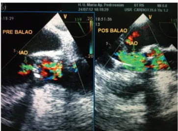
Aortic valvuloplasty
The procedure was performed in cases of valve stenosis affecting the native valve, after the positioning of a balloon
placed at aortic ring level. It was done after hypotension, with mean arterial pressure of 30mmHg, induced by increasing heart
rate to approximately 180 beats per minute through artiicial
cardiac pacing with external pacemaker. The balloon catheter
was placed inside the aortic valve ring and inlated with 2 ATM.
Next, TEE was used to evaluate and verify both the decrease in aortic transvalvular gradient and the onset or increase of
valve insuficiency. (Figure 4).
Valve prosthesis implantation
Implantation was performed after valvuloplasty with balloon catheter in patients with native valve stenosis and without valvuloplasty in patients with pre-existing bioprosthesis dysfunction. The implant was done after hypotension induced
Fig. 3 – Transcatheter aortic valve mounted and crimped onto latex balloon
Fig. 2 – Measurement of the aortic ring through transesophageal echocardiography
through tachycardia, which was controlled by a previously implanted temporary pacemaker. Mean arterial pressure was kept between 30 and 40 mmHg during release of the prosthesis. The positioning of the valve was guided by TEE and radioscopy so as to place one third of the length of the prosthesis inside the left ventricular cavity. Aortography was performed after the implant to assess for the incidence of regurgitation and the patency of the coronary ostia. In addition, ideal prosthetic
positioning was conirmed by TEE. Successful intervention was conirmed through: decrease in transvalvular gradient;
increase in valve area in case of stenosis; and improvement in
the level of insuficiency in case of bioprosthesis dysfunction.
The absence of considerable paraprosthetic regurgitation was also considered a measure of success.
Hospital follow-up
After the procedure, patients were taken to postoperative
ward for intensive care. From the irst postoperative on,
weight heparin. Success was deined as correct implantation
of the valve in the aortic ring, improvement in hemodynamic parameters previous to the echocardiogram, and absence
of signiicant valve and perivalve leakage, in the absence
of fatal complications. Evaluated outcomes were: mortality for any reason (30 days, global, and after discharge); major cardiovascular events; hospitalization due to either prosthesis dysfunction or clinical deterioration; functional class; stroke; vascular complication; renal failure, and major bleeding. Stroke and acute myocardial infarction were determined in accordance with the Valve Academic Research Consortium [16].
Statistical Analysis
Statistical analysis was performed using BioStat version
5.3.5. Conidence level of 0.05 was used as signiicant.
RESULTS
Six patients underwent transcatheter aortic valve implantation, 4 male and 2 female. Patients’ average age was 65.83 + 20.18 years old. Procedures were performed at the University Hospital of the Federal University of Mato Grosso do Sul, in a hybrid operating room. Four patients (66.6%) were candidates due to valve stenosis. One of them had early stenosis of aortic valve bioprosthesis implanted a year before. The other 2 patients (33.3%) were candidates due
to insuficiency of previously implanted bioprosthesis. Mean
STS score for mortality of these patients was 12.09 + 1.28%. Table 4 shows the valvular diseases treated, STS score for each patient, and preoperative echocardiographic parameters. Successful valve implantation was possible in six cases. In 4 patients, the implants were performed through the transfemoral artery; in 2, through the external iliac artery, since the femoral arteries were smaller than 7mm in diameter. There was no need of conversion to open surgery due to access limitation or graft migration. There were no intraoperative or hospital deaths.
The sizes of the devices used were: two 22mm, two 24mm, one 26mm, and one 28mm. In the valve-in-valve cases, two 22mm and one 24mm devices were used.
Hemodynamics as assessed through echocardiography was satisfactory. In patients with native aortic valve or
bioprosthesis stenosis, there was a signiicant decrease in the
mean gradient, from 66.84 ± 15.46 mmHg to 19.74 ±10.61 mmHg, in the postoperative (P=0.002), resulting in a reduction of 70.46%. Figure 5 shows pre and postoperative mean transvalvular gradient. Figure 6 shows pre and postoperative echocardiographic measurements of the transcatheter aortic
valve prosthesis. Subsequent examinations conirmed gradient decrease (Table 5). Patients with bioprosthesis insuficiency
showed discrete increase in the mean transvalvular gradient, no
stenosis with consequence to hemodynamics, and signiicant
Fig. 4 – Transesophageal echocardiography showing an increase in aortic insuficiency after a balloon valvuloplasty
Table 4. Aortic valvular disease, STS scores and preoperative echocardiographic parameters.
Patient 1 2 3 4 5 6
Disease AS AI Biop
AS AI Biop
AS AI Biop
STS score (%) 12 13 11.82
14 11.43
10.3
Mean Gd (mmHg) 47
9 81
9 77 62.39
LVEF (%) 25 40 50 50 41 70
LVDD (mm) 71 60 63 60 71 41
LVSD (mm) 51 52 46 43 56 25 AS = aortic stenosis; AI Biop = aortic insuficiency due bioprosthesis dysfunction; mean Gd = mean gradient; LVEF = left ventricular ejection fraction; LVDD = left ventricular diastolic diameter; LVSD = left ventricular systolic diameter; mm = millimeters; mmHg = millimeters of mercury
Echocardiographic parameters
improvement in the left ventricular systolic function (Figure 7). Generally, preoperative mean ejection fraction for all patients was 46.66 ± 15.2%, increasing to 61.16 ± 12.25% in the postoperative, an increase of 31.11% (P=0.0001).
There was incidence of discrete paravalvular insuficiency
in every patient who underwent intervention in native aortic valve whereas there was no residual regurgitation after intervention in bioprosthesis.
There was no need to implant more than one valve in the same patient.
Figure 8 shows angiographic aspect after a successful implantation, with positioning of the prosthesis and patency of coronary ostia.
Complications
The most serious vascular complication was rupture of the
right iliac artery in one of the patients, with signiicant bleeding. It was solved by inlating a balloon catheter in abdominal aorta with interruption of the distal blood low, followed by
exploratory laparotomy and iliac-femoral anastomosis with
Table 5. Pre and postoperative echocardiographic parameters after transcatheter aortic valve implantation.
Patient 1 2 3 4 5 6
Disease AS AI Biop
AS AI Biop
AS AI Biop
Pre mean Gd (mmHg) 47
9 81
9 77 62.39
Post mean Gd (mmHg) 13.8
22.3 19.61
18 10,85
34.7
Pre LVEF (%) 25 40 50 50 41 70
Post LVEF (%) 43 50 64 73 64 73
P
<0.05 <0.05 <0.05 <0.05 <0.05 <0.05 AS = aortic stenosis; AI Biop = aortic insuficiency due bioprosthesis dysfunction; mean Gd = mean gradient; LVEF = left ventricular ejection fraction; LVDD = left ventricular diastolic diameter; LVSD = left ventricular systolic diameter; mm = millimeters; mmHg = millimeters of mercury
Fig. 5 – Average transvalvular gradient before and after the
transcatheter aortic prosthesis implantation (Before, blue; after, red) Fig. 6 – Echocardiographic representation of the transvalvular gradients (Before, blue; after, red)
polytetraluorethylene (PTFE) graft. Furthermore, there was
a case of hematuria after vesical probing in a patient with prostatic hyperplasia, which was treated with non-surgical methods and solved spontaneously. There was no incidence of atrioventricular blocks needing definitive pacemaker implant. Thrombocytopenia was observed in 5 patients (83.3%), with a mean of 40000 platelets and minimum of 18000 platelets postoperative, with spontaneous recovery
during hospitalization. The cause was not identiied. One of
DISCUSSION
Since 1960, aortic valve replacement has been the only treatment option for severe aortic valve stenosis, increasing patient’s survival rate, regardless of age, and estimated 4% mortality [17]. As previously stated, approximately one third of patients with severe aortic stenosis are ineligible for conventional surgical treatment due to high surgical risk [8].
Drug treatment for these patients leads to a survival rate of 60% in 1 year and 32% in 5 years [5]. Transcatheter aortic valve implantation is a therapeutic option for critically ill patients and it has shown great results. After 1 year of follow-ups, it has demonstrated non-inferiority in relation to conventional aortic valve replacement in addition to a 20% reduction in absolute mortality compared to clinical treatment of inoperable patients, with NNT=5 [14].
In the present study, access was transarterial, femoral, or iliac. Several studies have shown higher mortality in cases of transapical intervention than in transfemoral access cases [18-21]. These results stem from higher severity in patients selected for the transapical intervention as well as a steeper learning curve for this kind of approach. However, it is believed that handling of the heart can lead to greater hemodynamic instability and bleeding. Additionally, respiratory dynamics can be impaired by the opening of the pleural cavity and result in worsening of postoperative respiratory function, especially in patients suffering from chronic obstructive pulmonary disease, since this comorbidity acts as an independent predictor of survival [19]. Besides demonstrating higher hospital mortality rates (13.9% versus 8.5%), a French multicenter
study [22] showed greater incidence of signiicant bleeding
(3.4% versus 1.5%) and longer hospitalization (13.3±7.8 days
versus 10.5±8.1 days) in patients treated via transapical access as opposed to those treated via transfemoral access.
In this initial series, the most serious complication was rupture of the right iliac artery with intraoperative bleeding,
Fig. 8 – Angiographic aspect after transcatheter valve prosthesis implantation
Fig. 7 – Transcatheter aortic valve implantation pre and postoperative
left ventricular ejection fraction
solved by inlating a balloon catheter in abdominal aorta
to interrupt blood flow, followed by right iliac-femoral anastomosis with PTFE graft. Main registries worldwide describe a higher incidence of vascular complications with transfemoral access [19,21,22]. However, the development and evolution of materials such as sheaths and prosthesis could make access through arteries that are smaller in diameter possible, and consequently, lower the incidence of vascular complications. The approach through the external iliac artery has been used in our series as an alternative to the small caliber of femoral arteries.
The Inovare prosthesis, currently designed for transapical
access, has presented proile characteristics in addition to the
ability to be crimped until the diameter needed for transplant through transfemoral access can be reached. It has good navigability through the aorta and appropriate echogenicity for the echocardiographic guidance of its positioning in the aortic ring.
There were no occurrences of transvalvular regurgitation in this group of patients. While three patients (50%) had discrete paravalvular regurgitation, there was none in the valve-in-valve patients, similar to results described by Gaia et al. [15]. Recently, moderate and significant valvular
regurgitation have been identiied as independent risk factors
for short and medium-term mortality after transcatheter aortic valve implantation [19,23]. The same can be observed in the long-term, with mortality being 21% in cases of moderate and
signiicant regurgitation and 6% in cases of discrete or absent
regurgitation [24]. In the present series, none of the patients
REFERENCES
1. Roberts WC, Ko JM. Frequency by decades of unicuspid, bicuspid, and tricuspid aortic valves in adults having isolated aortic valve replacement for aortic stenosis, with or without associated aortic regurgitation. Circulation. 2005;111(7):920-5.
pacemaker implant. Moreover, there was no hospital mortality. Despite this being a small sample, they were critically ill patients with mean STS score of 12.09 + 1.28%, results were deemed very satisfactory for an initial experience, taking into consideration hospital mortality of approximately 5% to 10%, as described by the following: Canadian registry, 10.4% [25]; SOURCE (SAPIEN Aortic Bioprosthesis European Outcome) registry, 8.5% [21]; FRANCE (French Aortic National CoreValve and Edwards) registry, 12.7% [22]; German registry, 8.2% [26]; and Italian registry, 5.4% [23]. In the PARTNER B cohort, mortality in 30 days was 5%, and 5.2% in PARTNER A cohort [20,27].
In our midst, Gaia et al. [28] described overall mortality of 42.42% and hospital mortality of 18.18% in 33 patients who underwent transcatheter aortic valve implantation through transapical access. The patients had mean EuroScore of 39.30% and mean STS score of 30.28%, which means that higher mortality rates than the ones described in major studies worldwide [20-26] can be explained by greater patient severity. A recent study in Italy [29] evaluated 165 patients who had undergone transcatheter aortic valve implantation. They were grouped according to EuroScore, above or below
20%. Hospital mortality was signiicantly higher in the group
with the highest score than in the group with the lowest score, 15.6% and 2.4%, respectively; late mortality (12 months) was 25.7% in more severe patients and 6.8% in the least severe group. This suggests that current mortality rates
described in the literature could be signiicantly inluenced
by the high risk of the population selected for this procedure. The indication of this procedure to lower risk patients could lead to lower mortality rates, perhaps even comparable to those of conventional surgery. New studies comparing the transcatheter intervention to conventional surgery in groups of low and intermediate risk patients have to be performed for comparative analysis.
CONCLUSION
Considering the limitations of the present study and the small sample, it can be concluded that the Inovare prosthesis, implanted through femoral or iliac access, proved to be technically feasible with satisfactory hemodynamic proile in the postoperative follow-up.
2. Selzer A. Changing aspects of the natural history of valvular aortic stenosis. N Engl J Med. 1987;317(2):91-8.
3. Stephan PJ, Henry AC 3rd, Hebeler RF Jr, Whiddon L, Roberts WC. Comparison of age, gender, number of aortic valve cusps, concomitant coronary artery bypass grafting, and magnitude of left ventricular-systemic arterial peak systolic gradient in adults having aortic valve replacement for isolated aortic valve stenosis. Am J Cardiol. 1997;79(2):166-72.
4. Otto CM, Lind BK, Kitzman DW, Gersh BJ, Siscovick DS. Association of aortic-valve sclerosis with cardiovascular mortality and morbidity in the elderly. N Engl J Med. 1999;341(3):142-7.
5. Varadarajan P, Kapoor N, Bansal RC, Pai RG. Clinical proile and natural history of 453 nonsurgically managed patients with severe aortic stenosis. Ann Thorac Surg. 2006;82(6):2111-5.
6. Leon MB, Smith CR, Mack M, Miller DC, Moses JW, Svensson LG, et al; PARTNER Trial Investigators. Transcatheter aortic-valve implantation for aortic stenosis in patients who cannot undergo surgery. N Engl J Med. 2010;363(17):1597-607.
7. Edwards FH, Peterson ED, Coombs LP, DeLong ER, Jamieson WR, Shroyer ALW, et al. Prediction of operative mortality after valve replacement surgery. J Am Coll Cardiol. 2001;37(3):885-92.
8. Iung B, Cachier A, Baron G, Messika-Zeitoun D, Delahaye F, Tornos P, et al. Decision-making in elderly patients with severe aortic stenosis: why are so many denied surgery? Eur Heart J. 2005;26(24):2714-20.
9. Cribier A, Savin T, Saoudi N, Rocha P, Berland J, Letac B. Percutaneous transluminal valvuloplasty of acquired aortic stenosis in elderly patients: an alternative to valve replacement? Lancet. 1986;1(8472):63-7.
10. Davidson CJ, Harrison JK, Leithe ME, Kisslo KB, Bashore TM. Failure of balloon aortic valvuloplasty to result in sustained clinical improvement in patients with depressed left ventricular function. Am J Cardiol. 1990;65(1):72-7.
11. Davies H. Catheter mounted valve for temporary relief of aortic insuficiency. Lancet. 1965;1:250.
12. Andersen HR, Knudsen LL, Hasenkam JM. Transluminal implantation of artiicial heart valves. Description of a new expandable aortic valve and initial results with implantation by catheter technique in closed chest pigs. Eur Heart J. 1992;13(5):704-8.
13. Cribier A, Eltchaninoff H, Bash A, Borenstein N, Tron C, Bauer F, et al. Percutaneous transcatheter implantation of an aortic valve prosthesis for calciic aortic stenosis: irst human case description. Circulation. 2002;106(24):3006-8.
The PARTNER Trial. Presented at: 2011 American College of Cardiology Annual Scientiic Session; April 2, 2011; New Orleans, LA.
15. Gaia DF, Palma JH, Ferreira CBND, Souza JAM, Agreli G, Guilhen JCS, et al. Implante transapical de valva aórtica: resultados de uma nova prótese brasileira. Rev Bras Cir Cardiovasc. 2010;25(3): 293-302.
16. Leon MB, Piazza N, Nikolsky E, Blackstone EH, Cutlip DE, Kappetein AP, et al. Standardized endpoint definitions for transcatheter aortic valve implantation clinical trials: a consensus report from the Valve Academic Research Consortium. Eur Heart J. 2011;32(2):205-17.
17. Kvidal P, Bergström R, Hörte LG, Ståhle E. Observed and relative survival after aortic valve replacement. J Am Coll Cardiol. 2000;35(3):747-56.
18. Thomas M, Schymik G, Walther T, Himbert D, Lefèvre T, Treede H, et al. Thirty-day results of the SAPIEN aortic Bioprosthesis European Outcome (SOURCE) Registry: a European registry of transcatheter aortic valve implantation using the Edwards SAPIEN valve. Circulation. 2010;122(1):62-9.
19. Moat NE, Ludman P, de Belder MA, Bridgewater B, Cunningham AD, Young CP, et al. Long-term outcomes after transcatheter aortic valve implantation in high-risk patients with severe aortic stenosis: the U.K. TAVI (United Kingdom Transcatheter Aortic Valve Implantation) Registry. J Am Coll Cardiol. 2011;58(20):2130-8.
20. Leon MB, Smith CR, Mack M, Miller DC, Moses JW, Svensson LG, et al; PARTNER Trial Investigators. Transcatheter aortic-valve implantation for aortic stenosis in patients who cannot undergo surgery. N Engl J Med. 2010;363(17):1597-607.
21. Thomas M, Schymik G, Walther T, Himbert D, Lefèvre T, Treede H, et al. One-year outcomes of cohort 1 in the Edwards SAPIEN Aortic Bioprosthesis European Outcome (SOURCE) Registry: the European registry of transcatheter aortic valve implantation using the Edwards SAPIEN valve. Circulation. 2011;124(4):425-33.
22. Gilard M, Eltchaninoff H, Iung B, Donzeau-Gouge P, Chevreul K, Fajadet J, et al; FRANCE 2 Investigators. Registry of transcatheter aortic-valve implantation in high-risk patients. N Engl J Med. 2012;366(18):1705-15.
23. Tamburino C, Capodanno D, Ramondo A, Petronio AS, Ettori F, Santoro G, et al. Incidence and predictors of early and late mortality after transcatheter aortic valve implantation in 663 patients with severe aortic stenosis. Circulation. 2011;123(3):299-308.
24. Gotzmann M, Korten M, Bojara W, Lindstaedt M, Rahlmann P, Mügge A, et al. Long-term outcome of patients with moderate and severe prosthetic aortic valve regurgitation after transcatheter aortic valve implantation. Am J Cardiol. 2012;110(10):1500-6.
25. Rodés-Cabau J, Webb JG, Cheung A, Ye J, Dumont E, Feindel CM, et al. Transcatheter aortic valve implantation for the treatment of severe symptomatic aortic stenosis in patients at very high or prohibitive surgical risk: acute and late outcomes of the multicenter Canadian experience. J Am Coll Cardiol. 2010;55(11):1080-90.
26. Zahn R, Gerckens U, Grube E, Linke A, Sievert H, Eggebrecht H, et al; German Transcatheter Aortic Valve Interventions-Registry Investigators. Transcatheter aortic valve implantation: irst results from a multi-centre real-world registry. Eur Heart J. 2011;32(2):198-204.
27. Smith CR, Leon MB, Mack MJ, Miller DC, Moses JW, Svensson LG, et al; PARTNER Trial Investigators. Transcatheter versus surgical aortic-valve replacement in high-risk patients. N Engl J Med. 2011;364(23):2187-98.
28. Gaia DF, Palma JH, Ferreira CBND, Souza JAM, Gimenes MV, Macedo MT, et al. Implante transcateter de valva aórtica: resultados atuais do desenvolvimento e implante de um nova prótese brasileira. Rev Bras Cir Cardiovasc 2011;26(3):338-47.