324
Revista da Sociedade Brasileira de Medicina Tropical 44(3):324-326, mai-jun, 2011
INTRODUCTION
1. Disciplina de Cirurgia do Aparelho Digestivo, Universidade Federal do Triângulo Mineiro, Uberaba, MG. 2. Curso de Medicina, Universidade Federal do Triângulo Mineiro, Uberaba, MG. 3. Disciplina de Imunologia, Universidade Federal do Triângulo Mineiro, Uberaba, MG.
Address to: Dr. Eduardo Crema. Rua Marcos Lombardi 305, Santos Dumont, 38050-170 Uberaba, MG, Brasil.
Phone: 55 34 7811-3830; 55 34 3318-5228 e-mail: [email protected] Received in 15/06/2010 Accepted in 08/02/2011
Prevalence of cholelithiasis in patients with chagasic megaesophagus
Prevalência de colelitíase em pacientes com megaesôfago chagásico
Eduardo Crema
1, Ellen Caroline Rosa Resende Silva
2, Priscila Melo Franciscon
2, Virmondes Rodrigues Júnior
3,
Aiodair Martins Júnior
1, Celso Júnior Oliveira Teles
2and Alex Augusto Silva
1ABSTACT
Introduction: he prevalence of cholelithiasis in the general population ranges from 9 to 18%. his prevalence is known to be higher in the presence of parasympathetic nerve damage of the biliary tract either due to surgery (vagotomy) or neuronal destruction (Chagas disease). he objective of this study was to evaluate the association of cholelithiasis and chagasic or idiopathic megaesophagus. Methods: The ultrasound scans of 152 patients with megaesophagus submited to cardiomyotomy and subtotal esophagectomy surgery were evaluated. he presence of cholelithiasis was compared between chagasic and idiopathic esophagopathy and ultrasound and clinical indings were correlated with age, sex and race. Results: A total of 152 cases of megaesophagus, including 137 with chagasic megaesophagus and 15 with idiopathic megaesophagus, were analyzed. he mean age was 56.7 years (45-67) in the 137 patients with chagasic megaesophagus and 35.6 years (27-44) in the 15 cases of idiopathic megaesophagus, with a signiicant diference between the two groups (p < 0.0001). he group with chagasic megaesophagus consisted of 59 (43%) women and 78 (56.9%) men, while the group with idiopathic megaesophagus consisted of 8 (53.3%) women and 7 (46.6%) men, showing no signiicant diference between the groups. Of the 137 patients with conirmed chagasic megaesophagus, 39 (28.4%) presented cholelithiasis versus one case (6.6%) in the 15 patients with idiopathic megaesophagus. Conclusions: he prevalence of cholelithiasis is high in patients with chagasic megaesophagus and preoperative ultrasound should be performed routinely in these patients in order to treat both conditions during the same surgical procedure.
Keywords: Chagas disease.Cholelithiasis. Megaesophagus. Gallbladder denervation.
RESUMO
Introdução: A prevalência de colelitíase observada na população em geral varia de 9 a 18%. Sabe-se que a prevalência de colelitíase é elevada quando existe lesão nervosa parassimpática das vias biliares, causada tanto por procedimentos cirúrgicos (vagotomias),quanto por destruição neuronal, como observado na forma digestiva da doença de Chagas. Propusemo-nos veriicar a associação entre megaesôfago de etiologia chagásica e a presença de colelitíase. Métodos:
Avaliou-se prospectivamente o exame ultrassonográico de 152 pacientes portadores de megaesôfago submetidos à cirurgia de cardiomiotomia e esofagectomia subtotal. Analisou-se comparativamente a esofagopatia chagásica e a idiopática com a preAnalisou-sença de colelitíaAnalisou-se, correlacionando os dados ultrassonográicos com os achados clínicos, idade, sexo e raça.
Resultados: Foram analisados 152 casos de megaesôfago, sendo 137 de etiologia chagásica e 15 idiopáticos. Entre os chagásicos, a idade média foi de 56,7 anos (45-67); e nos idiopáticos, a média de idade foi de 35,6 anos (27-44), veriicando-se diferença signiicativa (p < 0,0001) em relação à média de idade. Dos 137 chagásicos, 78 (56,9%) eram do sexo masculino; entre os 15 idiopáticos, 7 (46,6%) eram do sexo masculino. A comparação entre os grupos em relação ao gênero não mostrou diferença signiicativa. Dentre os 137 chagásicos detectou-se 39 (28,4%) casos de colelitíase e, dentre os 15 casos de megaesôfago idiopático, constatou-se colelitíase em apenas um (6,6%). Conclusões: Conclui-se que portadores de megaesôfago chagásico possuem alta prevalência de colelitíase, e que a ultrassonograia deve ser realizada rotineiramente no pré-operatório, a im de tratar ambas as afecções no mesmo ato cirúrgico.
Palavras-chaves: Doença de Chagas. Colelitíase. Megaesôfago. Desnervação da vesícula biliar.
he chronic digestive form of Chagas disease is characterized by the destruction of intramural ganglia and parasympathetic dener vation throughout the digestive tract, especially afecting the esophagus1 and rectosigmoid junction. However, studies investigating accessory organs and glands of the digestive tract in Chagas disease, particularly the gallbladder, are scarce2-4. The incidence of cholelithiasis is high in the presence of parasympathetic nerve damage of the biliary tract either due to surgery (vagotomy)5,6 or neuronal destruction1. Similarly, the incidence of cholelithiasis is higher in chagasic patients than in the general population2,4. The following factors have been highlighted as responsible for this increase: gallbladder denervation leading to alterations in organ tonus and contractility and consequently diminished biliary low and greater stasis2,4; hypostimulation of the gallbladder by cholecystokinin due to lower supply of foods to the stomach and duodenum as a result of poor swallowing in cases of megaesophagus and of anorexia in cases of megacolon; alterations in gastroduodenal function, such as vagal denervation of the stomach that diminishes intestinal motility and gastric juice acidity; elevated secretion of saliva and mucus in cases of megaesophagus that neutralize the gastric juice, reducing stimuli for cholecystokinin secretion2, have also been reported to contribute to the formation of cholelithiasis. Furthermore, in the case of patients with megaesophagus and megacolon, denervation of the small bowel probably reduces the absorption of bile salts and consequently triggers the formation of lithogenic bile2,4.
Given the scarcity of literature data, the aim of the present study was to evaluate the association between megaesophagus and cholelithiasis in chagasic patients atended at the University Hospital of the Universidade Federal do Triângulo Mineiro (UFTM).
Article/Artigo
325
METHODS
RESULTS
Crema E et al - Cholelithiasis in chagasic megaesophagus patients
Ultrasound scans of the gallbladder of 152 patients with megaesophagus treated at the Digestive Surgery Service of UFTM were analyzed. All patients were submitted to Heller cardiomyotomy or subtotal esophagectomy. he scans were analyzed regarding the presence of cholelithiasis, bile duct dilatation and/ or choledocholithiasis. Chagasic megaesophagus was considered when the patient presented a positive result in at least two of the three following tests: ELISA, passive hemagglutination and indirect immunoluorescence, or positive polymerase chain reaction (PCR) in a myotomy fragment or surgical specimen, associated with histopathological alterations characteristic of chagasic megaesophagus. Patients not presenting these conditions were classiied as presenting idiopathic megaesophagus. he presence of cholelithiasis was compared between chagasic and idiopathic esophagopathy. he ultrasound and clinical indings were correlated with age, sex and race.
he results were analyzed statistically by the x2 test, with the level of error probability set at 5%. Parametric analysis of variance (ANOVA, F test) was used to determine possible diferences in age, gender and race between the chagasic and idiopathic groups.
A total of 152 patients with megaesophagus submited to surgical treatment at the Digestive Surgery Service of UFTM were studied. Of these, 137 presented chagasic megaesophagus and 15, idiopathic megaesophagus.
The mean age of patients with chagasic megaesophagus was 56.7 (45-67) years-old and the mean age of patients with idiopathic megaesophagus was 35.6 (27-44) years-old, showing a significant difference between the groups. Seventy-eight (56.9%) of the 137 patients with chagasic megaesophagus were males and 59 (43.1%) were females. In the idiopathic group, 8 (53.3%) were men and 7 (46.7%) were women. Despite the predominance of male patients in the chagasic group, no signiicant diference was observed (Figures 1, 2 and 3).
56.7
35.6
0 10 20 30 40 50 60
Chagasic Idiopathic
Category
M
ean
age
FIGURE 1 -distribution of patients with chagasic (CH) and idiopathic (Id) megaesophagus according to age (n = 152).
56.9%
43.1% 53.3%
46.7%
0.0 10.0 20.0 30.0 40.0 50.0 60.0
Woman
V
a
lu
e
Gender
Chagasic
Idiopathic Man
%
FIGURE 2 - distribution of patients with chagasic (CH) and idiopathic (Id) megaesophagus according to gender (n = 152).
96
24 17
10 3 2
0 20 40 60 80 100 120
White Black Mulatto
Race
Chagasic
Idiopathic
Number of individuals
326
Rev Soc Bras Med Trop 44(3):324-326, mai-jun, 2011
DISCUSSION
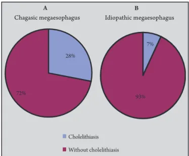
FIGURE 4 -A:Prevalence of cholelithiasis in patients with chagasic megaesophagus (n = 137), and (B): in patients with idiopathic megaesophagus (n = 15).
A predominance of white patients was observed in both groups, with 96 (70%) cases in the chagasic group and 10 (66.7%) in the idiopathic group, followed by blacks [24 (17.5%) chagasic and 3 (20%) idiopathic] and mulatos [17 (12.4%) chagasic and 2 (13.3%) idiopathic].
Cholelithiasis was present in 39 (28.4%) of the 137 patients with chagasic megaesophagus and in 1 (6.7%) of the 15 patients with idiopathic megaesophagus (Figures 4A and B). This diference was statistically signiicant. Despite the high incidence of cholelithiasis in patients with chagasic megaesophagus, the rates of choledocholithiasis observed in this series were not higher than those reported for the general population (9.2 to 18%).
7%
93%
A B
28%
72%
Cholelithiasis
Chagasic megaesophagus Idiopathic megaesophagus
Without cholelithiasis
Studies investigating the gallbladder in Chagas disease are scarce7.
herefore, the objective of the present study was to evaluate the incidence of cholelithiasis in patients with megaesophagus. According to the literature, the incidence of cholelithiasis is high in the presence of parasympathetic
nerve damage of the biliary tract either due to surgery (vagotomy)5,6,8
or to neuronal destruction, as observed in the chronic digestive form
of Chagas disease1,2,9. One study reported a prevalence of cholelithiasis
of 8.4% in patients with chagasic megaesophagus, but observed no case of cholelithiasis among patients with idiopathic megaesophagus. In the present study, the rate of cholelithiasis in patients with megaesophagus (28.4%) was much higher than that reported previously, probably because of the routine use of preoperative ultrasound in these patients.
No signiicant diference in sex or race was observed between patients with idiopathic and chagasic megaesophagus. However, a signiicant diference in age was observed between the two groups; patients with idiopathic megaesophagus were younger than those with chagasic megaesophagus.
In conclusion, the prevalence of cholelithiasis is high in patients with chagasic megaesophagus and it is good practice to perform a preoperative ultrasound routinely in these patients in order to treat both conditions during the same surgical procedure.
he authors declare that there is no conlict of interest. CONFLICT OF INTEREST
REFERENCES
1. Adad SJ, Resende AV, Jorge BH. Estudo sistematizado do plexo mientérico nos diferentes terços do esôfago de chagásicos crônicos com e sem megaesôfago. Rev Soc Bras Med Trop 1992; 25:101.
2. Pinotti HW, Raia A, Bettarello A, Conte VP. Ocorrência de colelitíase em portadores de megaesôfago chagásico. Estudo comparativo com não chagásicos. Rev Hosp Clin Fac Med S Paulo 1980; 35:21-24.
3. Pinoti HW, Felix VN, Zilberstein B, Cecconello I. Surgical complications of Chagas’ disease: megaesophagus, achalasia of the pylorus and cholelithiasis. World J Surg 1991; 15:198-204.
4. Oliveira LCM, Nascimento RS, Rocha A, Gonçalves EAG, Silva JMF, Oliveira VA, et al. Colelitíase em chagásicos crônicos. Arq Gastroenterol 1997; 34:222-226. 5. Diaconescu MR, Simon I, Costea I, Glod M, Terinte R. Cholelithiasis following
gastric surgery. Chirurgia 1997; 92:343-347.
6. Kinoshita H, Imayama H, Hashino K, Aoyagi S. Study of cholelithiasis ater gastrectomy. Kurume Med J 2000; 47:105-108.
7. Rocha A, Almeida H, Teixeira VPA, Silva AM. Prevalência da colelitíase em chagásicos crônicos necropsiados no Triângulo Mineiro - correlação com o megaesôfago, o megacólon e a insuiciência cardíaca. Arq Gastroenterl S Paulo 1985; 22:3-6.
8. Iorio A, Chatas E, Kesner L, Masciangioli G, Zampini E, Gandval C. Litiasis vesicular post-vagotomia y operaciones gastricas. Prensa Med Argent 1983; 70:113-116.