1 Ataturk University, Medical Faculty, Department of Neurosurgery, Erzurum, Turkey 2 Ataturk University, Medical Faculty, Department of Psychiatry, Erzurum, Turkey 3 Ataturk University, Medical Faculty, Department of Cardiology, Erzurum, Turkey 4 Ataturk University, Medical Faculty, Department of Anesthesiology, Erzurum, Turkey
5 Ataturk University, Medical Faculty, Department of Pathology, Erzurum, Turkey 6 Sakarya University, Medical Faculty, Department of Neurology, Sakarya, Turkey 7 Ataturk University, Medical Faculty, Department of Neurology Erzurum, Turkey
Yazışma Adresi /Correspondence: Dilcan Kotan, ORIGINAL ARTICLE / ÖZGÜN ARAŞTIRMA
Role of carotid body for neuronal protection in experimental subarachnoid
haemorrhage
Deneysel subaraknoid kanamada karotid cismin nöron korumasındaki rolü
Mehmet Dumlu Aydın1, Nazan Aydın2, Adnan Bayram3, Canan Atalay4, Sare Altaş5,
Dilcan Kotan6, Hızır Ulvi7
ÖZET
Amaç: Karotid cisimler, temel arteriyel kemoregulatuar üniteler olarak bilinirler. Karotid cisimlerin serebral sirkü-lasyonda ve kan pH regülasyonunda önemli bir rolü
oldu-ğu iyi bilinmesine rağmen, subaraknoid kanamadaki rol
-leri henüz araştırılmamıştır. Biz subaraknoid kanamada karotis cisim nöron yoğunluğunun beyin üzerinde nöron koruyucu etkisinin olup olmadığını araştırdık.
Yöntemler: Yirmi hibrit tavşan çalışmada kullanıldı. Bun
-ların dört tanesi (n=4) referans grup olarak kullanıldı ve kalanların (n=16) sisterna magna’ları içerisine otolog kan enjeksiyonu yapılarak subaraknoid kanama geliştirildi ve bir ay sonra hayvanların yaşam süresi sonlandırıldı. Tüm karotid cisim ve beyin dokuları, stereolojik metodlar kulla
-nılarak histopatolojik olarak incelendi. Karotid cisimdeki nöronal yoğunluk ile hipokampustaki dejenere nöron yo
-ğunluğu arasındaki ilişki istatistiksel olarak karşılaştırıldı.
Bulgular: Subaraknoid kanaması olan beş tavşan takip
süresi içerisinde öldü (n=5). Normal tavşan ailesinde ka
-rotid cisim ortalama nöronal hücre yoğunluğu 4500±500/
mm3 ve hipokampus ortalama nöronal hücre yoğunluğu 170,000±17,000/mm3 olarak saptandı. Karotid cisminde yüksek nöron yoğunluğu olan tavşanların hipokampusla
-rındaki dejenere nöron hücre yoğunluğu 20,000±3,000/
mm3 iken karotid cisminde düşük nöron yoğunluğu olan tavşanların hipokampuslarındaki dejenere nöron hücre yoğunluğu 65,000±8,000/mm3 saptandı. Karotid cismin nöronal yoğunluğu ve hipokampusun dejenere nöron sa
-yıları arasındaki farklılık istatistiksel olarak anlamlıydı.
Sonuç: Karotid cismin nöron yoğunluğu, subaraknoid he
-morajide beyin dokusunun korunmasında önemli bir rol
oynayabilir.
Anahtar kelimeler: Subaraknoid hemoraji, karotid cisim, hipokampüs, nörodejenerasyon, serebral iskemi
ABSTRACT
Objective: Carotid bodies are known as main arterial chemoregulatory units. Despite well known that carotid bodies have an important role in cerebral circulation and blood pH regulation, their roles has not been investigated in subarachnoid haemorrhage. We investigated whether
there is neuroprotective effect of neuron density of carotid
bodies on the brain in subarachnoid haemorrhage.
Methods: Twenty hybrid rabbits were studied. Four of
them were used as reference group (n=4) and the re -maining was obliged to subarachnoid haemorrhage by
in-jecting autologous blood into their cisterna magna (n=16) and sacriiced after one month. All carotid bodies and
brains examined histopathologically using by stereologic
methods. The relationship between the neuronal density of carotid body and degenerated neuron density of the
hippocampus were compared statistically.
Results: Five rabbits with subarachnoid haemorrhage
dead during the follow-up time (n=5). The average neu
-ronal density of carotid body was 4500±500 cells/mm3 and of hippocampus 170.000±17.000 cell/mm3 in nor-mal rabbit family. The degenerated neuron density of the hippocampus was 20.000±3.000 cells/mm3 in rabbits with have high neuron density of carotid body and was 65.000±8.000 cells/mm3 in rabbits with low neuron den-sity of carotid body. The differences between the neuronal density of carotid body and the degenerated neuron num
-bers of the hippocampus were signiicant.
Conclusion: The neuron density of carotid body may
play an important role on the protection of brain in sub -arachnoid haemorrhage.
INTRODUCTION
The carotid bodies (CB) are the most vascularised and chemosensitive structures in the body, CB are localised at the carotid bifurcation and supplied by mainly external and rarely internal carotid arteries. Cerebrovascular and cardiorespiratory autonomy are mainly regulated by neurochemical circuitry of CB. Glomus cells are chemosensitive units of the CB and they synaptically connected to glosso-pharyngeal nerve terminals. Glomus cells are very sensitive in the blood pH changes [1,2,3]. When the O2 difference in the arteriovenous blood is less than 1%, CB are stimulated [4]. CB dysfunction can result in cerebral circulation disorders and car-diorespiratory disturbances [1]. Severe vasospasm induced by subarachnoid haemorrhage (SAH) can
lead to decreased cerebral blood low, disordered
glucose metabolism, increased ischemia, decreased cerebral perfusion pressure, increased intracranial pressure, neuronal degeneration and early mortality [5,6,7]. If so, CB dysfunction can result in cerebral glucose metabolism disorders, cerebral circulation and cardio-respiratory disturbances and failure of
body luid pH regulation [8]. To examine whether
the neuron density of CB has a role in the progres-sion of SAH, neuron density of the CB and degen-erated neuron density of the CA1 (cornu Ammon)
region of the hippocampus were examined in SAH developed animals. The results shown that the low
neuron density of CB may have an important role on the development of hippocampal
neurodegenera-tion and worsened prognosis of SAH.
METHODS
Twenty hybrid rabbits were studied at two years old and weighing 3.5 ± 0.25 kg. Animal husbandry and the study design followed the guidelines of the National Istitudes of Health. The study design was
approved by the Committee on Animal Research at
our university. Four of them were used to examine
of normal stereologic anatomy of CB and
hippo-campus. The remainder animals (n=16) were an -aesthetized by subcutaneous injection of a mixture
of ketamine hydrochloride (25 mg/kg), lidocain hy
-drochloride (15 mg/kg), and acepromasine (1 mg/ kg). After preparing the occipito-cervical region, SAH was produced by the injection of 0.5cc blood
into cisterna magna taken from auricular veins. All animals were followed-up one month in the normal laboratory standarts without treatment and all of them were sacriied at the end of experiment. Their CB and brains were removed and preserved in 10%
formalin solution for seven days. The specimens
were embedded in parafin blocks and consecutive twenty sections of 5 µm of all preparations were taken for the stereological examinations. CB prepa
-rations were stained with hematoxylene and eo
-sin (H&E). Hippocampus slices were stained with
TUNEL staining for the detection of apoptosis. All
preparations were observed light microscope and stereologic method were used for the determination
of neuron numbers of the CB and CA1 regions of hippocampus.
Histopathologically, cytoplasmic condensation,
nuclear shrinking, cellular angulations and peri-cy -toplasmic halo formation secondary to cy-toplasmic
regression and Tunnel staining positivity were con -sidered as the criteria of neuronal degeneration.
Physical dissector method was used to evaluate
the numbers of neurons in CB and CA1. This meth-od can easily estimate the particle number, be read-ily performed, intuitively simple, free from assump-tions about particle shape, size and orientation, and
unaffected overprotection and truncation. Data were
obtained from dissector pairs, consisting of parallel
sections taken at known intervals. Two labeled con -secutive sections obtained from tissue samples
(dis-sector pairs) were mounted on each slide. Twenty dissector pairs were taken in each block for analyse of neurons. A counting frame was placed on con -secutive section photographs on screen of personal computer (PC) for counting of neurons. The bottom
and the left hand edges of the frame were exclud
-ed for counting (exclusion) lines together with the
extension lines. Other boundaries of the frame and
the top-right corner were considered to be inclusion points and any particle which hit these lines or was
located inside the frame counted as a dissector
par-ticle. Neurons of CB and CA1 regions were counted if they were visible in the reference section. Refer
-ence and look-up sections were reversed in order to double the number of dissector pairs without taking new sections (see Figure 1). The average numerical
Figure 1. Histopathological appearance of a brain with
subarachnoid haemorrhage (SAH) and is presented (BA: Basilar artery) (H&E x20, LM).
NvGN=ΣQN/txA
Where ΣQN is the total number of counted neu -rons appearing only in the reference sections; t is
the section thickness and A is the area of the count
-ing frame. Cavalieri volume estimation method was
used to obtain the total number of neurons in each
specimens. Total number of neurons was calculated
by multiplication of the volume (mm3) and
numeri-cal density of neurons in each CB or CA1 region.
To analyze the results, average neuronal nu-merical density of CB and CA1 region of all
ani-mals were accepted as mean values of normal rabbit family. The neuronal density of CB was higher than 5000 named as SAH-resistant group (GR) and less
ones non resistant group (GNR). GR is the group
of damages less, has more carotid body density and consequently has less damage of cisterna magna and hippocampus. GNR is the group of damages more, has less carotid body density and consequently has more damage of cisterna magna and hippocampus.
The all dead animals were also included into
the GNR. The neuron density of CB and
degener-ated neuron number of CA1 were compared statisti
-cally and results were analyzed with Mann-Whitney
U Test.
RESULTS
Four of the sixteen rabbits died in the study group because of cardio-respiratory arrest during the
fol-low-up period (n=4) and the others lived one month (n=12). The average neuronal density of CB was es
-timated as 4500±500 cell/mm3 (Figure 2 A-B) and those of hippocampus 170.000±17.000 cell/mm3 of normal rabbit family. In this classiication, the de
-generated neuron density of the CA1 was estimated as 20.000±3.000 cell/mm3 in the GR (Figure 3) and 65.000±8000 cells/mm3 in the GNR (Figure 4). The difference between the neuronal density of CB and degenerated neuron numbers of CA1 of the GR was signiicant (p<0.005). But, the difference was more signiicant in GNR (p<0.0001). The animals with
high neuron density in their CB have good
clini-cal outcome and low cerebral insult. The carotid
bodies are responsible from regulate of blood and
cerebrospinal luid (CSF) pH, by means of glosso -pharingeal nerve. High density of nerve regulates to blood chemistry better. A lot of carotid body nerve
remains healthy in ischemic injury, exactly like this,
increases to resistance of SAH.
Table 1. The average neuronal numerical density of neurons of CB and degenerated neuronal numerical density of
CA1 region of the hippocampus
GN GR GNR
Normal neuron density of B (cell/mm3) 4500±500 >5000 <4000
Normal neuron density of hippocampus (cell/mm3) 170.000±17.000 165.000±9.000 130.000±12.000 Degenerated neuron density of hippocampus (cell/mm3) 10±2 5.000±300 30.000±3.000
CB: Carotid body, GN: Ganglial neurons, GR: SAH-resistant group, GNR: SAH-non resistant group SAH: Subaracnoid
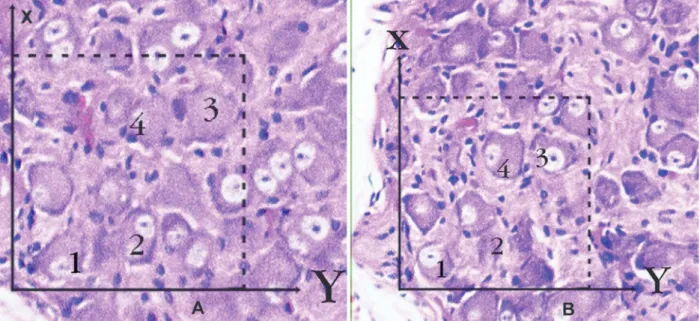
Figure 2 A-B. Application of the physical dissector method in which micrographs in same ields of view (A, B) are taken
from two parallel adjacent thin sections separated by a distance of 5 µm in a normal rabbit. Upper and right lines of unbiased counting frames represent the inclusion lines and the lower and left lines including the extensions are exclu
-sion lines. Any neuron nucleolus hitting the inclu-sion lines is excluded and nucleus proiles hitting the inclu-sion lines and located inside the frame are counted as dissector particles unless their proile extends up to the look-up section. The number of neurons from the two dissectors occurs in a volume given by the product of the counting frame area and distance between the sections. The numerical density of neurons is calculated from NvN=∑Q-N / t x A. In this applica
-tion, the nucleoli marked with ‘1, 3’ are not dissector particles on A section as it disappeared section B. The nucleoli marked with ‘2, 4’ are dissector particles on A section as it appeared section B (H&E x100, LM).
Figure 4. Considerable numbers of degenerated neurons
as appear in brown colored (DN) are observed among the normal neurons (NN) in CA1 regions of hippocampus of the GNR. Both the nucleus and the cell body of neurons were shrunken and had lost much of then morphologic details (Tunnel Staining, x100).
Figure 3. A histopathological appearance of CA1 region
DISCUSSION
The carotid body is the main arterial
chemorecep-tor with the characteristics of high blood low, el -evated metabolism, oxygen-sensing functions and susceptibility of arterial pH changes [4]. They have also vital functions for cerebrovascular and car-dio-respiratory autoregulation [1,2].
Chemosensi-tive units of the CB are glomoid structures which
formed by clusters of glomus cells located around the capillaries. Distal extentions of glomus cells are synaptically connected to petrosal ganglion neurons via glossopharyngeal nerve terminals and proxi-mal ends connected to cardiorespiratory centers in the paraventricular nucleus of the hypothalamus [6]. In response to hypoxia, hypercapnia and blood pH chances glomus cells release appropriate neu-rotransmitters and stimulate paraventricular nucleus
via neural networks of CB [3,4]. Vasoactive mol -ecules produced by CB modulate the chemosensory
processes body luids pH and cerebral autoregula -tion. Doux et al reported that CB dysfunctions can result in cerebrovascular and cardiorespiratory autoregulation disorders [1]. Even, panic disorder
and agoraphobia are presented with dyspnea and
hyperventilation are the cardinal signs of a panic
attack can be resulted from failure of CB alarm
system. SAH develops due to various etiologic factors leading to tear of blood vessels in the sub-arachnoid spaces. Severely vasospasm triggering by SAH lead to acute cerebral ischemia, brain edema, blood-brain barrier disruption, increased intracra-nial pressure, decreased cerebral perfusion
pres-sure and inally apoptotic degeneration of the brain.
Disordered cerebral autoregulation is the most im-portant dangerous factor in the progression of SAH [5]. Early cerebral vasoconstruction and diminished
cerebral blood low occur in the majority of subjects [9]. Severe SAH is associated with loss of cerebral
autoregulation and cardiorespiratory irregularities
[5]. Microvascular aggregation of red blood cells
has also been accused of acute ischemic damage. Decreased CBF and decreased CPP are the most
im-portant factors in early mortality [10]. The mortality rate of SAH is about 25% within 24 hours and 45% at 30 days [11]. Extensive global ischemic brain
damage can result in death shortly after severe SAH
[8]. Vasoconstruction can lead to decreased cerebral blood low and cerebral ischemia [12]. Profound el -evation of intracranial pressure is an important
fac-tor in the development of cerebral ischemic damage in SAH [13]. Experimental SAH can be induced by autolog blood injection into the cisterna magna in animal models [11]. We also used the same meth-od for observing clinical outcome and to examine
if there was a relationship between the neuronal
density of CB and degenerated neuron numbers of the CA1 region of hippocampus.
Histopathologi-cal analyses were done by using stereologic meth -ods [14,15,16]. Apoptotic degeneration at the CA1
region was determined by TUNEL staining [17].
Some treatment methods directed to CB have been applied for reduce the complication of SAH. That the ischemic neuronal damage of the CB and brain may be recovered via early revascularization of the CB via posterior cerebral circulation in ischemic
brain disease [18]. Intracerebral transplantation of CB can also ameliorate stroke-induced cerebral in -farction [19]. CB receptors have a glucose sensing role in the blood entering the brain and integrating information about blood glucose levels by CB is essential for central nervous system metabolism. The nucleus tractus solitarius is an important relay station in central metabolic control and receives signals from peripheral glucose sensitive afferents
from CB [20,21]. Chemoreceptors close to ventral
surfaces of the medulla are responive to CO2 level
and pH changes in the cerebrospinal luid. Chemical
informations coming from surfaces of medulla and CB are integrated together at the respiratory centers [22]. At this situation, sensibility of chemorecep-tors can be change and chaotic state occur at the pH regulating centers. If so, CB have less neuron does not regulate adequately glucose metabolism, pH
regulation and cerebral circulation. Insuficient CB
can aggravate the ischemic insult generating effect of SAH. To decrease nerve in carotid body is result from secondary denervation injury to ischemic inju-ry of glossopharingeal nerve. High nerve density is
regulates blood chemistry better. More carotid body remains healthy, exactly like this increases resis -tance of SAH. Ischemic injury of brain stem in SAH spoils to morphology and functions of carotid body.
Exactly like this, pH of blood and CSF is spoiled. Because of that, ischemic injury grows and arises nerve death and inally brain death. Separately, it
arises to vasodilatation of glossopharingeal nerve.
If there is a damage, cerebral vessels will arises to
In summary, this study in rabbits shows that the low neuron density of CB can be considered as a
cause of the severity of neuronal degeneration in the
hippocampus in SAH. Although the marked differ
-ence between the neuron density of CB and degen -erated neuron number of hippocampus is thus unex-plained, this difference may have important
impli-cations. CB have low neuron density may be impor -tant in both discontinuation of metabolic processes
responsible for pH regulation in the body luid and
important decrease in vasoactive neurotransmitter production by glomus cells essential for the mainta-nence of glucose metabolism, cerebrovascular auo-toregulation. For reducing the dangerous effects of SAH on the brain, supportive interventions might
be inquired toward to preserve of the CB structure
and functions.
REFERENCES
1. Doux JD, Yun AJ. The link between carotid artery disease and ischemic stroke may be partially attributable to au -tonomic dysfunction and failure of cerebrovascular auto-regulation triggered by Darwinian maladaptation of the ca -rotid baroreceptors and chemoreceptors. Med Hypotheses 2006;66:176-81.
2. Kusakabe T, Matsuda H, Hayashida Y. Hypoxic adaptation of the rat carotid body. Histol Histopathol 2005;20:987-97. 3. Rey S, Iturriaga R. Endothelins and nitric oxide: vasoactive
modulators of carotid body chemoreception. Curr Neuro-vasc Res 2004;1:465-73.
4. Oikawa S, Hirakawa H, Kusakabe T. Autonomic cardio -vascular responses to hypercapnia in conscious rats: the roles of the chemo- and baroreceptors. Auton Neurosci 2005;117:105-14.
5. Bederson JB, Germano IM, Guarino L. Cortical blood low and cerebral perfusion pressure in a new noncraniotomy model of subarachnoid haemorrhage in the rat. Stroke 1995; 26:1086-91.
6. Schultz HD, Sun SY. Chemorelex function in heart failure. Heart Fail Rev 2000;5:45-56.
7. De Toma G, Nicolanti V, Plocco M, et al. Barorelex failure syndrome after bilateral excision of carotid body tumors: an underestimated problem. J Vasc Surg 2000; 31:806-10. 8. Adams HPJ, Kassell NF, Torner JC. Early management of
subarachnoid haemorrhage: A report of the Cooperative Aneurysm Study. J Neurosurg 1981;46:454-66.
9. Naveri L, Stromberg C, Saavedra JM. Angiotensin IV re -verses the acute cerebral blood low reduction after experi -mental subarachnoid haemorrhage in the rat. J Cereb Blood Flow Metab 1994;14:1096-99.
10. Bederson JB, Guarino L, Germano IM. Failure of chang -es in cerebral perfusion pr-essure to account for ischemia caused by subarachnoid haemorrhage: A new experimental model. Soc Neurosci Abstr 1994;20:224-8.
11. Broderick JP, Brott TG, Duldner JE. Initial and recurrent bleeding are the major causes of death following subarach -noid haemorrhage. Stroke 1994;25:1342-47.
12. Delgado TJ, Brismar J, Svendgaard NA. Subarachnoid haemorrhage in the rat: Angiography and luorescence mi -croscopy of the major cerebral arteries. Stroke 1985;16:595-602.
13. Bederson JB, Levy AL, Ding WH, et al. Acute vasocon-striction after subarachnoid haemorrhage. Neurosurgery 1998;42:352-60.
14. Cruz-Orive LM, Weibel ER. Recent stereological methods for cell biology: a brief survey. Am J Physiol 1990;258:148-56.
15. Gundersen HJ, Bendtsen TF, Korbo L, et al. Some new, simple and eficient stereological methods and their use in pathological research and diagnosis. APMI 1988;96:379-94.
16. Sterio DC. The unbiased estimation of number and siz-es of arbitrary particlsiz-es using the disector. J Micros. 1984;134:127-36.
17. Ostrowski RP, Colohan RT. Mechanisms of hyperbaric ox -ygen-induced neuroprotection in a rat model of subarach-noid haemorrhage. J Cerebral Blood Flow & Metabolism 2005;25:554-71.
18. Aydin MD, Ozkan U. Protective effect of posterior cere -bral circulation on carotid body ischemia. Acta Neurochir 2002;144:369-72.
19. Yu G, Fournier C, Hess DC. Transplantation of carotid body cells in the treatment of neurological disorders. Neurosci Biobehav Rev, 2005;28:803-10.
20. Unur E, Aycan K: Arteries of the carotid body in rats. Anat Histol Embriol 1999;28:167-69.
21. Montero SA, Yarkov A, Lemus M, et al. Carotid Chemo -receptor Relex Modulation by Arginine-Vasopressin Mi -croinjected into the Nucleus Tractus Solitarius in Rats. Ar-chives of Medical Research 2006;37:709-16.